
Bone biology

Abstract
Bone metastases are a frequent complication of breast cancer, leading to reduced quality of life and increased morbidity. By intratibially injecting PyMT breast cancer cells in mice, we showed that the presence in the gut microbiome of segmented filamentous bacteria (SFB), a taxon that induces IL-17–producing Th17 cells and TNF-α (TNF)–producing T cells (TNF+ T cells), accelerated intraosseous tumor growth. SFB increased the activity of parathyroid hormone–related protein (PTHrP), a known driver of breast cancer bone metastasis produced by breast cancer cells. SFB promoted the PTHrP-induced expansion of intestinal Th17 cells and TNF+ T cells, their migration to bone marrow (BM) in tumor-bearing bones, and the BM expression of TNF, IL-17, and RANKL, cytokines that promote bone resorption. Demonstrating the functional relevance of IL-17 and TNF, silencing of either IL-17 or TNF blunted the growth of bone lesions in SFB+ mice. Thus, the abundance of SFB in the microbiome of mice and of SFB-like taxa in humans may determine the potency of PTHrP and the rate of growth of bone metastasis in breast cancer. Targeting the gut microbiota may represent a novel therapeutic strategy for breast cancer bone metastasis.
Authors
Nand K. Roy, Subhashis Pal, Hamid Y. Dar, Sanchiti Patil, M. Neale Weitzmann, Roberto Pacifici
Abstract
Bone morphogenetic protein-2 (BMP2) is used clinically to promote bone regeneration, but dose-limiting side effects restrict its clinical utility. Canonical Wnt signaling is essential for bone formation, and R-spondin-2 (RSPO2) is a secreted Wnt agonist highly expressed by osteoprogenitors. We found that BMP2 strongly induced RSPO2 during early human mesenchymal stem cell (hMSC) osteoblast differentiation. Recombinant RSPO2 synergized with BMP2 in both hMSCs and MC3T3-E1 preosteoblasts, whereas RSPO2 silencing impaired BMP2-driven osteogenic responses. In a murine critical-size calvarial defect model, co-delivery of RSPO2 and a suboptimal dose of BMP2 yielded significantly more new bone than BMP2 alone, while RSPO2 alone was ineffective. Enhanced canonical Wnt activation with BMP2-RSPO2 co-treatment was shown with an in vivo Wnt reporter mouse. Bulk RNA sequencing of MC3T3-E1 cells revealed that BMP2-RSPO2 co-treatment differentially activated osteogenesis-related genes and pathways compared to BMP2 alone. The guanine exchange factor (GEF), Prex1, was selectively upregulated by BMP2-RSPO2 co-treatment, and was required for RSPO2-mediated enhancement of BMP2-induced osteoblastogenesis. These findings identify RSPO2 as an enhancer of BMP2-driven osteogenesis, amplifying Wnt activation and involving PREX1-dependent signaling. RSPO2 or PREX1 activation may represent a co-therapy that could enhance BMP2 efficacy while mitigating the adverse effects of high-dose BMP2.
Authors
Buchan Jiang, Erin B. Meymarian, Shije Shahin, Jack T. Wilkening, Alexander J. Knights, Archana Sanjay, Tristan Maerz, Yadav Wagley, Kurt D. Hankenson
Abstract
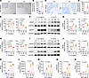
Fractures heal by rapid formation of mineralized callus, a process requiring periosteal cell proliferation and differentiation. Our objective was to dissect the contribution of proliferating osteoblast lineage cells to fracture callus formation. First, mice expressing thymidine kinase (TK) in 3.6Col1a1-lineage cells were treated with ganciclovir (GCV) to ablate proliferating osteolineage cells for 5 or 10 days. Immunostaining demonstrated that this approach specifically depleted TK+ proliferating cells in the bony regions of the callus, while sparing other proliferating cells. Single-cell RNA-seq of callus cells revealed that GCV-treated Col1-TK mice had fewer osteoblasts and chondrocytes than controls, with more myofibroblasts and immune cells, consistent with fibrous nonunion. In controls, 15-30% of callus cells expressing the early osteoblast markers osterix (Sp7) and the late marker osteocalcin (Bglap) were in the cell cycle. Next, we targeted proliferating osteolineage cells at different stages of differentiation by crossing Osx-CreERT2, Ocn-Cre and Dmp1-CreERT2 mice with ROSA-TK mice. Following fracture, each Cre;ROSA-TK mouse line exhibited decreased callus bone volume and a shift from callus bone to fibrous tissue. Therefore, during fracture repair, proliferation of callus cells at early and mature stages of osteoblast differentiation is critical to the formation of a mineralized callus that is essential for healing.
Authors
Nicole R. Gould, Andre F. Coello, Jennifer A. McKenzie, Mariam Obaji, Tiandao Li, Katherine R. Hixon, Leyi Chen, Kristen Barwick, Tiffany Lee, Bo A. Zhang, David Ornitz, Matthew J. Silva
Abstract
Inflammation driven by the innate immune response plays a crucial role in osteoarthritis (OA) pathogenesis, yet the underlying mechanisms remain incompletely understood. Moreover, current antiinflammatory therapies primarily offer symptomatic relief without altering disease progression. Nucleotide-binding oligomerization domain 2 (NOD2) is an intracellular pattern recognition receptor that detects a broad range of microbial and damage-associated stimuli and has been implicated in several inflammatory conditions. In this study, we investigated the role of NOD2 in OA-associated inflammation and cartilage degradation. Elevated NOD2 expression was observed in both human and mouse osteoarthritic cartilage. Conditional KO of Nod2 in chondrocytes suppressed inflammation-induced catabolic responses in vitro and protected against cartilage degradation in mouse OA models. Mechanistically, we identified tumor necrosis factor receptor–associated factor 6 (TRAF6) as a key downstream mediator through which NOD2 promotes chondrocyte catabolism. Furthermore, we showed that pharmacological inhibition of NOD2 using 2 independent small-molecule inhibitors significantly attenuated OA progression in vivo. Collectively, these findings establish NOD2 as a critical regulator of OA-associated inflammation and cartilage degradation, and they highlight its potential as a therapeutic target for disease-modifying OA treatment.
Authors
Yuting Wang, Song Li, Yonghui Dong, Jiaming Zhang, Jian Liu, Zhenggang Wang, Shuang Liang, Nathan R. Martinez, Hongxu Pu, Peng Cheng, Anmin Chen, Qing Yang, Charles K.F. Chan, Wen Jiang, Jun Xiao, Fengjing Guo, Liming Zhao
Abstract
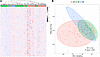
BACKGROUND Elucidating immune signals through well-defined cohorts of pediatric acute pancreatitis (AP) and chronic pancreatitis (CP) patients is critical. This study aimed to evaluate plasma chemokine and cytokine levels in pediatric participants with CP, compared with AP and healthy controls (HCs), to identify unique biomarkers of CP.METHODS Individuals were identified from a prospectively collected pediatric cohort (n = 146). Immunoproteins (n = 247) were measured using the NULISAseq platform on samples from individuals with CP (n = 71), AP (n = 55), and HCs (n = 20).RESULTS The measured analytes showed separation among the 3 groups (R2 = 0.13, P < 0.001). In the CP group, TRANCE, TWEAK, FLT-1, HGF, and TRAIL were increased when compared with HC and AP patients (FDR-corrected P <0.05). A multivariable logistic regression model including all 5 proteins provided an AUC of 0.94 (0.93–0.96) for differentiating samples from CP versus AP or HC samples. In the AP group, CRP, IL-6, CD3E, ENRAGE, and MIF were elevated compared with HCs, while FGF-2, TAFA-5, IL-33, TRANCE, and CXCL12 were downregulated in the same acute time period (FDR-corrected P < 0.05). In the 21 patients with AP for whom follow-up samples were obtained, there was a notable decrease in sequential expression of IL-6 and CRP over 12 months and increased expression of CCL25, TAFA-5, and TRANCE proteins. Additionally, TRANCE was expressed on CP pancreatic tissue.CONCLUSIONS TRANCE was increased in pediatric patients with CP and decreased in those with AP during a flare. Future studies are needed to investigate the role of TRANCE and other analytes in the pathogenesis of CP.
Authors
Peter R. Farrell, Bomi Lee, Faizan Ahmed, Maria E. Moreno-Fernandez, Ajay Dixit, Juan Pablo Gurria, Nicholas J. Ollberding, Qing Duan, Vineet Garlapally, Phoebe Christian, Sohail Z. Husain, Maisam Abu-El-Haija

Abstract
Methyltransferase-like 5 (METTL5) is a methyltransferase responsible for rRNA N6-methyladenosine (m6A) modification, mutations in which are associated with skeletal abnormalities and cognitive deficits. Despite METTL5’s clinical relevance, the molecular mechanisms underlying METTL5-related genetic disorders remain poorly understood. In this study, we demonstrated that Mettl5 KO led to reduced bone mass and smaller body size in mice and impaired the osteogenic differentiation of mesenchymal stem cells. Mechanistically, Mettl5 deficiency decreased the translation efficiency of oxidative stress–responsive serine-rich protein 1 mRNA, downregulated the expression of key antioxidant genes, and diminished antioxidant capacity. Importantly, administration of the antioxidant N-acetylcysteine (NAC) partially rescued skeletal defects in Mettl5-KO mice. These findings reveal a critical role for METTL5 in antioxidant defense and suggest that NAC supplementation may represent a promising therapeutic strategy for METTL5-related disorders.
Authors
Kexin Lei, Qi Yin, Qiwen Li, Qian Wang, Zhong Zhang, Fei Xue, Ruoshi Xu, Xinyi Zhou, Lin Peng, Shoichiro Kokabu, Shuibin Lin, Quan Yuan
Abstract
Fibroblast-like synoviocytes (FLSs) are crucial in driving synovial inflammation and joint damage in rheumatoid arthritis (RA). This study explored the functions and underlying mechanisms of GALNT1-mediated O-glycosylation, which is markedly upregulated in RA FLSs, in synovial aggression and subsequent experimental joint damage. Targeted suppression of GALNT1 effectively curtailed migration and invasion in RA FLSs and mitigated arthritis severity in a rat collagen-induced arthritis (CIA) model. Mechanistically, NEK9 was identified as a pivotal substrate and downstream effector of GALNT1, affecting the aggressive phenotype of RA FLSs. In vitro experiments further demonstrated that O-glycosylation of NEK9, mediated by GALNT1, promotes the pathogenic phenotype of RA FLSs by promoting cytoskeleton reorganization and restraining excessive endoplasmic reticulum (ER) stress activation. Our study provides mechanistic insights into the activation of RA FLSs and identifies GALNT1 as a potential therapeutic target for RA.
Authors
Yaoyao Zou, Haobo Lin, Jianling Su, Jieying Wang, Qin Zeng, Tianxiao Feng, Yunxia Lei, Jianda Ma, Hudan Pan, Hanshi Xu, Lie Dai, Yang Li

Abstract
Temporomandibular joint osteoarthritis (TMJOA), a prevalent subtype of temporomandibular disorders, is characterized by progressive cartilage degradation and subchondral bone destruction. Despite advancements in understanding TMJOA pathogenesis, the molecular mechanisms underlying its progression remain unclear. In this study, elevated Slit guidance ligand 2 (SLIT2) expression was observed in TMJ tissues of unilateral anterior crossbite–induced TMJOA mice and synovial fluid from patients with TMJOA, correlating with disease severity. Furthermore, SLIT2 overexpression in transgenic mice intensified TMJOA progression, whereas heterozygous deletion of roundabout guidance receptor 1/2 (ROBO1/2) alleviated cartilage and bone damage. Mechanistically, SLIT2 promoted ROBO1-LRP6 complex formation, facilitating LRP6 phosphorylation and β-catenin nuclear translocation. This cascade upregulated matrix-degrading enzymes while downregulating cartilage structural proteins, exacerbating cartilage destruction and subchondral bone loss. These findings suggest that the SLIT2/ROBO1/LRP6 axis may represent a potential therapeutic target for TMJOA and provide mechanistic insights into disease progression.
Authors
Guan Luo, Baoyi Chen, Wenjun Chen, Huiyi Lin, Weiqi Guo, Qingbin Zhang, Jiang Li, Lijing Wang, Janak Lal Pathak, Yuhui Yang, Weijun Zhang, Xiaoyu Zhang, Beining Zheng, Ziyi Wang, Shiting Wei, Jiaxin He, Wei-Jie Zhou, Chang Liu
Abstract
Adaptive remodeling of retrodiscal tissue following anterior disc displacement (ADD) of the temporomandibular joint (TMJ) has been recognized for decades, yet the underlying cellular dynamics and molecular mechanisms remain unclear. Using a porcine ADD model, this study investigated the cellular and molecular basis driving retrodiscal tissue adaptation. Histological staining revealed adaptive remodeling of retrodiscal tissue after ADD induction, with dense connective tissue and cartilaginous masses replacing loose connective tissue. Furthermore, single-cell RNA sequencing (scRNA-seq) captured pronounced fibroblast expansion during tissue remodeling, notably the FB2 subcluster with high developmental potential, and the emergence of a mural cell subcluster MC4 associated with extracellular matrix (ECM) remodeling. CellChat analysis highlighted MC4-FB2 crosstalk via FGF2 and BMP5 signaling. The combination of pathway-aware multi-layered hierarchical network (P-NET) and Seurat with drug database screening identified five promising compounds. Among them, Zaprinast demonstrated the most robust effects by enhancing the remodeling capability of fibroblasts in vitro, and also alleviated TMJ deformation in vivo. Collectively, fibroblast activation is pivotal for early retrodiscal tissue adaptation following ADD, which is driven by MC4-derived FGF2/BMP5 signaling. Zaprinast treatment potentiates this remodeling process. These findings provide new insights into cellular basis of TMJ adaptation and identify potential therapeutic targets for ADD management.
Authors
Wenlin Yuan, Yilin Chen, Ruojin Yan, Wei Liu, Chenyu Wang, Ying Wang, Qiaoli Dai, Wen Li, Mengqi Zhu, Xiao Chen, Jiejun Shi
Abstract
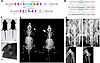
The TRPV4 skeletal dysplasias are characterized by short stature, short limbs with prominent large joints, and progressive scoliosis. They result from dominant missense mutations that activate the TRPV4 calcium permeable ion channel. As a platform to understand the mechanism of disease and to test the hypothesis that channel inhibition could treat these disorders, we developed a knock-in mouse that conditionally expresses the p.R594H Trpv4 mutation. Embryonic, chondrocyte-specific induction of the mutation using Col2a1-Cre resulted in a skeletal dysplasia affecting the long bones, spine, and craniofacial skeletal elements, consistent with the human skeletal dysplasia phenotypes produced by TRPV4 mutations. Cartilage growth plate histological abnormalities included disorganized proliferating chondrocyte columns and reduced hypertrophic chondrocyte development, reflecting abnormal endochondral ossification. In vivo treatment with the TRPV4-specific inhibitor GSK2798745 markedly improved the radiographic skeletal phenotype and rescued the growth plate histological abnormalities. ScRNA-Seq of chondrocyte transcripts from affected mice identified calcium-mediated effects on multiple signaling pathways as potential mechanisms underlying the defects in linear and cartilage appositional growth observed in both mutant mice and patients. These results provide preclinical evidence demonstrating TRPV4 inhibition as a rational, mechanism-based therapeutic strategy to ameliorate disease progression and severity in the TRPV4 skeletal dysplasias.
Authors
Lisette Nevarez, Taylor K. Ismaili, Jennifer Zieba, Jorge Martin, Davis Wachtell, Derick Diaz, Jocelyn A. Ramirez, Valeria Aceves, Joshua Ito, Ryan S. Gray, David Goldstein, Sunil Sahdeo, Deborah Krakow, Daniel H. Cohn
No posts were found with this tag.


