
Research ArticleImmunologyPulmonology
Open Access |  10.1172/jci.insight.189205
Cyclooxygenase-2 negatively regulates innate lymphoid cell type 2 differentiation and function during allergic lung inflammation
Hong Li, Matthew L. Edin, Daniel Menendez, J. Alyce Bradbury, Joan P. Graves, Artiom Gruzdev, Gregory S. Whitehead, Maria I. Sifre, Laura M. DeGraff, and Darryl C. Zeldin
Division of Intramural Research, National Institute of Environmental Health Sciences, National Institutes of Health, Research Triangle Park, North Carolina, USA.
Address for correspondence. Darryl C. Zeldin, Division of Intramural Research, NIH/NIEHS, 111 T.W. Alexander Drive, Building 101, Room A214, Research Triangle Park, North Carolina, 27709, USA. Phone: 984.287.3641; Email: zeldin@niehs.nih.gov.
Find articles by Li, H. in: PubMed | Google Scholar
Division of Intramural Research, National Institute of Environmental Health Sciences, National Institutes of Health, Research Triangle Park, North Carolina, USA.
Address for correspondence. Darryl C. Zeldin, Division of Intramural Research, NIH/NIEHS, 111 T.W. Alexander Drive, Building 101, Room A214, Research Triangle Park, North Carolina, 27709, USA. Phone: 984.287.3641; Email: zeldin@niehs.nih.gov.
Find articles by
Edin, M.
in:
PubMed
|
Google Scholar
|

Division of Intramural Research, National Institute of Environmental Health Sciences, National Institutes of Health, Research Triangle Park, North Carolina, USA.
Address for correspondence. Darryl C. Zeldin, Division of Intramural Research, NIH/NIEHS, 111 T.W. Alexander Drive, Building 101, Room A214, Research Triangle Park, North Carolina, 27709, USA. Phone: 984.287.3641; Email: zeldin@niehs.nih.gov.
Find articles by Menendez, D. in: PubMed | Google Scholar
Division of Intramural Research, National Institute of Environmental Health Sciences, National Institutes of Health, Research Triangle Park, North Carolina, USA.
Address for correspondence. Darryl C. Zeldin, Division of Intramural Research, NIH/NIEHS, 111 T.W. Alexander Drive, Building 101, Room A214, Research Triangle Park, North Carolina, 27709, USA. Phone: 984.287.3641; Email: zeldin@niehs.nih.gov.
Find articles by Bradbury, J. in: PubMed | Google Scholar
Division of Intramural Research, National Institute of Environmental Health Sciences, National Institutes of Health, Research Triangle Park, North Carolina, USA.
Address for correspondence. Darryl C. Zeldin, Division of Intramural Research, NIH/NIEHS, 111 T.W. Alexander Drive, Building 101, Room A214, Research Triangle Park, North Carolina, 27709, USA. Phone: 984.287.3641; Email: zeldin@niehs.nih.gov.
Find articles by Graves, J. in: PubMed | Google Scholar
Division of Intramural Research, National Institute of Environmental Health Sciences, National Institutes of Health, Research Triangle Park, North Carolina, USA.
Address for correspondence. Darryl C. Zeldin, Division of Intramural Research, NIH/NIEHS, 111 T.W. Alexander Drive, Building 101, Room A214, Research Triangle Park, North Carolina, 27709, USA. Phone: 984.287.3641; Email: zeldin@niehs.nih.gov.
Find articles by Gruzdev, A. in: PubMed | Google Scholar
Division of Intramural Research, National Institute of Environmental Health Sciences, National Institutes of Health, Research Triangle Park, North Carolina, USA.
Address for correspondence. Darryl C. Zeldin, Division of Intramural Research, NIH/NIEHS, 111 T.W. Alexander Drive, Building 101, Room A214, Research Triangle Park, North Carolina, 27709, USA. Phone: 984.287.3641; Email: zeldin@niehs.nih.gov.
Find articles by
Whitehead, G.
in:
PubMed
|
Google Scholar
|
Division of Intramural Research, National Institute of Environmental Health Sciences, National Institutes of Health, Research Triangle Park, North Carolina, USA.
Address for correspondence. Darryl C. Zeldin, Division of Intramural Research, NIH/NIEHS, 111 T.W. Alexander Drive, Building 101, Room A214, Research Triangle Park, North Carolina, 27709, USA. Phone: 984.287.3641; Email: zeldin@niehs.nih.gov.
Find articles by Sifre, M. in: PubMed | Google Scholar
Division of Intramural Research, National Institute of Environmental Health Sciences, National Institutes of Health, Research Triangle Park, North Carolina, USA.
Address for correspondence. Darryl C. Zeldin, Division of Intramural Research, NIH/NIEHS, 111 T.W. Alexander Drive, Building 101, Room A214, Research Triangle Park, North Carolina, 27709, USA. Phone: 984.287.3641; Email: zeldin@niehs.nih.gov.
Find articles by DeGraff, L. in: PubMed | Google Scholar
Division of Intramural Research, National Institute of Environmental Health Sciences, National Institutes of Health, Research Triangle Park, North Carolina, USA.
Address for correspondence. Darryl C. Zeldin, Division of Intramural Research, NIH/NIEHS, 111 T.W. Alexander Drive, Building 101, Room A214, Research Triangle Park, North Carolina, 27709, USA. Phone: 984.287.3641; Email: zeldin@niehs.nih.gov.
Find articles by Zeldin, D. in: PubMed | Google Scholar
Published September 22, 2026 - More info
JCI Insight. 2026;11(21):e189205. https://doi.org/10.1172/jci.insight.189205.
© 2026 Li et al. This work is licensed under the Creative Commons Attribution 4.0 International License. To view a copy of this license, visit http://creativecommons.org/licenses/by/4.0/.
Received: November 14, 2024; Accepted: September 15, 2026


-
Results
COX-2–/– mice have increased lung ILC2 cells during OVA-induced allergic inflammation in vivo. To investigate the role of COX-2 on lung ILC2 cells in vivo, we first compared lung ILC2 cell numbers after OVA-induced allergic lung inflammation between COX-2+/+ and COX-2–/– mice. Mice were sensitized with OVA (20 μg/mouse/day) in the presence of adjuvant (Alum) on days 0 and 1. After 14 days, mice were exposed to vehicle or 1% OVA/PBS via inhalation for 5 consecutive days (30 min per day). Twenty-four hours after the last exposure, mouse lungs, BALF, blood, and lymph nodes were collected and analyzed (Figure 1A). ILC2 cells (defined as CD45+Lin– CD90.2+CD161–RORγt–CD11b–) in the lung, BALF, blood, and lymph nodes were quantified as a percentage of CD45+Lin– cells as previously described (Supplemental Figure 1A; supplemental material available online with this article; https://doi.org/10.1172/jci.insight.189205DS1) (33). Compared with vehicle, OVA sensitization/exposure significantly increased lung ILC2 cells (Figure 1B), as well as the percentage of ILC2 cells expressing the cytokines IL-5 and IL-13 and the transcription factor GATA3 (Figure 1D and Supplemental Figure 2A). Importantly, ILC2 cells were significantly increased in COX-2–/– lungs (9.3% ± 0.7%) compared with COX-2+/+ lungs (4.7% ± 0.4%) after OVA challenge (Figures 1, B and C). ILC2 cells from lungs of COX-2–/– mice also had a significantly higher percentage of cells expressing IL-5, IL-13, and GATA3 than ILC2 cells from lungs of COX-2+/+ mice (Figure 1D and Supplemental Figure 2B). ILC2 cells in BALF, blood, and lymph nodes were not significantly different between COX-2+/+ and COX-2–/– mice after OVA exposure (Supplemental Figure 1B). Together, these results indicate that disruption of COX-2 results in increased lung ILC2 cells with increased IL-5, IL-13, and GATA3 expression during OVA-induced allergic inflammation in vivo.
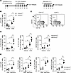 Figure 1
COX-2–/– lungs have increased ILC2 cells during allergic inflammation induced by OVA and Alternaria in vivo. (A) Schematic of the in vivo allergic lung inflammation models used. COX-2+/+ and COX-2–/– mice were sensitized to OVA in the presence of adjuvant (Alum) via i.p. injection (Days 0 and 1) and then exposed to 1% OVA via aerosol for 5 consecutive days (Day 14 to 18). Alternatively, mice were exposed to Alternaria extract by i.n. injection (20 μg/mouse/day) for 4 days. Mice were sacrificed 24 hours after the last exposure and lungs were collected for analysis. (B) ILC2 cells as a percentage of CD45+Lin– cells were quantified by FACS in lungs from vehicle- and OVA-treated COX-2+/+ and COX-2–/– mice (n = 5 per group, *P < 0.05). (C) Representative FACS plots of OVA-treated COX-2+/+ and COX-2–/– lungs showing ILC2 cells (GATA3+ CD11b–). (D) IL-5+, IL-13+, and GATA3+ ILC2 cells as a percentage of CD90+KLRG1+ cells in lungs from OVA-treated COX-2+/+ and COX-2–/– mice were quantified by FACS analysis. (E) After PBS or Alternaria exposure, ILC2 cells as a percentage of CD45+Lin– cells, or IL-5+, IL-13+, and GATA3+ ILC2 cells as a percentage of CD90+KLRG1+ cells in lungs from COX-2+/+ and COX-2–/– mice were quantified by FACS analysis. n = 3–5/group, *P < 0.05 by 2-way ANOVA.
COX-2–/– mice have increased lung ILC2 cells during Alternaria-induced allergic inflammation in vivo. COX-2 disruption also resulted in increased ILC2 cells in response to Alternaria. COX-2+/+ and COX-2–/– mice were exposed to Alternaria extract (20 μg/mouse/day i.n.) for 4 days (Figure 1A) similar to previous experiments (34). Twenty-four hours after the last exposure, ILC2 cells from mouse lungs, BALF, blood, and lymph nodes were analyzed. Compared with vehicle, Alternaria exposure increased lung ILC2 cells, and the percentage of ILC2 cells that express IL-5, IL-13, and GATA3 (Figure 1E). Importantly, COX-2–/– lungs had increased ILC2 cells and increased ILC2 cells expressing IL-5, IL-13, and GATA3 compared with COX-2+/+ lungs after Alternaria exposure (Figure 1E). ILC2 cells in BALF, blood, and lymph nodes were also higher in COX-2–/– mice compared with COX-2+/+ mice after Alternaria (Supplemental Figure 1C). Together, these results indicate that COX-2 disruption results in increased ILC2 cells with increased expression of IL-5, IL-13, and GATA3 during Alternaria-induced allergic inflammation in vivo.
COX-2 disruption increases ILC2 cell differentiation from CLP cells in vitro. To examine the role of COX-2 in ILC2 cell differentiation in vitro, we isolated CD127+CD135+Sca-1 CLP cells from bone marrows (Supplemental Figure 3A) of COX-2+/+ and COX-2–/– mice. These CLP cells were then differentiated to ILC2 cells via coculture with OP9-DL1 feeder cells (35, 36) over 10 days and stimulation with SCF, IL-7, and IL-33 (37, 38) as depicted in Supplemental Figure 3B. Treatment with SCF, IL-7, and IL-33 increased the level of COX-2 (Ptgs2) mRNA by ~50% relative to vehicle (Figure 2A). Importantly, a significantly higher percentage of CLP cells from COX-2–/– mice differentiated to ILC2 cells than CLP cells from COX-2+/+ mice (1.350 ± 0.13% versus 0.6 ± 0.12%), respectively; P < 0.05) (Figure 2B). Moreover, a higher percentage of COX-2–/– CLP cells differentiated with SCF, IL-7, and IL-33 expressed IL-5, IL-13, and GATA3 compared with COX-2+/+ CLP cells (Figure 2C). In agreement with these observations, CLP cells treated with the selective COX-2 inhibitor NS-398 expressed higher levels of IL-5, IL-13, and GATA3 after SCF, IL-7, and IL-33 treatment (Figure 2D). Together, these data suggest that COX-2 is induced after SCF, IL-7, and IL-33 treatment and that COX-2 disruption increases ILC2 cell differentiation from CLP cells in vitro.
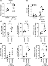 Figure 2
COX-2 disruption increases ILC2 cell differentiation from bone marrow–derived CLP cells in vitro. CLP cells were isolated from bone marrows of COX-2+/+ and COX-2–/– mice and treated with either vehicle or SCF + IL-7 + IL-33 in vitro to induce ILC2 cell differentiation. (A) COX-2 (Ptgs2) mRNA levels measured by qPCR in CLP cells from COX-2+/+ mice treated with either vehicle or SCF + IL-7 + IL-33 in vitro. (B) FACS analysis of GATA3+ cells from COX-2+/+ and COX-2-/- CLP cells treated with either vehicle or SCF + IL-7 + IL-33 in vitro. (C) IL-5+, IL-13+, and GATA3+ ILC2 cells as a percentage of CD90+KLRG1+ cells derived from COX-2+/+ and COX-2–/– CLP cells. (D) IL-5+, IL-13+, and GATA3+ ILC2 cells as a percentage of CD90+KLRG1+ cells derived from COX-2+/+ CLP cells differentiated in the presence of vehicle or NS398. n = 3–5 per group, *P < 0.05 by t test (A and C) or 1-way ANOVA (B and D).
COX-2 products inhibit ILC2 cell differentiation from bone marrow–derived CLP cells in vitro. We hypothesized that COX-2 negatively regulates ILC2 cell differentiation by generating prostanoids, which act through their cognate receptors (39). To define which prostanoids are able to regulate ILC2 cell differentiation of CLP cells, we first examined the expression of the prostanoid receptors during ILC2 differentiation in vitro. SCF, IL-7, and IL-33 treatment of CLP cells significantly induced expression of EP2 (Ptger2), EP3 (Ptger3), IP (Ptgir), and TP (Tbxa2r) receptors (Supplemental Figure 4A). The increases in expression of EP2, EP3, IP, and TP receptors were confirmed by surface staining and FACS analysis (Supplemental Figure 4B). EP1 (Ptger1), EP4 (Ptger4), and FP (Ptgfr) receptors were expressed but were either decreased or unchanged during ILC2 cell differentiation. DP (Ptgdr) and DP2/CRTH2 (Ptgdr2) receptors were expressed at or below the limit of detection (Supplemental Figure 4A). This suggests that PGE2-EP1-4, PGF2α-FP, PGI2-IP, and/or TXA2-TP signaling might regulate CLP cell differentiation to ILC2 cells.
To assess which prostanoids regulate ILC2 cell differentiation, COX-2+/+ CLP cells were differentiated to ILC2 cells in vitro with SCF, IL-7, and IL-33 in the presence or absence of vehicle (ethanol) or 5–1,000 nM PGE2, PGD2, PGI2, PGF2α, or cTXA2. While high concentrations of several prostanoids reduced ILC2 cell differentiation, only PGE2 suppressed IL-5, IL-13, and GATA3 protein expression and Il5, Il13, and Gata3 mRNA levels at biologically relevant concentrations as low as 50 nM (Figure 3A and Supplemental Figures 5 and 6). These data suggest that PGE2 is likely the most relevant COX-2–derived prostanoid, which inhibits ILC2 cell differentiation. Therefore, we focused on the role of PGE2-EP receptor signaling in regulating ILC2 cell differentiation.
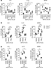 Figure 3
Effect of prostanoids and receptor modulators on ILC2 cell differentiation in vitro. CLP cells were isolated from bone marrows of COX-2+/+ mice and treated with either vehicle or SCF + IL-7 + IL-33 in vitro to induce ILC2 cell differentiation. (A) The percentage of IL-5+, IL-13+, and GATA3+ ILC2 cells was quantified by FACS in COX-2+/+ CLP cells differentiated in vitro in the presence of vehicle or 100 nM of PGD2, PGI2, PGF2α, PGE2, or TXA2. (B) The percentage of IL-5+, IL-13+, and GATA3+ ILC2 cells was quantified by FACS in COX-2+/+ or COX-2–/– CLP cells differentiated in vitro in the presence of vehicle or 100 nM of PGE2. (C) The percentage of IL-5+, IL-13+, and GATA3+ ILC2 cells was quantified by FACS in COX-2+/+ CLP cells differentiated in vitro in the presence of vehicle, 100 nM PF-04418948 (EP2 antagonist), or 100 nM MF498 (EP4 antagonist). n = 3–5 per group, *P < 0.05 by 1-way ANOVA (A and C) or multiple t tests with Holm Šídák correction (B).
PGE2-EP2 signaling is critical in regulating lung ILC2 cell differentiation in vitro and in vivo. To further examine the role of PGE2 in suppression of ILC2 cell differentiation, we differentiated CLP cells isolated from COX-2+/+ and COX-2–/– mice to ILC2 cells in the presence or absence of PGE2 in vitro. PGE2 suppressed differentiation of COX-2+/+ CLP cells to ILC2 cells as determined by the percentage of ILC2 cells expressing IL-5, IL-13, or GATA3 protein (Figure 3B). PGE2 also suppressed the enhanced differentiation of COX-2–/– CLP cells to ILC2 cells (Figure 3B). To examine the role of PGE2-EP2 receptor signaling in ILC2 cell differentiation, we treated COX-2+/+ CLP cells with the selective EP2 agonist butaprost. The EP2 agonist significantly inhibited ILC2 cell differentiation (Supplemental Figure 7). Conversely, treatment with an EP2 antagonist (PF-04418948) increased ILC2 cell differentiation (Figure 3C). In contrast, the EP4 antagonist MF498 suppressed ILC2 cell differentiation (Figure 3C). These data suggest that PGE2 inhibits ILC2 cell differentiation primarily through EP2 receptor activation in vitro.
To examine the role of PGE2 in regulating lung ILC2 cell differentiation in vivo, PGE2 was delivered by osmotic minipumps during OVA-induced allergic lung inflammation (Figure 4A). While vehicle-treated COX-2–/– mouse lungs had increased ILC2 cells compared with vehicle-treated COX-2+/+ mouse lungs, PGE2 treatment decreased COX-2–/– mouse lung ILC2 cells to levels that were similar to those observed in COX-2+/+ mice (Figure 4B). Consistent with these data, the increased expression of Il5 and Il13 mRNAs in COX-2–/– lungs relative to COX-2+/+ lungs were also attenuated by administration of PGE2 (Figure 4C). These data suggest that the lung ILC2 cell phenotype during OVA-induced allergic lung inflammation in COX-2–/– mice can be rescued by PGE2 treatment in vivo.
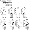 Figure 4
PGE2, EP2 agonist, EP2 antagonist, and COX-2 inhibitor regulate ILC2 cells during allergic lung inflammation in vivo. (A) COX-2+/+ and COX-2–/– mice were treated with either vehicle, PGE2, butaprost, PF-04418948, or CAY10404 via Alzet minipump during OVA-induced allergic lung inflammation. (B) ILC2 cells were quantified by FACS in lungs from allergic COX-2+/+ and COX-2–/– mice treated with vehicle and COX-2–/– mice treated with PGE2. n = 5–7 per group, *P < 0.05 by ANOVA. (C) Il5, Il13, and Il33 mRNA levels were quantified by qPCR in lungs of allergic COX-2+/+ and COX-2–/– mice treated with vehicle and allergic COX-2–/– mice treated with PGE2. (D and E) ILC2 cells (D) and IL-5+, IL-13+, and GATA3+ ILC2 cells (E) in lungs of COX-2+/+ mice treated with butaprost (EP2 agonist), PF-04418948 (EP2 antagonist), or CAY10404 (COX-2 inhibitor) during OVA-induced allergic lung inflammation. n = 6–9 per group, *P < 0.05 by 1-way ANOVA.
We next tested the effects of the selective EP2 receptor agonist (butaprost), the selective EP2 receptor antagonist (PF-04418948), and the selective COX-2 inhibitor (CAY10404) on regulation of lung ILC2 cells during OVA-induced allergic lung inflammation in vivo. The EP2 receptor agonist significantly decreased lung ILC2 cells after OVA exposure (Figure 4D) and tended to reduce IL-5+ and IL-13+ ILC2 cells. Conversely, administration of either the EP2 receptor antagonist or the COX-2 inhibitor significantly increased lung ILC2 cells and increased IL-5+, IL-13+, and GATA3+ ILC2 cells (Figure 4, D and E).
To confirm the function of the EP2 receptor in the regulation of ILC2 cells in vivo, we induced allergic lung inflammation in EP2+/+ and EP2–/– mice. After OVA treatment, lungs from allergic EP2–/– mice had significantly increased ILC2 cells compared with lungs from allergic EP2+/+ mice (12.8 ± 4.2 versus 5.4 ± 2.3, n = 6; P < 0.05) (Figure 5A). In addition, lungs from OVA-treated EP2–/– mice had increased IL-5+, IL-13+, and GATA3+ ILC2 cells than lungs from OVA-treated EP2+/+ mice (Figure 5B). Similarly, lungs from Alternaria-treated EP2–/– mice had significantly increased ILC2 cells compared with lungs from Alternaria-treated EP2+/+ mice (12.4 ± 2.2 versus 4.5 ± 1.8, n = 3–5; P < 0.05) (Figure 5C). Lungs from Alternaria-treated EP2–/– mice also had increased IL-5+, IL-13+, and GATA3+ ILC2 cells than lungs from Alternaria-treated EP2+/+ mice (Figure 5C). Furthermore, in vitro differentiation of EP2–/– bone marrow–derived CLP cells to ILC2 cells was significantly greater than differentiation of EP2+/+ bone marrow–derived CLP cells as measured by increased expression of Il5, Il13, and Gata3 mRNAs (Figure 5D). Together these data indicate that COX-2–derived PGE2 inhibits lung ILC2 cells via signaling through the EP2 receptor in vitro and in vivo.
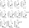 Figure 5
EP2–/– mice have increased ILC2 cells during allergic lung inflammation in vivo and in vitro. (A) Lungs from OVA-treated EP2–/– mice have increased ILC2 cells compared with lungs from OVA-treated EP2+/+ mice as determined by FACS. n = 6 per group, *P < 0.05. (B) IL-5+, IL-13+, and GATA3+ ILC2 cells were quantified by FACS in lungs from OVA-treated EP2+/+ and EP2–/– mice. n = 6 per group, *P < 0.05. (C) Lungs from Alternaria-treated EP2–/– mice have increased ILC2 cells compared with lungs from Alternaria-treated EP2+/+ mice as determined by FACS. n = 3–5 per group, *P < 0.05. (D) After in vitro differentiation with SCF + IL-7 + IL-33, bone marrow-derived CLP cells from EP2–/– mice have increased Il5, Il13, and Gata3 mRNA levels compared with bone marrow–derived CLP cells from EP2+/+ mice. n = 3 per group, *P < 0.05 by t test (A and B) or 2-way ANOVA (C and D).
Allergic COX-2–/– mice have altered lung ILC2 cell gene expression. To examine whether COX-2 disruption alters ILC2 cell gene expression during allergic lung inflammation, we performed single-cell RNA-seq on isolated lung cells from COX-2+/+ and COX-2–/– mice exposed to OVA as described in Figure 1A. Uniform manifold approximation and projection (UMAP) dimensional reduction of 6,955 lymphoid T cells identified 18 distinct cell clusters (Figure 6A). These clusters, including the ILC2 cell cluster (red, #8), were identified by characteristic lineage specific markers (Supplemental Figure 8A). After OVA exposure, the number of ILC2 cells in COX-2–/– mice was increased relative to COX-2+/+ mice (Figure 6B and Supplemental Figure 8B). OVA exposure caused changes in expression of 323 genes in ILC2 cells from COX-2+/+ mice (155 genes downregulated, 168 genes upregulated), including induction of IL-2, Th2, and Th17 signaling pathways (Supplemental Figure 8C, Supplemental Figure 9A, and Supplemental Table 2). By comparison, OVA exposure caused changes in expression of 1,386 genes in ILC2 cells from COX-2–/– mice (612 genes downregulated, 774 genes upregulated), including induction of T cell migration and cytokine stimulus pathways (Supplemental Figure 8C and 9B). Relative to ILC2 cells from allergic COX-2+/+ mice, ILC2 cells from allergic COX-2–/– mice had differential expression of 260 genes (118 genes downregulated, 142 genes upregulated) (Figure 6C). Pathway analysis suggested that ILC2 cells from allergic COX-2–/– mice had upregulation of multiple pathways involved in leukocyte migration and proliferation (Figure 6D and Supplemental Table 2). Significantly increased expression of Ccl5 (RANTES) in COX-2–/– ILC2 cells is a key component of many of these pathways. ILC2 cells from allergic COX-2–/– mice had downregulation of several pathways involved in apoptosis, telomere maintenance, and both lipid and protein phosphorylation (Figure 6D and Supplemental Table 2). Together, these data suggest that COX-2 disruption, in addition to increasing the number of ILC2 cells in allergic lungs, also alters the ability of ILC2 cells to proliferate, migrate, and produce or respond to cytokine signals.
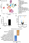 Figure 6
COX-2 disruption alters ILC2 gene expression profiles during allergic lung inflammation. Lung cells were isolated from vehicle and OVA-exposed COX-2+/+ and COX-2–/– mice as shown in Figure 1A. Cells from 3 mice per treatment group were pooled and single-cell RNA-seq was performed. (A) Uniform manifold approximation and projection (UMAP) of 6,955 lymphoid cells identified 18 cell clusters, including ILC2 cells (red, #8). (B) The percentage of lymphoid cells identified as ILC2 cells in allergic COX-2+/+ and COX-2–/– mice is shown. (C) Volcano plot (–Log10 [P value] versus Log2 FC) indicates genes differentially expressed between OVA-exposed COX-2+/+ and COX-2–/– ILC2 cells. (D) Normalized enrichment scores (NES) from GSEA indicate the top 5 pathways significantly increased (orange) or decreased (blue) in OVA-exposed COX-2–/– ILC2 cells versus OVA-exposed COX-2+/+ ILC2 cells.
COX-2–derived PGE2 inhibits lung epithelial cytokine expression in vitro and in vivo. Activated epithelial cells produce IL-33, TSLP, and IL-25 to induce lung ILC2 cell differentiation during OVA-induced allergic lung inflammation (40). While vehicle-treated COX-2–/– mouse lungs had increased Il33 mRNA compared with vehicle-treated COX-2+/+ mouse lungs, PGE2 treatment decreased COX-2–/– mouse lung Il33 to levels that were similar to those observed in COX-2+/+ mice (Figure 4C). To further investigate whether COX-2–derived PGE2 regulates these epithelial cytokines in the allergic lung, we examined IL-33, TSLP, and IL-25 expression in frozen lung tissue sections after OVA sensitization/exposure by confocal microscopy using immunofluorescence conjugated IL-33 and TSLP antibodies (Figure 7A and Supplemental Figure 11). Intensity quantification of the resulting confocal images indicates that COX-2–/– lung epithelial cells have significantly higher IL-33 and TSLP expression compared with COX-2+/+ lung epithelial cells (Figure 7A). We consistently observed increased Il33 and/or Tslp mRNA levels in OVA-treated COX-2–/– lungs compared with OVA-treated COX-2+/+ lungs (Figure 4C and Figure 7B). Similarly, Alternaria exposure increased Il33 and Tslp mRNA levels in COX-2+/+ lungs. This induction was further enhanced in Alternaria-treated COX-2–/– lungs (Figure 7C). In most of our experiments, Il25 mRNA levels were either unchanged or at the limit of detection (not shown). Together, these data suggest that COX-2 negatively regulates lung epithelial cytokines during OVA- and Alternaria-induced allergic lung inflammation.
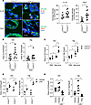 Figure 7
COX-2 suppresses airway epithelial IL-33 and TSLP expression in vivo and in vitro. (A) After OVA-induced allergic inflammation, lung epithelial IL-33 and TSLP were visualized by confocal microscopy using anti-mouse antibodies to EpCAM (green), IL-33 or TSLP (red), and DAPI (blue), and the average pixel intensity in Epcam+ cells was quantified. Epithelial expression of both IL-33 and TSLP are increased in COX-2–/– mice relative to COX-2+/+ mice. (B and C) Levels of Il33 and Tslp mRNAs in lungs from OVA-treated (B), and PBS or Alternaria-treated (C), COX-2+/+ and COX-2–/– mice were measured by qPCR. (D) Purified COX-2–/– airway epithelial cells have increased levels of Il33 and Tslp after LPS treatment. In contrast, levels of Il25 remain unchanged. (E) PGE2 significantly inhibits Il33 and Tslp expression in purified airway epithelial cells from COX-2+/+ mice treated with LPS. n = 3–12 per group, *P < 0.05 by t test (A and B), multiple t tests (C and D) or 1-way ANOVA (E).
To quantify the effect of COX-2 on epithelial cytokine production, we purified airway epithelial cells from COX-2+/+ and COX-2–/– lungs and examined epithelial cytokine production after a 4-hour treatment with bacterial LPS in vitro. LPS treatment increased Il33 and Tslp mRNA levels in both COX-2+/+ and COX-2–/– epithelial cells; however, COX-2–/– epithelial cells had a significantly greater increase in Il33 and Tslp expression than COX-2+/+ epithelial cells (Figure 7D). Importantly, treatment of purified airway epithelial cells with PGE2 attenuated LPS-induced Il33 and Tslp expression (Figure 7E). We also examined prostanoid receptor expression in lung epithelial cells after LPS treatment. While all receptors were detectable, LPS significantly induced lung epithelial expression of the IP (Ptgir) and EP2 (Ptger2) receptors (Supplemental Figure 10). Taken together, these data suggest that COX-2–derived PGE2 can also inhibit expression of the lung epithelial cytokines (IL-33 and TSLP), which promote ILC2 cell differentiation.
IL-33 increases lung ILC2 cells in both COX-2+/+ and COX-2–/– mice in vivo. COX-2–derived PGE2 downregulates both the epithelial production of ILC2-inducing IL-33 and TSLP, and the ability of CLP cells to differentiate to ILC2 cells in response to those epithelial cytokines. We sought to determine whether exogenous IL-33 would restore the ILC2 cell phenotype in COX-2–/– lungs in vivo. We gave IL-33 intranasally to COX-2+/+ and COX-2–/– mice for 5 consecutive days and analyzed ILC2 cell numbers and function 24 hours after the fifth dose (Figure 8A). Both COX-2+/+ and COX-2–/– lungs exhibit very low ILC2 cell numbers with vehicle treatment (1.03 ± 0.50 versus 0.87 ± 0.25, n = 3). In contrast, IL-33 treatment significantly increased lung ILC2 cells in both COX-2+/+ and COX-2–/– lungs to a comparable degree (3.2 ± 1.1 versus 3.3 ± 0.6, n = 6) (Figure 8B). IL-33 also induced comparable amounts of Il5 and Il13 mRNAs in COX-2+/+ and COX-2–/– mice (Figure 8C). One interpretation is that exogenous IL-33 normalized the ILC2 cell phenotype in COX-2–/– mice, which would suggest a major effect of COX-2 disruption was to increase epithelial cytokines during lung inflammation. An alternative explanation may be that the exogenous IL-33 instillation overwhelmed the ILC2 cell differentiation or recruitment such that the resulting ILC2 cell responses were no longer modulable by changes in PG signaling.
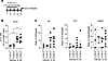 Figure 8
COX-2+/+ and COX-2–/– lungs have similar ILC2 cell numbers after IL-33–induced inflammation in vivo. (A) Schematic of the IL-33–induced lung inflammation model. Mice were given 1.5 μg of IL-33 in 50 μL of PBS i.n. for 5 consecutive days. Lungs were collected and analyzed 24 hours after the last IL-33 dose. (B) Lung ILC2 cell numbers were analyzed by FACS. (C) Levels of Il5, Il13, and Gata3 mRNAs during IL-33–induced lung inflammation were quantified by qPCR (n = 3–6 per group, *P < 0.05 by 1-way ANOVA).
Effect of PGE2 and EP receptor modulators on human ILC2 cell differentiation in vitro. To determine whether PGE2 similarly regulates human ILC2 cell differentiation, lymphocytes isolated from peripheral blood of healthy human donors were cultured with IL-33 and IL-2 to induce differentiation toward an ILC2 cell phenotype. Stimulation with IL-33 and IL-2 significantly increased the percentage of IL-5+, IL-13+, and GATA3+ ILC2 cells compared with vehicle-treated controls (Figure 9A). Importantly, treatment with either PGE2 or the selective EP2 receptor agonist butaprost significantly attenuated this increase. Conversely, treatment with the selective EP2 receptor antagonist PF-04418948 significantly increased the percentage of IL-5+, IL-13+, and GATA3+ ILC2 cells, whereas the selective EP4 receptor antagonist MF498 had no significant effect (Figure 9B). Consistent with our observations in mouse ILC2 cells, these findings indicate that PGE2 negatively regulates human ILC2 cell differentiation, predominantly through PGE2 activation of the EP2 receptor.
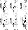 Figure 9
Effect of prostanoids and receptor modulators on human ILC2 cell differentiation in vitro. CLP cells were isolated from peripheral blood of normal healthy human donors and treated with either vehicle or IL-33 + IL-2 in vitro to induce ILC2 cell differentiation. (A) IL-5+, IL-13+, and GATA3+ ILC2 cells as a percentage of CD90+ cells were quantified by FACS in ILC2 cells differentiated from CLP cells in vitro in the presence of vehicle, 100 nM butaprost, or 100 nM PGE2. (B) IL-5+, IL-13+, and GATA3+ ILC2 cells as a percentage of CD90+ cells were quantified by FACS in ILC2 cells differentiated from CLP cells in vitro in the presence of vehicle, 100 nM PF-04418948 (EP2 antagonist), or 100 nM MF498 (EP4 antagonist). n = 5 per group, *P < 0.05 by paired 1-way ANOVA. Lines connect samples from individual patients.
-
Discussion
COX-2 and its eicosanoid products play important roles in regulating inflammation, innate immunity, and adaptive immunity (41). We previously reported that COX-2–derived eicosanoids regulate Th9 and Th17 cell differentiation and function during allergic lung inflammation (27, 29). ILC2 cells are non-B/non-T cells that secrete Th2-like cytokines including IL-5 and IL-13 that play critical roles in allergic lung disease pathogenesis. Therefore, we hypothesized that COX-2 and its products may also regulate lung ILC2 cells during allergic lung inflammation. We used COX-2–/– mice, EP2–/– mice, selective COX-2 inhibitors, and selective EP2 receptor agonists and antagonists to examine lung ILC2 cell differentiation and function during OVA- and Alternaria-induced allergic lung inflammation. We found the following: (a) allergic COX-2–/– mice have increased lung ILC2 cells, more cells expressing the ILC2 cytokines IL-5 and IL-13, and the ILC2 transcription factor GATA3, and differential ILC2 gene expression profiles compared with COX-2+/+ mice in vivo; (b) these effects are observed in both OVA- and Alternaria-induced models of allergic airway inflammation; (c) differentiation of CLP cells to ILC2 cells in vitro is accompanied by increased COX-2 expression; (d) CLP cells from COX-2–/– mice or from COX-2+/+ mice treated with a selective COX-2 inhibitor exhibit increased ILC2 cell differentiation and function in vitro; (e) COX-2–/– epithelial cells produce increased amounts of the ILC2-inducing cytokines IL-33 and TSLP in vitro and in vivo; (f) treatment with PGE2 or a selective EP2 receptor agonist suppresses ILC2 cell differentiation and function in vitro and in vivo; (g) allergic EP2–/– mice have increased lung ILC2 cells and increased ILC2 cell expression of IL-5, IL-13, and GATA3 compared with EP2+/+ mice in vivo; (h) PGE2 suppresses lung epithelial expression of Il33 and Tslp; (i) IL-33 can restore the lung ILC2 cell phenotype in COX-2–/– mice; and (j) PGE2 and EP2 receptor activation similarly suppress IL-33/IL-2-induced differentiation of human peripheral blood-derived ILC2 cells. Together, these findings indicate that COX-2–derived PGE2 regulates lung ILC2 cell differentiation and function during allergic inflammation via a mechanism that involves the EP2 receptor, IL-33 and TSLP.
NSAIDs, including COX-2 selective inhibitors, are the most commonly prescribed antiinflammatory medications in the world. COX-2 catalyzes the committed step in the biosynthesis of PGE2, PGD2, PGF2α, PGI2, and TXA2 (42). COX-2–derived prostanoids regulate the functions of immune and other cell types through specific G-protein coupled receptors including the EP, DP, FP, IP, and TP receptors (43, 44). In addition to the relatively uncommon but severe condition of aspirin-exacerbated respiratory disease (AERD), NSAID use is more generally associated with increased risk of asthma exacerbation (45). Although the precise molecular mechanisms that underlie this association remain enigmatic, several possibilities have been suggested, including increased production of proinflammatory leukotrienes via “shunting” of AA down the lipoxygenase pathway and decreased production of antiinflammatory PGs within the airway (46). Nevertheless, it is widely accepted that the acute and transient antiinflammatory, antipyretic, and antinociceptive benefits of NSAIDs in asthmatic patients are more than outweighed by their prolonged and pronounced effects on the immune system, namely effects on T cell differentiation and function leading to increased production of proinflammatory cytokines such as IL-4, IL-5, and IL-13 that initiate and perpetuate the allergic asthmatic response.
COX-derived prostanoids regulate many immune cell types during allergic lung inflammation. For example, PGE2, PGF2α and PGI2 increase Th17 cell differentiation (47), while PGE2 and PGD2 suppress Th9 cell differentiation (27). TXA2 inhibits both Th2 and Th9 cell differentiation (47), possibly by inhibiting the immune synapse between antigen-presenting cells and naive CD4+ T cells (48). PGD2 recruits eosinophils and mast cells to the lung during allergic inflammation and potentiates their degranulation. Conversely, PGE2 attenuates eosinophilic and mast cell–induced inflammation (49). PGI2 inhibits DC activation and cytokine production, which impairs antigen presentation to naive CD4+ T cells (50).
In our experiments, multiple PGs were capable of suppressing ILC2 cell differentiation and function in vitro; however, only PGE2 had effects on ILC2 cells at physiologically relevant concentrations as low as 50 nM. Interestingly, we found that while ILC2 cells expressed multiple prostanoid receptors, a selective EP2 receptor agonist inhibited and a selective EP2 receptor antagonist enhanced ILC2 cell differentiation and function in vitro and in vivo. Importantly, systemic administration of PGE2 via osmotic minipump rescued the lung ILC2 cell phenotype in allergic COX-2–/– mice in vivo. Moreover, EP2–/– mice had significantly increased lung ILC2 cells during allergic lung inflammation in vivo and EP2–/– bone marrow–derived CLP cells exhibited increased ILC2 cell differentiation compared with EP2+/+ CLP cells in vitro. Notably, these findings were not restricted to the OVA model. We observed similar increases in lung ILC2 cells and IL-5+, IL-13+, and GATA3+ ILC2 cells in both COX-2–/– and EP2–/– mice following Alternaria extract exposure. Furthermore, Alternaria-induced expression of the epithelial cytokines Il33 and Tslp was enhanced in COX-2–/– lungs, paralleling our observations in OVA-treated mice. The consistency of these findings across 2 distinct models of allergic lung inflammation suggests that COX-2–derived PGE2 functions as a broadly acting negative regulator of ILC2 cell responses rather than a pathway-specific regulator. Our evidence supports the concept that COX-2 negatively regulates ILC2 cells via PGE2-EP2 signaling by multiple pathways. Importantly, PGE2 and EP2 agonism or antagonism similarly regulate human ILC2 cell differentiation.
Our results support and expand upon prior studies showing that COX-derived prostanoids are important regulators of ILC2 cell differentiation, activation, and effector function. Maric and coworkers reported that PGE2 suppressed blood and tonsillar ILC2 cell activation via EP2 and EP4 receptors in vitro (30). Consistent with our findings, Zhou and coworkers demonstrated that pharmacologic COX inhibition enhanced Alternaria-induced pulmonary ILC2 cell responses and IL-33 release in vivo, while both PGE2 and PGI2 suppressed epithelial IL-33 release in vitro (34). Zhou et al. also showed that PGI2 inhibited ILC2 cell function via the IP receptor in an in vivo mouse model of fungal allergen exposure (32). In addition, PGD2 was shown to be essential for cytokine-induced ILC2 cell activation via the CRTH2 receptor in vitro (31, 51). Thus, COX-derived prostanoids can have both ILC2-promoting and ILC2-inhibiting effects. Since COX-2–/– mice have higher levels of ILC2 cells in vivo, our data suggest that, on balance, COX-2–derived prostanoids inhibit ILC2 differentiation, activation, and/or recruitment during allergic lung inflammation.
It is interesting to note that allergic COX-2–/– mice exhibit a 1.5-fold increase in lung ILC2 cells compared with COX-2+/+ mice but a much more pronounced 6- to 20-fold increase in lung expression of Il5 and Il13 in vivo. Moreover, we observed a 2-fold increase in differentiation of COX-2–/– CLP cells to ILC2 cells compared with COX-2+/+ and a similar 2-fold increase in expression of Il5 and Il13 in vitro. This suggests that other non-ILC2 cells may be contributing to the increased lung expression of Il5 and Il13 after allergen challenge in vivo. In this regard, we have previously shown that allergic COX-2–/– mice exhibit increased Th2 cell differentiation and function (27). Thus, the effects of COX-2 products on proinflammatory cytokine levels in vivo are complex and likely involve influences on multiple different immune cell types.
Pathway analysis of single-cell RNA-seq data suggests that COX-2–/– ILC2 cells have enhanced signaling in pathways that promote T cell chemotaxis and T cell/lymphocyte migration after OVA-induced allergic lung inflammation. In particular, Ccl5 was the most significantly induced gene in COX-2–/– ILC2 cells compared with COX-2+/+ ILC2 cells. CCL5 (RANTES) is increased in asthmatics and acts as a pulmonary chemoattractant for multiple cell types, including DCs, T cells, and eosinophils during allergic inflammation (52, 53). Thus, COX-2–/– lungs have both an increased number of ILC2 cells and ILC2 cells that more strongly recruit other cells types that exacerbate allergic inflammation.
Allergen-activated airway epithelial cells secrete IL-33, IL-25, and TSLP, which are known to stimulate ILC2 cell differentiation (54). We found that COX-2–/– airway epithelial cells have increased IL-33 and TSLP expression in vivo after OVA challenge or Alternaria-induced inflammation compared with COX-2+/+. Moreover, LPS treatment of purified airway epithelial cells from COX-2–/– lungs in vitro resulted in increased Il33 and Tslp expression compared with COX-2+/+. Furthermore, PGE2 significantly attenuated airway epithelial cell induction of Il33 and Tslp following LPS treatment in vitro. These results indicate that COX-2-derived PGE2 inhibits IL-33 and TSLP production by airway epithelial cells. Therefore, increased levels of IL-33 and TSLP produced by airway epithelial cells in allergic COX-2–/– mice likely contribute to the ILC2 cell phenotype. The fact that IL-33 administration was able to induce similar numbers of ILC2 cells in lungs of both COX-2+/+ and COX-2–/– mice further suggests a central role of this epithelial-derived cytokine in the observed ILC2 cell phenotype.
Our results are consistent with a model that involves effects of COX-2–derived PGE2 on both epithelial cells and CLP cells (Figure 10). OVA exposure leads to upregulation of COX-2 in both epithelial cells and CLP cells. In COX-2+/+ epithelial cells, increased COX-2 results in increased PGE2 production and increased signaling through the EP2 receptor which results in decreased production of IL-33 and TSLP, leading to reduced differentiation of CLP cells to ILC2 cells. The increased PGE2 also has a direct effect on CLP cells by suppressing their ability to differentiate to ILC2 cells in response to epithelial-derived IL-33 and TSLP. In contrast, COX-2–/– epithelial and CLP cells are unable to upregulate COX-2 and, therefore, produce less PGE2 in response to OVA. This results in reduced signaling through the EP2 receptor, leading to increased production of IL-33 and TSLP and to increased differentiation of CLP cells to ILC2 cells.
 Figure 10.
Proposed model for the regulation of lung ILC2 cells by COX-2 derived PGE2 during allergic lung inflammation. In COX-2+/+ mice (left), OVA or Alternaria induces upregulation of COX-2, resulting in increased PGE2 signaling to airway epithelial and CLP cells. Enhanced PGE2-EP2 signaling reduces production of IL-33 and TSLP from airway epithelial cells and inhibits ILC2 cell differentiation from CLP cells. In COX-2–/– mice (right), OVA or Alternaria cannot upregulate COX-2 resulting in lower PGE2 levels. Decreased PGE2-EP2 signaling results in higher IL-33 and TSLP production in airway epithelial cells and reduces the suppressive effect of PGE2-EP2 signaling during ILC2 cell differentiation.
An important limitation of the current study is that the cellular source and precise mechanism of PGE2-mediated regulation of ILC2 cell responses in vivo remain uncertain. In previous studies, COX-2–/– BALF had ~10%–40% lower PGE2 levels, though these results were not statistically significant (27, 55, 56). Important changes in autocrine or paracrine PG signaling may not be captured in total BALF analyses. While our in vitro findings support the capacity for autocrine PGE2 signaling in ILC2 cells, they do not establish whether epithelial cells or ILC2 cells are a major source of PGE2 in vivo. Similarly, our data do not conclusively establish whether autocrine PGE2/EP2 signaling regulates epithelial IL-33 and TSLP production in vivo. Future studies with cell-selective disruption of COX-2 and/or EP2 in ILC2 and epithelial cells will be required to address these possibilities.
In conclusion, we show that COX-2–derived prostanoids negatively regulate lung ILC2 cell differentiation and function during allergic lung inflammation through 2 separate pathways. PGE2 signaling via the EP2 receptor inhibits airway epithelial cell formation of IL-33 and TSLP to limit the major epithelial cytokine drivers of ILC2 cell differentiation. PGE2 also directly suppresses CLP cell differentiation into ILC2 cells. While multiple prostanoids have been shown to regulate ILC2 cell differentiation and function, our studies suggest that COX-2–derived PGE2 is the predominant PG involved during OVA- and Alternaria-induced allergic lung inflammation. Our findings also suggest the possibility that COX-2–derived PGE2 may represent a potentially new therapeutic target for allergic lung disease in humans.


