
Research ArticleMetabolismMuscle biology
Open Access |  10.1172/jci.insight.196134
Omega-3 fatty acid supplementation improves skeletal muscle mitochondrial function in a model of Barth syndrome
Katharina B. Kuentzel,1 Ana Vranešević,1 Samuel A.J. Trammell,1 Fabian Finger,2,3 Jesper F. Havelund,4 Yvette L. Schooneveldt,1 Ivan Bradić,4 Nicoline R. Andersen,1 Anna S. Hassing,1 Katja T. Michler,1 Martin R. Larsen,4 Zachary Gerhart-Hines,2 Steven M. Claypool,5 Jonas T. Treebak,2 Andreas M. Fritzen,1 Matthew P. Gillum,1 Steen Larsen,1,6,7 Nils Færgeman,4 and Trisha J. Grevengoed1
1Department of Biomedical Sciences, and
2Novo Nordisk Foundation Center for Metabolic Research, University of Copenhagen, Copenhagen, Denmark.
3Center for Adipocyte Signaling (ADIPOSIGN), and
4Department of Biochemistry and Molecular Biology, University of Southern Denmark, Odense, Denmark.
5Johns Hopkins University School of Medicine, Baltimore, Maryland, USA.
6Institute of Sports Medicine Copenhagen, Department of Orthopedic Surgery M, Copenhagen University Hospital – Bispebjerg and Frederiksberg, Denmark.
7Clinical Research Centre, Medical University of Bialystok, Bialystok, Poland.
Address correspondence to: Trisha J. Grevengoed, Blegdamsvej 3B, 2200, Copenhagen, Denmark. Phone: 45.8171.6121; Email: grevengoed@sund.ku.dk.
Find articles by Kuentzel, K. in: PubMed | Google Scholar
1Department of Biomedical Sciences, and
2Novo Nordisk Foundation Center for Metabolic Research, University of Copenhagen, Copenhagen, Denmark.
3Center for Adipocyte Signaling (ADIPOSIGN), and
4Department of Biochemistry and Molecular Biology, University of Southern Denmark, Odense, Denmark.
5Johns Hopkins University School of Medicine, Baltimore, Maryland, USA.
6Institute of Sports Medicine Copenhagen, Department of Orthopedic Surgery M, Copenhagen University Hospital – Bispebjerg and Frederiksberg, Denmark.
7Clinical Research Centre, Medical University of Bialystok, Bialystok, Poland.
Address correspondence to: Trisha J. Grevengoed, Blegdamsvej 3B, 2200, Copenhagen, Denmark. Phone: 45.8171.6121; Email: grevengoed@sund.ku.dk.
Find articles by Vranešević, A. in: PubMed | Google Scholar
1Department of Biomedical Sciences, and
2Novo Nordisk Foundation Center for Metabolic Research, University of Copenhagen, Copenhagen, Denmark.
3Center for Adipocyte Signaling (ADIPOSIGN), and
4Department of Biochemistry and Molecular Biology, University of Southern Denmark, Odense, Denmark.
5Johns Hopkins University School of Medicine, Baltimore, Maryland, USA.
6Institute of Sports Medicine Copenhagen, Department of Orthopedic Surgery M, Copenhagen University Hospital – Bispebjerg and Frederiksberg, Denmark.
7Clinical Research Centre, Medical University of Bialystok, Bialystok, Poland.
Address correspondence to: Trisha J. Grevengoed, Blegdamsvej 3B, 2200, Copenhagen, Denmark. Phone: 45.8171.6121; Email: grevengoed@sund.ku.dk.
Find articles by
Trammell, S.
in:
PubMed
|
Google Scholar
|

1Department of Biomedical Sciences, and
2Novo Nordisk Foundation Center for Metabolic Research, University of Copenhagen, Copenhagen, Denmark.
3Center for Adipocyte Signaling (ADIPOSIGN), and
4Department of Biochemistry and Molecular Biology, University of Southern Denmark, Odense, Denmark.
5Johns Hopkins University School of Medicine, Baltimore, Maryland, USA.
6Institute of Sports Medicine Copenhagen, Department of Orthopedic Surgery M, Copenhagen University Hospital – Bispebjerg and Frederiksberg, Denmark.
7Clinical Research Centre, Medical University of Bialystok, Bialystok, Poland.
Address correspondence to: Trisha J. Grevengoed, Blegdamsvej 3B, 2200, Copenhagen, Denmark. Phone: 45.8171.6121; Email: grevengoed@sund.ku.dk.
Find articles by Finger, F. in: PubMed | Google Scholar
1Department of Biomedical Sciences, and
2Novo Nordisk Foundation Center for Metabolic Research, University of Copenhagen, Copenhagen, Denmark.
3Center for Adipocyte Signaling (ADIPOSIGN), and
4Department of Biochemistry and Molecular Biology, University of Southern Denmark, Odense, Denmark.
5Johns Hopkins University School of Medicine, Baltimore, Maryland, USA.
6Institute of Sports Medicine Copenhagen, Department of Orthopedic Surgery M, Copenhagen University Hospital – Bispebjerg and Frederiksberg, Denmark.
7Clinical Research Centre, Medical University of Bialystok, Bialystok, Poland.
Address correspondence to: Trisha J. Grevengoed, Blegdamsvej 3B, 2200, Copenhagen, Denmark. Phone: 45.8171.6121; Email: grevengoed@sund.ku.dk.
Find articles by Havelund, J. in: PubMed | Google Scholar
1Department of Biomedical Sciences, and
2Novo Nordisk Foundation Center for Metabolic Research, University of Copenhagen, Copenhagen, Denmark.
3Center for Adipocyte Signaling (ADIPOSIGN), and
4Department of Biochemistry and Molecular Biology, University of Southern Denmark, Odense, Denmark.
5Johns Hopkins University School of Medicine, Baltimore, Maryland, USA.
6Institute of Sports Medicine Copenhagen, Department of Orthopedic Surgery M, Copenhagen University Hospital – Bispebjerg and Frederiksberg, Denmark.
7Clinical Research Centre, Medical University of Bialystok, Bialystok, Poland.
Address correspondence to: Trisha J. Grevengoed, Blegdamsvej 3B, 2200, Copenhagen, Denmark. Phone: 45.8171.6121; Email: grevengoed@sund.ku.dk.
Find articles by Schooneveldt, Y. in: PubMed | Google Scholar
1Department of Biomedical Sciences, and
2Novo Nordisk Foundation Center for Metabolic Research, University of Copenhagen, Copenhagen, Denmark.
3Center for Adipocyte Signaling (ADIPOSIGN), and
4Department of Biochemistry and Molecular Biology, University of Southern Denmark, Odense, Denmark.
5Johns Hopkins University School of Medicine, Baltimore, Maryland, USA.
6Institute of Sports Medicine Copenhagen, Department of Orthopedic Surgery M, Copenhagen University Hospital – Bispebjerg and Frederiksberg, Denmark.
7Clinical Research Centre, Medical University of Bialystok, Bialystok, Poland.
Address correspondence to: Trisha J. Grevengoed, Blegdamsvej 3B, 2200, Copenhagen, Denmark. Phone: 45.8171.6121; Email: grevengoed@sund.ku.dk.
Find articles by Bradić, I. in: PubMed | Google Scholar
1Department of Biomedical Sciences, and
2Novo Nordisk Foundation Center for Metabolic Research, University of Copenhagen, Copenhagen, Denmark.
3Center for Adipocyte Signaling (ADIPOSIGN), and
4Department of Biochemistry and Molecular Biology, University of Southern Denmark, Odense, Denmark.
5Johns Hopkins University School of Medicine, Baltimore, Maryland, USA.
6Institute of Sports Medicine Copenhagen, Department of Orthopedic Surgery M, Copenhagen University Hospital – Bispebjerg and Frederiksberg, Denmark.
7Clinical Research Centre, Medical University of Bialystok, Bialystok, Poland.
Address correspondence to: Trisha J. Grevengoed, Blegdamsvej 3B, 2200, Copenhagen, Denmark. Phone: 45.8171.6121; Email: grevengoed@sund.ku.dk.
Find articles by Andersen, N. in: PubMed | Google Scholar
1Department of Biomedical Sciences, and
2Novo Nordisk Foundation Center for Metabolic Research, University of Copenhagen, Copenhagen, Denmark.
3Center for Adipocyte Signaling (ADIPOSIGN), and
4Department of Biochemistry and Molecular Biology, University of Southern Denmark, Odense, Denmark.
5Johns Hopkins University School of Medicine, Baltimore, Maryland, USA.
6Institute of Sports Medicine Copenhagen, Department of Orthopedic Surgery M, Copenhagen University Hospital – Bispebjerg and Frederiksberg, Denmark.
7Clinical Research Centre, Medical University of Bialystok, Bialystok, Poland.
Address correspondence to: Trisha J. Grevengoed, Blegdamsvej 3B, 2200, Copenhagen, Denmark. Phone: 45.8171.6121; Email: grevengoed@sund.ku.dk.
Find articles by Hassing, A. in: PubMed | Google Scholar
1Department of Biomedical Sciences, and
2Novo Nordisk Foundation Center for Metabolic Research, University of Copenhagen, Copenhagen, Denmark.
3Center for Adipocyte Signaling (ADIPOSIGN), and
4Department of Biochemistry and Molecular Biology, University of Southern Denmark, Odense, Denmark.
5Johns Hopkins University School of Medicine, Baltimore, Maryland, USA.
6Institute of Sports Medicine Copenhagen, Department of Orthopedic Surgery M, Copenhagen University Hospital – Bispebjerg and Frederiksberg, Denmark.
7Clinical Research Centre, Medical University of Bialystok, Bialystok, Poland.
Address correspondence to: Trisha J. Grevengoed, Blegdamsvej 3B, 2200, Copenhagen, Denmark. Phone: 45.8171.6121; Email: grevengoed@sund.ku.dk.
Find articles by Michler, K. in: PubMed | Google Scholar
1Department of Biomedical Sciences, and
2Novo Nordisk Foundation Center for Metabolic Research, University of Copenhagen, Copenhagen, Denmark.
3Center for Adipocyte Signaling (ADIPOSIGN), and
4Department of Biochemistry and Molecular Biology, University of Southern Denmark, Odense, Denmark.
5Johns Hopkins University School of Medicine, Baltimore, Maryland, USA.
6Institute of Sports Medicine Copenhagen, Department of Orthopedic Surgery M, Copenhagen University Hospital – Bispebjerg and Frederiksberg, Denmark.
7Clinical Research Centre, Medical University of Bialystok, Bialystok, Poland.
Address correspondence to: Trisha J. Grevengoed, Blegdamsvej 3B, 2200, Copenhagen, Denmark. Phone: 45.8171.6121; Email: grevengoed@sund.ku.dk.
Find articles by
Larsen, M.
in:
PubMed
|
Google Scholar
|
1Department of Biomedical Sciences, and
2Novo Nordisk Foundation Center for Metabolic Research, University of Copenhagen, Copenhagen, Denmark.
3Center for Adipocyte Signaling (ADIPOSIGN), and
4Department of Biochemistry and Molecular Biology, University of Southern Denmark, Odense, Denmark.
5Johns Hopkins University School of Medicine, Baltimore, Maryland, USA.
6Institute of Sports Medicine Copenhagen, Department of Orthopedic Surgery M, Copenhagen University Hospital – Bispebjerg and Frederiksberg, Denmark.
7Clinical Research Centre, Medical University of Bialystok, Bialystok, Poland.
Address correspondence to: Trisha J. Grevengoed, Blegdamsvej 3B, 2200, Copenhagen, Denmark. Phone: 45.8171.6121; Email: grevengoed@sund.ku.dk.
Find articles by Gerhart-Hines, Z. in: PubMed | Google Scholar
1Department of Biomedical Sciences, and
2Novo Nordisk Foundation Center for Metabolic Research, University of Copenhagen, Copenhagen, Denmark.
3Center for Adipocyte Signaling (ADIPOSIGN), and
4Department of Biochemistry and Molecular Biology, University of Southern Denmark, Odense, Denmark.
5Johns Hopkins University School of Medicine, Baltimore, Maryland, USA.
6Institute of Sports Medicine Copenhagen, Department of Orthopedic Surgery M, Copenhagen University Hospital – Bispebjerg and Frederiksberg, Denmark.
7Clinical Research Centre, Medical University of Bialystok, Bialystok, Poland.
Address correspondence to: Trisha J. Grevengoed, Blegdamsvej 3B, 2200, Copenhagen, Denmark. Phone: 45.8171.6121; Email: grevengoed@sund.ku.dk.
Find articles by Claypool, S. in: PubMed | Google Scholar
1Department of Biomedical Sciences, and
2Novo Nordisk Foundation Center for Metabolic Research, University of Copenhagen, Copenhagen, Denmark.
3Center for Adipocyte Signaling (ADIPOSIGN), and
4Department of Biochemistry and Molecular Biology, University of Southern Denmark, Odense, Denmark.
5Johns Hopkins University School of Medicine, Baltimore, Maryland, USA.
6Institute of Sports Medicine Copenhagen, Department of Orthopedic Surgery M, Copenhagen University Hospital – Bispebjerg and Frederiksberg, Denmark.
7Clinical Research Centre, Medical University of Bialystok, Bialystok, Poland.
Address correspondence to: Trisha J. Grevengoed, Blegdamsvej 3B, 2200, Copenhagen, Denmark. Phone: 45.8171.6121; Email: grevengoed@sund.ku.dk.
Find articles by
Treebak, J.
in:
PubMed
|
Google Scholar
|
1Department of Biomedical Sciences, and
2Novo Nordisk Foundation Center for Metabolic Research, University of Copenhagen, Copenhagen, Denmark.
3Center for Adipocyte Signaling (ADIPOSIGN), and
4Department of Biochemistry and Molecular Biology, University of Southern Denmark, Odense, Denmark.
5Johns Hopkins University School of Medicine, Baltimore, Maryland, USA.
6Institute of Sports Medicine Copenhagen, Department of Orthopedic Surgery M, Copenhagen University Hospital – Bispebjerg and Frederiksberg, Denmark.
7Clinical Research Centre, Medical University of Bialystok, Bialystok, Poland.
Address correspondence to: Trisha J. Grevengoed, Blegdamsvej 3B, 2200, Copenhagen, Denmark. Phone: 45.8171.6121; Email: grevengoed@sund.ku.dk.
Find articles by Fritzen, A. in: PubMed | Google Scholar
1Department of Biomedical Sciences, and
2Novo Nordisk Foundation Center for Metabolic Research, University of Copenhagen, Copenhagen, Denmark.
3Center for Adipocyte Signaling (ADIPOSIGN), and
4Department of Biochemistry and Molecular Biology, University of Southern Denmark, Odense, Denmark.
5Johns Hopkins University School of Medicine, Baltimore, Maryland, USA.
6Institute of Sports Medicine Copenhagen, Department of Orthopedic Surgery M, Copenhagen University Hospital – Bispebjerg and Frederiksberg, Denmark.
7Clinical Research Centre, Medical University of Bialystok, Bialystok, Poland.
Address correspondence to: Trisha J. Grevengoed, Blegdamsvej 3B, 2200, Copenhagen, Denmark. Phone: 45.8171.6121; Email: grevengoed@sund.ku.dk.
Find articles by Gillum, M. in: PubMed | Google Scholar
1Department of Biomedical Sciences, and
2Novo Nordisk Foundation Center for Metabolic Research, University of Copenhagen, Copenhagen, Denmark.
3Center for Adipocyte Signaling (ADIPOSIGN), and
4Department of Biochemistry and Molecular Biology, University of Southern Denmark, Odense, Denmark.
5Johns Hopkins University School of Medicine, Baltimore, Maryland, USA.
6Institute of Sports Medicine Copenhagen, Department of Orthopedic Surgery M, Copenhagen University Hospital – Bispebjerg and Frederiksberg, Denmark.
7Clinical Research Centre, Medical University of Bialystok, Bialystok, Poland.
Address correspondence to: Trisha J. Grevengoed, Blegdamsvej 3B, 2200, Copenhagen, Denmark. Phone: 45.8171.6121; Email: grevengoed@sund.ku.dk.
Find articles by Larsen, S. in: PubMed | Google Scholar
1Department of Biomedical Sciences, and
2Novo Nordisk Foundation Center for Metabolic Research, University of Copenhagen, Copenhagen, Denmark.
3Center for Adipocyte Signaling (ADIPOSIGN), and
4Department of Biochemistry and Molecular Biology, University of Southern Denmark, Odense, Denmark.
5Johns Hopkins University School of Medicine, Baltimore, Maryland, USA.
6Institute of Sports Medicine Copenhagen, Department of Orthopedic Surgery M, Copenhagen University Hospital – Bispebjerg and Frederiksberg, Denmark.
7Clinical Research Centre, Medical University of Bialystok, Bialystok, Poland.
Address correspondence to: Trisha J. Grevengoed, Blegdamsvej 3B, 2200, Copenhagen, Denmark. Phone: 45.8171.6121; Email: grevengoed@sund.ku.dk.
Find articles by
Færgeman, N.
in:
PubMed
|
Google Scholar
|
1Department of Biomedical Sciences, and
2Novo Nordisk Foundation Center for Metabolic Research, University of Copenhagen, Copenhagen, Denmark.
3Center for Adipocyte Signaling (ADIPOSIGN), and
4Department of Biochemistry and Molecular Biology, University of Southern Denmark, Odense, Denmark.
5Johns Hopkins University School of Medicine, Baltimore, Maryland, USA.
6Institute of Sports Medicine Copenhagen, Department of Orthopedic Surgery M, Copenhagen University Hospital – Bispebjerg and Frederiksberg, Denmark.
7Clinical Research Centre, Medical University of Bialystok, Bialystok, Poland.
Address correspondence to: Trisha J. Grevengoed, Blegdamsvej 3B, 2200, Copenhagen, Denmark. Phone: 45.8171.6121; Email: grevengoed@sund.ku.dk.
Find articles by
Grevengoed, T.
in:
PubMed
|
Google Scholar
|
Published June 9, 2026 - More info
JCI Insight. 2026;11(15):e196134. https://doi.org/10.1172/jci.insight.196134.
© 2026 Kuentzel et al. This work is licensed under the Creative Commons Attribution 4.0 International License. To view a copy of this license, visit http://creativecommons.org/licenses/by/4.0/.
Received: May 28, 2025; Accepted: June 5, 2026


-
Results
DHA is preferentially incorporated into CL in cells lacking TAZ. HEK293 cells lacking TAZ (31) displayed an increase in CL synthase (CRLS1) gene expression (Figure 1A) and malondialdehyde (MDA) levels (Figure 1B). Elevated lipid peroxidation upon TAZ deficiency might be associated with increased cellular stress (32, 33); thus, our cells resemble a valid in vitro model. Without TAZ, cells cannot remodel CL to incorporate LA, leading to higher MLCL and CL that contains less LA (4), but the extent to which other fatty acids are incorporated into CL to compensate for the reduced LA remains poorly understood. Thus, we investigated how different fatty acids are incorporated and turned over into phospholipids. We used a relatively physiologic mixture of saturated, mono-, and polyunsaturated fatty acids with a [14C]-labeled fatty acid tracer to follow its incorporation or breakdown. While the incorporation of oleic acid (OA, C18:1) was unchanged in TAZ-knockout (TAZ KO) cells (Figure 1C), the use of polyunsaturated fatty acids was altered (Figure 1, D–F). As anticipated (34), TAZ KO cells incorporated less LA into CL, but increased incorporation of other fatty acids, especially DHA (Figure 1, D–F), indicating a potential compensation with these fatty acids. Without TAZ, less DHA was incorporated into phosphatidylcholine (PC), a major donor of acyl chains for CL (Figure 1F), suggesting a shift in DHA utilization in TAZ-deficient cells. Additionally, we observed more MLCL with OA and LA in TAZ KO cells, but were unable to detect any MLCL with labeled arachidonic acid (ARA, C20:4) or DHA (Figure 1, C and E). Higher amounts of ARA were incorporated into phosphatidylethanolamine (PE) and phosphatidylinositol (PI) in TAZ KO cells (Figure 1E), showing how altered CL remodeling has a wide range of effects on the different phospholipids.
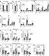 Figure 1
DHA is preferentially incorporated into CL in cells lacking TAZ. (A) Gene expression in HEK293 cells (n = 3). (B) Lipid peroxidation malondialdehyde (MDA) measured by TBARS assay (n = 6). (C–F) HEK293 cells were incubated with a mix of fatty acids and 1-[14C]–labeled fatty acid for 24 hours. Experiments were performed in triplicate in 2 or 3 independent assays. (G–L) HEK293 cells were incubated with a mix of fatty acids and 1-[14C]–labeled fatty acid for 24 hours, 4 hours (pulse), or 4 hours and then 20 hours with only unlabeled fatty acid (chase). Experiments were performed in triplicate in 2–3 independent assays. (M) Acid-soluble metabolites in media after 4 hours of incubation (pulse). Measurement was performed in 6 wells/experiment in 2–3 independent assays. Statistical significance was determined by unpaired Student’s t test (B) or multiple t tests with the Holm-Šídák method for multiple comparison testing (A and C–M) where appropriate. *P < 0.05; **P ≤ 0.01.
In a pulse-chase experiment, we observed that control cells had a time-dependent incorporation of LA, which could be consistent with LA being incorporated into one phospholipid before being transferred to CL (Figure 1G and Supplemental Figure 1, A–E; supplemental material available online with this article; https://doi.org/10.1172/jci.insight.196134DS1). In the TAZ KO cells, we observed decreased incorporation of LA into CL at 24 hours and after chase, the time points where this transacylase activity would be important (Figure 1G). The incorporation of [14C]DHA or [14C]ARA in CL was elevated in TAZ KO cells (Figure 1, H and I) after the pulse, but not after the chase, indicating a higher turnover. The highest turnover was observed with DHA, indicating that a steady supply of DHA is required to maintain the high DHA content in CL in TAZ KO cells. Corresponding levels of DHA in PC, a major acyl-chain donor for CL, were also lower in the TAZ KO cells, whereas the other fatty acids displayed less clear effects (Figure 1, J–L). Additionally, we observed larger release of partially degraded [14C]DHA into the media as acid-soluble metabolites from TAZ KO cells (Figure 1M). Higher oxidative stress and dysfunctional mitochondria (32) may contribute to turnover of the highly oxidizable DHA in TAZ-deficient cells, thus making a continuous supply of these molecules potentially beneficial.
Muscle cells lacking TAZ incorporate DHA and EPA into CL. Because BTHS strongly affects specific cell types, such as muscle, we tested whether knocking out TAZ in the myoblast-like cell line C2C12 would alter the incorporation of exogenously provided omega-3 fatty acids into CL. Differentiated WT and TAZ KO C2C12 cells were treated for 2 days with a mixture of DHA and EPA at a ratio similar to menhaden fish oil (FO). Of note, TAZ KO cells do not differentiate as well as the WT controls (35).
DHA and EPA treatment led to high incorporation into CL of these fatty acids (Figure 2, A and B), as well as changes in various lipid classes (Supplemental Figure 2, A–D) in WT and TAZ KO cells. Genotype was a major driver of CL levels and species, with TAZ KO cells displaying reduced total CL and elevated MLCL (Figure 2, C–E). DHA and EPA treatment did not alter total CL or MLCL in the TAZ KO cells (Figure 2, C and D), but did change the CL species containing DHA or EPA (Figure 2F), and normalized several CL species containing EPA, LA, or ARA to WT levels (Figure 2G and Supplemental Figure 2E). Of note, TAZ KO cells incorporated less DHA and EPA into CL than WT controls (Figure 2F). Thus, providing DHA and EPA to muscle cells lacking TAZ leads to their incorporation into CL and may normalize some CL species.
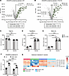 Figure 2
Muscle cells lacking TAZ incorporate DHA and EPA into CL. Volcano plots of DHA- and EPA-containing lipids in (A) WT and (B) TAZ KO C2C12 cells. (C) Total cardiolipin (CL). (D) Total monolyso-cardiolipin (MLCL). (E) MLCL-to-CL ratio. (F) All DHA- or EPA-containing CL species. (G) Heatmap of selected CL species. n = 5–6. Statistical significance was determined by 2-way ANOVA with the Holm-Šídák method for multiple comparison testing (C–F). ***P ≤ 0.001; ****P ≤ 0.0001.
Mice lacking TAZ in skeletal muscle display impaired mitochondrial respiration and lower lean mass. To determine whether the high DHA or EPA incorporation and turnover in TAZ-deficient cells (Figure 1I and Figure 2F) could be functionally relevant in vivo, we generated a skeletal muscle–specific Taz-KO mouse line (TazloxP/YxHSA-cre; TAZ MKO), using previously generated TazloxP/Y mice (8). The TAZ MKO mice were viable and fertile and displayed the expected skeletal muscle loss of Taz expression (Figure 3A) and elevated MLCL:CL ratio (Figure 3B) compared with littermate controls (TazloxP/Y). Additionally, mitochondrial respiration was impaired in the TAZ MKO mice in the highly oxidative soleus muscle (Figure 3C), but mitochondrial respiration was preserved in the more glycolytic extensor digitorum longus (EDL) muscle (Figure 3D). The lack of TAZ also caused an expected decrease (36) in lean mass (Figure 3E) and a reduction in voluntary running distance (Figure 3F), which fits well with the human phenotype (1, 37). In summary, the skeletal muscle–specific TAZ-deficient mouse represents a valid in vivo model resembling the human disease for further studying our hypothesis that supplementation with omega-3 fatty acids could improve the BTHS muscle phenotype.
 Figure 3
Mice lacking TAZ in skeletal muscle display impaired mitochondrial respiration and lower lean mass. (A) Gene expression in gastrocnemius (n = 5). (B) Ratio of total MLCL to total CL area (n = 7–9). (C and D) Mitochondrial respiration in soleus and EDL (n = 5). (E) Body composition by MRI in chow-fed mice (n = 35–48). (F) Voluntary wheel running (n = 7). Statistical significance was determined by t tests (A, B, and F) or multiple t tests (C–E) with the Holm-Šídák method for multiple comparison testing where appropriate. *P < 0.05; ***P ≤ 0.001; ****P ≤ 0.0001.
Omega-3 fatty acid supplementation prevents HFD-induced loss of lean mass in TAZ MKO mice. To examine the effects of elevated dietary omega-3 fatty acids, male control and TAZ MKO littermates were placed on a high-fat Western diet (HFD) with or without 10% kcal from menhaden FO for 10 weeks. DHA and EPA accounted for approximately 6% of fatty acids or 2%–3% of kcal in the FO diet. Male mice were used because BTHS occurs nearly exclusively in males (38). An HFD was chosen to induce skeletal muscle mitochondrial impairment and stress, and to ensure mice would eat the FO-containing diet. The diet did not affect Taz or Alcat1 expression, and Crls1 was elevated in TAZ MKO mice fed FO (Figure 4A). Over the 10-week feeding study, TAZ MKO mice gained less body weight and fat mass regardless of diet (Figure 4, B and C). Despite having lower baseline lean mass (Figure 3E), TAZ MKO mice fed the normal HFD lost more lean mass over the 10-week feeding period (Figure 4C). Importantly, TAZ MKO mice supplemented with FO maintained lean mass, similar to control mice (Figure 4C). Surprisingly, no substantial impairment in the endurance exercise capacity for forced treadmill running was observed with loss of TAZ in skeletal muscle (Figure 4D), indicating that the observed major defect in exercise capacity in the whole-body TAZ KO (11) may be due to defects in heart function. Additionally, excess adiposity may hamper exercise capacity in the control mice fed the HFD (39), and counter potential effects of lacking TAZ in skeletal muscle per se on exercise capacity. Expression of myogenesis- and atrophy-related genes revealed no interaction between diet and genotype, but Myog expression was elevated and Gdf11 was reduced in TAZ MKO gastrocnemius regardless of diet (Figure 4E). The gastrocnemius of TAZ MKO HFD-fed mice displayed slightly smaller cross-sectional area of the myocytes, which was normalized with FO feeding (Figure 4, F and G). Protein analysis of different fiber types revealed no changes in the subtype-determining markers with the FO diet or genotype, and no pattern towards a drastic change in a specific fiber subtype enrichment (Figure 4H). Together, these data indicate that omega-3 fatty acid supplementation prevents lean mass loss and normalized muscle fiber myocyte size, several of the impaired phenotypes central to BTHS (37, 38, 40).
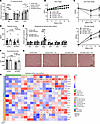 Figure 4
Omega-3 fatty acid supplementation prevents HFD-induced loss of lean mass in TAZ MKO mice. (A) Gene expression for CL metabolism–related markers in gastrocnemius (n = 5–6). (B) Body weight over 10 weeks of diet feeding (n = 15–21). (C) Change in fat and lean mass after 6 and 10 weeks of diet feeding (n = 15–21). (D) Exercise capacity on treadmills (n = 10–12/group). (E) Gene expression for myogenesis and atrophy markers in gastrocnemius (n = 5). (F) Average cross-sectional area of fibers in gastrocnemius (n = 3–5). (G) Representative H&E-stained gastrocnemius. Scale bar: 100 μm. (H) Heatmap of fiber type markers of proteomics analysis of gastrocnemius (n = 4–6). *Significant difference with the genotypte. #Significant difference with the diet. Statistical significance was determined by 2-way ANOVA with the Holm-Sidak method for multiple comparison testing where appropriate. *P < 0.05; **P ≤ 0.01; ***P ≤ 0.001; ****P ≤ 0.0001.
Impaired mitochondrial respiration in TAZ MKO soleus can be rescued by omega-3 fatty acid supplementation. Soleus was chosen for further analysis due to its high mitochondrial content and dependence on oxidative metabolism, as well as its apparent mitochondrial respiratory defects when lacking TAZ (Figure 3C). FO feeding of control mice increased mitochondrial content and fatty acid oxidative capacity, as indicated by citrate synthase (CS) and β-hydroxy acyl-CoA dehydrogenase (HAD) activity, respectively (Figure 5, A and B). Interestingly, HFD-fed TAZ MKO mice had higher mitochondrial content than control mice (Figure 5A), suggesting potential compensation for impaired mitochondrial function. This parameter was normalized with FO feeding in the TAZ MKO mice, which importantly means that potential changes to respiration are not due to FO-fed TAZ MKO mice having more dysfunctional mitochondria. Expression of transcription factors related to mitochondrial biogenesis was not affected by diet, but a general elevation in TAZ MKO mice was observed for Nrf1 (Figure 5C), indicating that a reduction in mitochondrial content in FO-fed TAZ MKO soleus was not due to impaired mitochondrial biogenesis signaling. No differences were seen in complex IV activity in TAZ MKO mice (Figure 5D), but HFD-fed TAZ MKO mice had impaired ADP-stimulated respiration (Figure 5E). Importantly, when fed FO to increase dietary omega-3 fatty acids, ADP- and succinate-stimulated respiration were normalized in TAZ MKO mice to levels of HFD-fed control mice (Figure 5E), indicating a rescue of mitochondrial respiratory capacity. At an earlier time point, after 5 weeks of FO feeding, we already observed a slight improvement in TAZ MKO ADP- and succinate-stimulated respiration without affecting respiration in the control mice (Supplemental Figure 3).
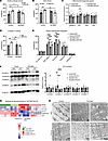 Figure 5
Impaired mitochondrial respiration in TAZ MKO soleus can be rescued by omega-3 fatty acid supplementation. (A and B) Citrate synthase and β-hydroxy acyl-CoA dehydrogenase (HAD) activity in soleus after 10 weeks of diet feeding (n = 5–6). (C) Expression of genes related to mitochondrial biogenesis in gastrocnemius (n = 5). (D and E) High-resolution respirometry in soleus after 10 weeks of diet feeding (n = 4–5). (F) Representative image of mitochondrial oxidative phosphorylation complexes and densitometry (n = 7–8). (G) Heatmap of rescued proteins of the gastrocnemius in the TAZ MKO proteome fed fish oil. *Significant difference with the genotype. #Significant difference with the diet. (H) Electron microscopy to show mitochondrial structure in soleus. Scale bars: 1 μm (4 left images) and 0.5 μm (2 right images) (n = 1). Statistical significance was determined by 2-way ANOVA with the Holm-Šídák method for multiple comparison testing where appropriate. *P < 0.05; **P ≤ 0.01; ***P ≤ 0.001; ****P ≤ 0.0001.
CL is important for the structure of the inner mitochondrial membrane and protein incorporation, especially for the electron transport chain (41–43). Protein levels of mitochondrial complex I and II were lower in TAZ MKO HFD-fed mice compared with controls, and FO feeding normalized complex I levels in the TAZ MKO soleus (Figure 5F), indicating one potential mechanism for the improved respiration with FO feeding in TAZ MKO mice. A proteome assessment of the gastrocnemius revealed that the genotype drives changes in the protein distribution (Supplemental Figure 4, A–D). However, several proteins in the gastrocnemius of TAZ MKO mice fed FO were normalized to the control condition. These proteins included 3 mitochondria-associated proteins: MFF, TMEM126A, and BCS1L (Figure 5G), supporting the observed functional rescue.
When comparing structures of interfibrillar mitochondria, we observed highly disrupted cristae with more open areas in TAZ MKO mice fed HFD, and unusual cristae patterns in TAZ MKO mice fed HFD plus FO (Figure 5H). These images indicate that, while FO feeding improved function, it did not fully normalize the mitochondrial structure, suggesting an abnormal compensation of membrane lipids. In summary, dietary FO supplementation appears to restore mitochondrial respiratory capacity in TAZ MKO mice without increasing mitochondrial number.
TAZ deficiency affects cellular health and FO modulates only some markers of stress and damage. In accordance with impaired muscle mitochondrial function in TAZ MKO mice fed HFD, the stress response seemed highly activated in the gastrocnemius muscle, a mixed-fiber type muscle (Figure 6A), with some indication that FO feeding improved this response indicated by the observed 37% reduction in Fgf21 gene expression. BTHS patients have elevated plasma FGF21 protein (44), and in line with this, the TAZ MKO HFD-fed mice also displayed elevated plasma FGF21 levels, which was normalized with FO feeding (Figure 6B). Because the liver retains functional TAZ in this model and the muscle expression of Fgf21 is highly elevated in TAZ MKO mice, it is likely that the elevation in circulating FGF21 levels is due to secretion from mitochondrially compromised muscle cells, as evident in muscle-specific mouse models with mitochondrial deficiencies (45–47). Interestingly, FO feeding lowered plasma FGF21 in both genotypes, indicating a reduced systemic cellular stress (48) in addition to those effects occurring in the skeletal muscle.
 Figure 6
TAZ deficiency affects cellular health and fish oil modulates only some markers of stress and damage. (A) Gene expression of stress-response markers in gastrocnemius (n = 5). (B) Plasma FGF21 (n = 5–6). Statistical significance was determined by 2-way ANOVA with the Holm-Šídák method for multiple comparison testing where appropriate. *P < 0.05; **P ≤ 0.01; ***P ≤ 0.001; ****P ≤ 0.0001.
Loss of TAZ in skeletal muscle prevented diet-induced changes in CL species. As we observed abnormal muscle mitochondrial structure despite improved mitochondrial function upon FO feeding in TAZ MKO mice (Figure 5G), we hypothesized that structural lipids might be altered. Therefore, we analyzed the whole-cell lipids of the soleus by LC-MS and observed that TAZ MKO muscle had low CL content, regardless of diet (Figure 7A). FO feeding of control mice shifted the composition of CL species towards longer, more unsaturated species (Figure 7, B and C). Interestingly, FO feeding did not change the CL composition of the TAZ MKO mice (Figure 7, B and D), indicating that even the nascent CL was likely unaffected. Additionally, MLCL was elevated in TAZ MKO mice, regardless of diet (Figure 7E). Therefore, the observed improvements in lean mass and mitochondrial respiration in TAZ MKO mice with FO feeding were not due to normalization of CL or reduction in MLCL in the soleus.
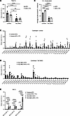 Figure 7
Loss of TAZ in skeletal muscle prevented diet-induced changes in cardiolipin species. (A) Total area assigned to cardiolipin (CL) in soleus (n = 6–9). (B) Unsaturation of acyl chains relative to area of CL (n = 6–9). (C and D) CL species in control (C) and TAZ MKO (D) soleus after 10 weeks of diet feeding (n = 6–9). (E) Monolyso-CL (MLCL) species (n = 6–9). Statistical significance was determined by 2-way ANOVA (A, B, and E) or multiple unpaired t tests (C and D) with the Holm-Šídák method for multiple comparison testing where appropriate. *P < 0.05; **P ≤ 0.01; ****P ≤ 0.0001.
TAZ deficiency alters many phospholipids, with differing responses to FO. We further investigated the other phospholipids to better understand the effects on membrane lipids. After removal of CL and MLCL from the analysis, the lipidome of the 4 different groups remained distinct (Figure 8A), emphasizing the broader impact of TAZ deficiency beyond its typical effects on CL, in line with our hypothesis that additional membrane lipids are altered. Both diet and lack of TAZ in skeletal muscle altered the lipidome, and while we cannot determine causality in the current model, several differences were of note. Most phospholipids were changed only at the species composition, but total phosphatidylserine (PS) was higher in TAZ MKO mice and was normalized to control levels with FO supplementation (Supplemental Figure 5A). The degree of unsaturation of the different phospholipid classes was largely affected by diet, with the CL species in TAZ MKO mice being a notable exception (Figure 8B and Supplemental Figure 5B). Several ceramides had a strong genotype effect, with partial normalization by FO feeding in the TAZ MKO mice (Supplemental Figure 5C). Consistent with this, the abundance of the hexosylceramide species HexCer (44:0; O2) was elevated in TAZ MKO mice but normalized to a similar level as seen in control groups with FO feeding of TAZ MKO mice (Supplemental Figure 5D). Additionally, PI (38:5) was detected at low levels in control mice, but was abundant in all TAZ MKO muscle. Interestingly, this PI was reduced by FO feeding in the TAZ MKO mice (Figure 8B and Supplemental Figure 5D). These results provide clues to the mechanisms behind the rescue of mitochondrial respiration in TAZ MKO mice as well as a resource to understand both the effects of loss of TAZ and omega-3 fatty acid supplementation in skeletal muscle.
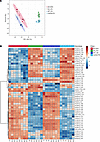 Figure 8
TAZ deficiency affects many phospholipids with differing responses to fish oil. (A) Principal component analysis (PCA) plot after removing cardiolipin (CL) and monolyso-CL (MLCL) species from the lipidomics data (n = 6–9). (B) Heatmap of the top 50 changes in phospholipid species (n = 6–9). Analyzed with MetaboAnalyst.


-
Methods
Sex as a biological variable. Our study exclusively examined male mice because the disease modeled (BTHS) is primarily relevant in males (2, 38, 69), as female carriers are mostly asymptomatic (49, 70). Investigated improvements are potentially relevant for the target group of males suffering from BTHS, but may not be irrelevant for affected females.
Cell culture. HEK293 Flp and TazTALEN.19 (TAZ KO) (31) cells were cultured to confluence in 25 mM glucose DMEM with 10% FBS. Cells were incubated with a relatively physiologic mixture of 25 μM oleate,15 μM palmitate, 5 μM linoleate, 5 μM arachidonate or 5 μM docosahexaenoic acid bound to bovine serum albumin (BSA). For labeling, 0.5 μCi of [1-14C]oleate, [1-14C]linoleate, [1-14C]arachidonate, or [1-14C]docosahexaenate was substituted for the appropriate amount of unlabeled fatty acid. Incubations lasted for 24 hours and then cells were washed twice with 0.1% BSA. For pulse-chase experiments, cells were incubated with the fatty acid mix and labeled fatty acid media for 4 hours (pulse), washed once with 0.1% BSA, and incubated with media containing an unlabeled fatty acid mixture for 20 hours. For oxidation measurements, 0.4 mL media was collected in a tube containing 20 μL 15% BSA and then incubated with 100 μL 20% perchloric acid overnight at 4°C. The acidified media was centrifuged at 20,000g for 5 minutes, and radioactivity in the supernatant was counted to determine acid-soluble metabolites. Lipids were extracted from cells using chloroform and methanol (71), and phospholipids were separated by thin layer chromatography (TLC) in chloroform/ethanol/water/triethylamine (30:35:7:35; v/v) with authentic standards (72). Radioactivity was quantified using a TLC scanner (AR-2000, Eckert-Ziegler).
TAZKO and WT control C2C12 cells (35) were a gift from Miriam Greenberg (Wayne State University, Detroit, Michigan, USA), and were maintained in high-glucose (25 mM) DMEM supplemented with 10% FBS and 1% penicillin/streptomycin. Differentiation of myoblasts was induced by switching the media to high-glucose DMEM without sodium pyruvate, supplemented with 2% horse serum and 1% penicillin/streptomycin. Media were refreshed every second day during the 7-day differentiation period. Differentiated cells were treated with either 50 μM BSA-conjugated DHA+EPA–containing fatty acid mix (7.5 μM DHA, 13.4 μM EPA, 29.1 μM oleate according to Banic et al. (73), with a ratio of EPA to DHA of 1.8:1 as in menhaden FO) or 50 μM BSA-conjugated oleate from day 7 to day 9, and the medium was replaced every day. Cells were washed with PBS containing 1% fatty acid–free BSA, harvested in 140 μL of 150 mM NH4OAc, snap-frozen in dry ice, and stored at –80°C until analysis.
Mouse model. All mouse protocols were approved by the Danish Animal Experiments Inspectorate and performed according to Animal Research: Reporting of In Vivo Experiments (ARRIVE) standards. Mice were housed in a specific pathogen–free environment on a 12-hour light/12-hour dark cycle and were maintained on a chow diet with ad libitum access to food and water unless otherwise specified. Skeletal muscle–specific TAZ MKO mice were generated by crossing the TazloxP strain (8) (provided by Douglas Strathdee, Cancer Research UK, London, United Kingdom) with a constitutively HSA-cre–expressing line (74). All mice were fully floxed and on a C57BL/6Ntac background. TAZ MKO mice were viable and fertile. Male mice were used for experiments, unless otherwise specified, as BTHS primarily occurs in male humans (2, 38, 69). At 8–12 weeks of age, male mice were placed on either an HFD containing 10% menhaden FO (4.7 kcal/g; 40% kcal fat, 17% kcal protein, 43% kcal carbohydrate, D22060303, Research Diets) or matched control diet (D22060302, Research Diets). DHA and EPA content in the HFD+FO diet was approximately 6% of total fatty acids, comparable to the approximately 3% of added fatty acids in the cell culture experiments. Following a 1-week acclimation period to individual housing with a running wheel, voluntary running was measured daily for 1 week and expressed as the average of the 7 days. Body composition was measured by MRI (Bruker LF90II MRI scanner) at baseline, 6 weeks, and 10 weeks of the diet study. Tissues and blood were collected from pentobarbital-anesthetized mice after 10 weeks and either snap frozen or processed further.
TBARS assay. Malondialdehyde content was determined in cell lysates and measured as thiobarbituric acid–reactive substance (TBARS) according to the manufacturer’s recommendation (10009055, Cayman Chemical).
Exercise capacity testing. Exercise capacity was measured as previously described (75) after 10 weeks on respective diets. Briefly, mice were acclimatized to the treadmill and running for 3 days and then rested for 1 day. Exercise capacity testing was conducted with 5% incline, starting at 6 m/min with increases in speed every 2 minutes until max speed of 16 m/min, with a maximum running time of 60 minutes. Exhaustion was defined as refusal to run for 5 seconds while in contact with the shock grid or after 3 shocks.
Histology. Gastrocnemius muscle was fixed in 4% paraformaldehyde and then embedded in paraffin, cut cross-sectionally, and stained with hematoxylin and eosin (H&E). The slides were visualized with an AXIO Lab A1 Light Microscope (Zeiss), bright-field, 20× objective magnification. Ten images of each sample were taken with AxioCam ICc 5 (Zeiss). The cross-sectional area of cells was measured using FIJI (ImageJ).
Mitochondrial function measurements. Mitochondrial respiratory capacity was measured in permeabilized skeletal muscle fibers of soleus (Figure 3C, Figure 5E, and Supplemental Figure 3) and EDL (Figure 3D) using high-resolution respirometry (Oxygraph-2k, Oroboros). Skeletal muscle tissue was placed in BIOPS buffer (76) after dissection and kept on ice. The skeletal muscle tissue was mechanically dissected on ice using sharp forceps, chemically permeabilized in BIOPS buffer containing saponin (50 μg/mL) for 30 minutes, and then washed twice for 10 minutes in MiR05 buffer. Skeletal muscle fibers were weighed and added to the respirometry chamber containing MiR05 buffer.
The following protocol was performed in duplicate at 37°C after hyperoxygenation (450–200 nmol/mL) to avoid potential oxygen limitation. Pyruvate (5 mM), malate (2 mM), and glutamate (10 mM) (LEAK) were added to each chamber, followed by ADP (5 mM) and MgCl2 (3 mM) to determine maximal complex I respiration (CIP). Cytochrome c (0.01 mM) was added to verify the integrity of the outer mitochondrial membrane. Succinate (10 mM) was applied to assess the maximal respiration in both complex I and II (CI+IIP). Subsequently, titration of rotenone (0.001 mM) and antimycin A (0.005 mM) was performed to determine the residual oxygen consumption respiration. The chambers were reoxygenated before addition of ascorbate (2 mM) and TMPD (0.5 mM) to assess cytochrome c oxidase (CIV) activity. The oxygen concentration at the time point, after the respiratory flux rate started to decline, was noted. Azid (100 mM) was applied, and the oxygen concentration was raised to the same level as noted in the previous step to correct for the autoxidation of TMPD and ascorbate under the given oxygen pressure.
CS and HAD activity was measured as previously described (77) with some minor changes. Five milligrams of wet weight skeletal muscle was homogenized in 800 μL homogenization buffer with 8 μL 10% Triton X-100. CS activity was used to estimate mitochondrial number.
Electron microscopy. Mice were perfused through the left ventricle with 2% v/v glutaraldehyde in 0.05 M sodium phosphate buffer (pH 7.2) and stored in this fixation mixture. Following isolation of suitable specimen blocks, the samples were rinsed 3 times in 0.15 M sodium phosphate buffer (pH 7.2) and subsequently postfixed in 1% w/v OsO4 with 0.05 M K3Fe(CN)6 in 0.12 M sodium phosphate buffer (pH 7.2) for 2 hours. The specimens were dehydrated in graded series of ethanol, transferred to propylene oxide, and embedded in Epon according to standard procedures. Sections, approximately 60 nm thick, were cut with an Ultracut 7 (Leica) and collected on 1-hole copper grids with Formvar supporting membranes, stained with uranyl acetate and lead citrate, and subsequently examined with a Philips CM 100 transmission electron microscope, operated at an accelerating voltage of 80 kV. Digital images were recorded with an OSIS Veleta digital slow scan 2k × 2k CCD camera and the ITEM software package.
Western blot analysis. Soleus was homogenized in ice-cold homogenization buffer (10% glycerol, 1% NP-40, 20 mM sodium pyrophosphate, 150 mM NaCl, 50 mM HEPES [pH 7.5], 20 mM β-glycerophosphate, 10 mM NaF, 2 mM phenylmethylsulfonyl fluoride [PMSF], 1 mM EDTA [pH 8.0], 1 mM EGTA [pH 8.0], 2 mM Na3VO4, 10 μg/mL leupeptin, 10 μg/mL aprotinin, 3 mM benzamidine) using a Qiagen Tissue-Lyser II (60 seconds at 30 Hz), after which the homogenate was placed end-over-end for 30 minutes at 5°C. The samples were then centrifuged at 10,000g for 15 minutes at 4°C and the supernatant (lysate) was collected discarding the remaining pellet and frozen at –80°C until further analyses. Lysate protein concentration was determined using the bicinchoninic acid (BCA) method. Soleus lysate (8 μg protein/lane) was separated using SDS-PAGE on a 4%–15% gradient gel and transferred to a PVDF membrane under standard conditions. The membrane was blocked in 3% BSA in TBST and then incubated with a primary antibody against OXPHOS complexes (Abcam, ab110413; 1:5,000) in TBST with 3% BSA, overnight at 4°C. After washing, the membrane was incubated with secondary antibody rabbit anti-mouse–HRP (Jackson ImmunoResearch, 115-035-062; 1:5,000 in TBST with 3% BSA) for 1 hour at room temperature. Bands were visualized using chemiluminescence using a Bio-Rad ChemiDoc MP Imaging System and enhanced chemiluminescence (ECL). Densitometry was analyzed with Image Lab software.
Proteomics. Approximately 10 μg of gastrocnemius muscle was lysed in 50 mM HEPES pH 8.5 containing 5% sodium deoxycholate, supplemented with cOmplete protease inhibitor (EDTA-free 118735800001, Roche). Samples were sonicated for 30 seconds on ice, and afterward, proteins were denatured at 98°C for 5 minutes. Samples were then centrifuged at 20,000g for 20 minutes at 4°C, and the supernatant was transferred to a new tube. Protein concentrations were determined by BCA. Samples were reduced with 10 mM dithiothreitol and alkylated with 20 mM iodoacetamide for 20 minutes each. An aliquot of 20 μg of protein was digested at 37°C after preincubation with LysC (0.04 AU/mg protein), then 5% trypsin was added overnight. Sodium deoxycholate was precipitated by adding TFA to a final concentration of 1% and then centrifuged at 20,000g for 10 minutes. Afterward, the supernatant was collected, peptides were purified using C18 stage tips, and the samples were then vacuum-dried. Finally, peptides were resuspended in 0.1% formic acid.
Approximately 1,000 ng of peptides, dissolved in solvent A (0.1% formic acid), was loaded onto a 20-cm analytical column (100-μm inner diameter) packed with ReproSil-Pur C18 AQ 1.9-μm RP material using a Thermo Scientific Vanquish Neo UHPLC system coupled to an Orbitrap Astral mass spectrometer. Elution of peptides was performed at a flow rate of 300 μL/min by increasing the percentage of solvent B (95% acetonitrile and 0.1% formic acid) from 0.3% to 38% over 40 minutes, then to 90% over 2 minutes, and holding at 90% for 5 more minutes. During the 30-minute gradient, the mass spectrometer operated in positive mode with Orbitrap resolution set at 240,000, a scan range of 400–1000 m/z, and a maximum injection time of 5 ms. Data-independent acquisition (DIA) was performed with higher-energy collision-induced dissociation set to 25%, a scan range of 150–2000 m/z, a maximum injection time of 3.5 ms, and a cycle time of 0.6 seconds. Fragment ions were detected using the Astral detector.
Raw data files were searched with Spectronaut 20.0 (https://biognosys.com/software/spectronaut/), using the Mus musculus reference proteome FASTA file, ID UP000000589, containing 21,757 genes (downloaded on 27.01.2025) (https://www.uniprot.org/proteomes/UP000000589). BGS factory settings were used for the search, with minor modifications. Most search settings were kept at their defaults. In brief, search settings included trypsin/P-digested peptides with a maximum of 2 missed cleavages, carbamidomethyl (C) as a fixed modification, and a maximum of 5 variable modifications, including acetyl (protein N-term) and oxidation (M). The Deep directDIA+ workflow was used for identification. For quantification, precursor filtering was set to “Identified (Qvalue),” and no imputation was performed. The proteotypicity filter was set to “Only protein group specific,” and cross-run normalization was used.
Bioinformatic analyses were performed in R (4.5.0) (https://cran.r-project.org) within a reproducible environment managed by the renv package. Protein intensities obtained after Spectronaut search were imported with readr. The dataset was filtered to retain only proteins with at least 3 valid values in each experimental group (WT_HFD, WT_FO, KO_HFD, KO_FO), and intensities were log2-transformed by DEP:make_se(). Missing values were imputed using the MinProb method (quantile q = 0.01) from the DEP package with a fixed random seed [123] set before imputation to ensure reproducibility. Differential protein abundance was determined using the limma package and a 2 × 2 factorial experimental design (genotype: WT/KO; diet: HFD/FO). Five contrasts were defined to test the genotype effect within each diet (KO − WT in HFD; KO − WT in FO), the diet effect within each genotype (HFD − FO in WT; HFD − FO in KO), and the genotype × diet interaction. Empirical Bayes moderation was applied to the model to increase statistical power, and P values were adjusted for multiple testing using the Benjamini-Hochberg false discovery rate (FDR) procedure. Proteins with an FDR of less than 5% and log2(fold change) of greater than 0.5 were considered statistically significant. Volcano plots were generated with ggplot2, and heatmaps were generated with pheatmap.
FGF21 measurement. Blood was centrifuged 8,000g for 10 minutes at 4°C to separate plasma. FGF21 was measured in plasma using the Mouse FGF-21 ELISA (Abcam, ab212160), according to the manufacturer’s instructions.
Lipidomics. Lipidomics analyses were performed essentially as described previously (78). Briefly, cell or tissue lipids were extracted using Folch extraction (71). Prior to tissue lysis, SPLASH mix (Merck) was added to the extraction solvent, and tissue samples were lysed by beat beating in a FastPrep-24 homogenizer. After centrifugation and phase separation, the apolar and polar phases were transferred to separate tubes, and the apolar phase dried under N2. Samples were resuspended in 30 μL methanol/chloroform (1:1) and centrifuged (5 minutes, 16,000g, 22°C) before transferring to HPLC vials. A quality control sample was constructed by pooling 3 μL of each sample. Samples (0.5 μL) were injected using a Vanquish Horizon UPLC (Thermo Fisher Scientific) equipped with a Waters ACQUITY Premier CSH (2.1 × 100 mm, 1.7 μm) column operated at 55°C. The analytes were eluted using a flow rate of 400 μL/min and the following composition of eluent A (acetonitrile/water [60:40], 10 mM ammonium formate, 0.1% formic acid) and eluent B (isopropanol/acetonitrile [90:10], 10 mM ammonium formate, 0.1% formic acid): 40% B from 0 to 0.5 minutes, 40%–43% B from 0.5 to 0.7 minutes, 43%–65% B from 0.7 to 0.8 minutes, 65%–70% B from 0.8 to 2.3 minutes, 70%–99% B from 2.3 to 6 minutes, 99% B from 6–6.8 minutes, 99%–40% B from 6.8–7 minutes before equilibration for 3 minutes with the initial conditions. The flow from the UPLC was coupled to a TimsTOF Flex (Bruker) instrument for mass spectrometric analysis. Compounds were annotated in Metaboscape (Bruker) using both an in-built rule-based annotation approach and using the LipidBlast MS2 library (79). Features were removed if their average signal were not greater than 5 times more abundant in the QC samples than blanks (water extraction). The signals were normalized to internal standards in the SPLASH mix before correction for signal drift, blank and QC filtration using Metabolink (80).
The identified lipids were outputted as unique ions as well as ions with multiple adduct forms. Identified adducts with retention times of 0.2 minutes or less apart were combined by summing the raw area counts. The new collapsed adducts were identified by the lipid family name followed by the total number of carbons and double bonds. The QC pool injections were used to calculate the relative standard deviations of the raw area counts for each lipid. Identified lipids with a relative standard deviation of 0.25 or less were uploaded to MetaboAnalyst 6.0 (81) for 1-factor and 2-factor statistical analyses. Missing values were replaced with one-fifth of the lowest positive area count within each feature row. Data lying outside of the interquartile range were removed (n = 11 removed features). The data were then quantile normalized, log10 transformed, and scaled using mean-centering. One-factor and 2-factor analysis of variance (ANOVAs) were performed with FDR corrections. An α level of 0.05 was considered significant. Data were analyzed, and heatmaps and PCA plots were created using MetaboAnalyst (81).
For the cell lipid analysis, pairwise comparisons were performed on 772 lipid species against either genotype or treatment condition using linear regression models. Lipid concentrations were log10 transformed prior to analysis. Lipid species with 3 or more missing values were excluded, while samples with singular missing values were omitted from the analysis for the respective lipid. Regression coefficients and 95% confidence intervals were converted to log2(fold change) for interpretation of results. P values were corrected for multiple comparisons using the Benjamini-Hochberg method.
Gene expression. Total RNA was isolated from approximately 50 mg of pulverized gastrocnemius muscle tissue using TRIzol reagent (Invitrogen, Thermo Fisher Scientific). cDNA was synthesized from 2 μg of isolated RNA (High-Capacity cDNA Reverse Transcription Kit, Thermo Fisher Scientific). qPCR was performed with 6 ng of cDNA in white 384-well plates using PowerUp SYBR Green PCR Master Mix (Applied Biosystems) and the LightCycler 480 II (Roche) instrument. Threshold cycle (Ct) values were recorded and analyzed using the 2–ΔΔCt method with the expression of Actb or GAPDH used as a housekeeping gene. Primer sequences used are listed in Table 1.
Statistics. Statistical analysis of the data was done using GraphPad Prism 10.1.1 software. Results are presented as mean values ± SEM. Unpaired 2-tailed t test, multiple t test, 2- or 1-way ANOVA was performed depending on the type of data, followed by Holm-Šídák post hoc analysis unless otherwise stated. Statistical significance was considered when the P value was less than 0.05 as follows.
Study approval. All murine protocols were approved by the Danish Animal Experiments Inspectorate and performed according to Animal Research: Reporting of In Vivo Experiments (ARRIVE) standards.
Data availability. Proteomics data have been deposited at the ProteomeXchange Consortium via the PRIDE partner repository (82) with the dataset identifier PXD077710. Lipidomics data have been deposited at the following repository: https://doi.org/10.6084/m9.figshare.32105305 All deposited data are publicly available as of the date of publication. Values for all data points in graphs are reported in the Supporting Data Values file. Other data will be shared upon reasonable request to the corresponding author.


