
Research ArticleMetabolismNeuroscience
Open Access |  10.1172/jci.insight.198842
Fatty acid amide hydrolase inhibition for treatment of amyotrophic lateral sclerosis
Daisuke Ito,1,2 Madoka Iida,1 Yohei Iguchi,1 Atsushi Hashizume,2 Shinichiro Yamada,1 Yoshiyuki Kishimoto,1 Shota Komori,1 Kazuki Obara,1 Shuto Nishisaki,3 Satoshi Yokoi,1,3 Teppei Shimamura,4 Yuto Takemoto,5 Masahiro Nakatochi,5 Tomohiro Akashi,4 Kunihiko Hinohara,6,7 Hyeon-Cheol Lee-Okada,8 Yohei Okada,9 Junichi Niwa,10 Gen Sobue,11 Shinji Tanaka,12 Ken Takashina,12 Takehiko Yokomizo,8 and Masahisa Katsuno1
1Department of Neurology,
2Department of Clinical Research Education,
3Department of Pathophysiological Laboratory Sciences,
4Division of Systems Biology,
5Public Health Informatics Unit, Department of Integrated Health Sciences, and
6Department of Immunology, Nagoya University Graduate School of Medicine, Nagoya, Aichi, Japan.
7Institute for Advanced Research, Nagoya University, Nagoya, Aichi, Japan.
8Department of Biochemistry, Juntendo University Graduate School of Medicine, Bunkyo-ku, Tokyo, Japan.
9Department of Neural iPSC Research Institute for Medical Science of Aging, Aichi Medical University, Nagakute, Aichi, Japan.
10Department of Neurology and
11Aichi Medical University School of Medicine, Nagakute, Aichi, Japan.
12Sohyaku, Innovative Research Division, Mitsubishi Tanabe Pharma Corporation, Yokohama, Japan.
Address correspondence to: Masahisa Katsuno or Daisuke Ito, Department of Neurology, Nagoya University Graduate School of Medicine, 65 Tsurumai-cho, Showa-ku, Nagoya 466-8550, Japan. Phone: 81.52.744.2389; Email: katsuno.masahisa.i1@f.mail.nagoya-u.ac.jp (MK). Phone: 81.52.7442390; Email: ito.daisuke.k4@f.mail.nagoya-u.ac.jp (DI).
Find articles by Ito, D. in: PubMed | Google Scholar
1Department of Neurology,
2Department of Clinical Research Education,
3Department of Pathophysiological Laboratory Sciences,
4Division of Systems Biology,
5Public Health Informatics Unit, Department of Integrated Health Sciences, and
6Department of Immunology, Nagoya University Graduate School of Medicine, Nagoya, Aichi, Japan.
7Institute for Advanced Research, Nagoya University, Nagoya, Aichi, Japan.
8Department of Biochemistry, Juntendo University Graduate School of Medicine, Bunkyo-ku, Tokyo, Japan.
9Department of Neural iPSC Research Institute for Medical Science of Aging, Aichi Medical University, Nagakute, Aichi, Japan.
10Department of Neurology and
11Aichi Medical University School of Medicine, Nagakute, Aichi, Japan.
12Sohyaku, Innovative Research Division, Mitsubishi Tanabe Pharma Corporation, Yokohama, Japan.
Address correspondence to: Masahisa Katsuno or Daisuke Ito, Department of Neurology, Nagoya University Graduate School of Medicine, 65 Tsurumai-cho, Showa-ku, Nagoya 466-8550, Japan. Phone: 81.52.744.2389; Email: katsuno.masahisa.i1@f.mail.nagoya-u.ac.jp (MK). Phone: 81.52.7442390; Email: ito.daisuke.k4@f.mail.nagoya-u.ac.jp (DI).
Find articles by Iida, M. in: PubMed | Google Scholar
1Department of Neurology,
2Department of Clinical Research Education,
3Department of Pathophysiological Laboratory Sciences,
4Division of Systems Biology,
5Public Health Informatics Unit, Department of Integrated Health Sciences, and
6Department of Immunology, Nagoya University Graduate School of Medicine, Nagoya, Aichi, Japan.
7Institute for Advanced Research, Nagoya University, Nagoya, Aichi, Japan.
8Department of Biochemistry, Juntendo University Graduate School of Medicine, Bunkyo-ku, Tokyo, Japan.
9Department of Neural iPSC Research Institute for Medical Science of Aging, Aichi Medical University, Nagakute, Aichi, Japan.
10Department of Neurology and
11Aichi Medical University School of Medicine, Nagakute, Aichi, Japan.
12Sohyaku, Innovative Research Division, Mitsubishi Tanabe Pharma Corporation, Yokohama, Japan.
Address correspondence to: Masahisa Katsuno or Daisuke Ito, Department of Neurology, Nagoya University Graduate School of Medicine, 65 Tsurumai-cho, Showa-ku, Nagoya 466-8550, Japan. Phone: 81.52.744.2389; Email: katsuno.masahisa.i1@f.mail.nagoya-u.ac.jp (MK). Phone: 81.52.7442390; Email: ito.daisuke.k4@f.mail.nagoya-u.ac.jp (DI).
Find articles by Iguchi, Y. in: PubMed | Google Scholar
1Department of Neurology,
2Department of Clinical Research Education,
3Department of Pathophysiological Laboratory Sciences,
4Division of Systems Biology,
5Public Health Informatics Unit, Department of Integrated Health Sciences, and
6Department of Immunology, Nagoya University Graduate School of Medicine, Nagoya, Aichi, Japan.
7Institute for Advanced Research, Nagoya University, Nagoya, Aichi, Japan.
8Department of Biochemistry, Juntendo University Graduate School of Medicine, Bunkyo-ku, Tokyo, Japan.
9Department of Neural iPSC Research Institute for Medical Science of Aging, Aichi Medical University, Nagakute, Aichi, Japan.
10Department of Neurology and
11Aichi Medical University School of Medicine, Nagakute, Aichi, Japan.
12Sohyaku, Innovative Research Division, Mitsubishi Tanabe Pharma Corporation, Yokohama, Japan.
Address correspondence to: Masahisa Katsuno or Daisuke Ito, Department of Neurology, Nagoya University Graduate School of Medicine, 65 Tsurumai-cho, Showa-ku, Nagoya 466-8550, Japan. Phone: 81.52.744.2389; Email: katsuno.masahisa.i1@f.mail.nagoya-u.ac.jp (MK). Phone: 81.52.7442390; Email: ito.daisuke.k4@f.mail.nagoya-u.ac.jp (DI).
Find articles by Hashizume, A. in: PubMed | Google Scholar
1Department of Neurology,
2Department of Clinical Research Education,
3Department of Pathophysiological Laboratory Sciences,
4Division of Systems Biology,
5Public Health Informatics Unit, Department of Integrated Health Sciences, and
6Department of Immunology, Nagoya University Graduate School of Medicine, Nagoya, Aichi, Japan.
7Institute for Advanced Research, Nagoya University, Nagoya, Aichi, Japan.
8Department of Biochemistry, Juntendo University Graduate School of Medicine, Bunkyo-ku, Tokyo, Japan.
9Department of Neural iPSC Research Institute for Medical Science of Aging, Aichi Medical University, Nagakute, Aichi, Japan.
10Department of Neurology and
11Aichi Medical University School of Medicine, Nagakute, Aichi, Japan.
12Sohyaku, Innovative Research Division, Mitsubishi Tanabe Pharma Corporation, Yokohama, Japan.
Address correspondence to: Masahisa Katsuno or Daisuke Ito, Department of Neurology, Nagoya University Graduate School of Medicine, 65 Tsurumai-cho, Showa-ku, Nagoya 466-8550, Japan. Phone: 81.52.744.2389; Email: katsuno.masahisa.i1@f.mail.nagoya-u.ac.jp (MK). Phone: 81.52.7442390; Email: ito.daisuke.k4@f.mail.nagoya-u.ac.jp (DI).
Find articles by Yamada, S. in: PubMed | Google Scholar
1Department of Neurology,
2Department of Clinical Research Education,
3Department of Pathophysiological Laboratory Sciences,
4Division of Systems Biology,
5Public Health Informatics Unit, Department of Integrated Health Sciences, and
6Department of Immunology, Nagoya University Graduate School of Medicine, Nagoya, Aichi, Japan.
7Institute for Advanced Research, Nagoya University, Nagoya, Aichi, Japan.
8Department of Biochemistry, Juntendo University Graduate School of Medicine, Bunkyo-ku, Tokyo, Japan.
9Department of Neural iPSC Research Institute for Medical Science of Aging, Aichi Medical University, Nagakute, Aichi, Japan.
10Department of Neurology and
11Aichi Medical University School of Medicine, Nagakute, Aichi, Japan.
12Sohyaku, Innovative Research Division, Mitsubishi Tanabe Pharma Corporation, Yokohama, Japan.
Address correspondence to: Masahisa Katsuno or Daisuke Ito, Department of Neurology, Nagoya University Graduate School of Medicine, 65 Tsurumai-cho, Showa-ku, Nagoya 466-8550, Japan. Phone: 81.52.744.2389; Email: katsuno.masahisa.i1@f.mail.nagoya-u.ac.jp (MK). Phone: 81.52.7442390; Email: ito.daisuke.k4@f.mail.nagoya-u.ac.jp (DI).
Find articles by Kishimoto, Y. in: PubMed | Google Scholar
1Department of Neurology,
2Department of Clinical Research Education,
3Department of Pathophysiological Laboratory Sciences,
4Division of Systems Biology,
5Public Health Informatics Unit, Department of Integrated Health Sciences, and
6Department of Immunology, Nagoya University Graduate School of Medicine, Nagoya, Aichi, Japan.
7Institute for Advanced Research, Nagoya University, Nagoya, Aichi, Japan.
8Department of Biochemistry, Juntendo University Graduate School of Medicine, Bunkyo-ku, Tokyo, Japan.
9Department of Neural iPSC Research Institute for Medical Science of Aging, Aichi Medical University, Nagakute, Aichi, Japan.
10Department of Neurology and
11Aichi Medical University School of Medicine, Nagakute, Aichi, Japan.
12Sohyaku, Innovative Research Division, Mitsubishi Tanabe Pharma Corporation, Yokohama, Japan.
Address correspondence to: Masahisa Katsuno or Daisuke Ito, Department of Neurology, Nagoya University Graduate School of Medicine, 65 Tsurumai-cho, Showa-ku, Nagoya 466-8550, Japan. Phone: 81.52.744.2389; Email: katsuno.masahisa.i1@f.mail.nagoya-u.ac.jp (MK). Phone: 81.52.7442390; Email: ito.daisuke.k4@f.mail.nagoya-u.ac.jp (DI).
Find articles by Komori, S. in: PubMed | Google Scholar
1Department of Neurology,
2Department of Clinical Research Education,
3Department of Pathophysiological Laboratory Sciences,
4Division of Systems Biology,
5Public Health Informatics Unit, Department of Integrated Health Sciences, and
6Department of Immunology, Nagoya University Graduate School of Medicine, Nagoya, Aichi, Japan.
7Institute for Advanced Research, Nagoya University, Nagoya, Aichi, Japan.
8Department of Biochemistry, Juntendo University Graduate School of Medicine, Bunkyo-ku, Tokyo, Japan.
9Department of Neural iPSC Research Institute for Medical Science of Aging, Aichi Medical University, Nagakute, Aichi, Japan.
10Department of Neurology and
11Aichi Medical University School of Medicine, Nagakute, Aichi, Japan.
12Sohyaku, Innovative Research Division, Mitsubishi Tanabe Pharma Corporation, Yokohama, Japan.
Address correspondence to: Masahisa Katsuno or Daisuke Ito, Department of Neurology, Nagoya University Graduate School of Medicine, 65 Tsurumai-cho, Showa-ku, Nagoya 466-8550, Japan. Phone: 81.52.744.2389; Email: katsuno.masahisa.i1@f.mail.nagoya-u.ac.jp (MK). Phone: 81.52.7442390; Email: ito.daisuke.k4@f.mail.nagoya-u.ac.jp (DI).
Find articles by Obara, K. in: PubMed | Google Scholar
1Department of Neurology,
2Department of Clinical Research Education,
3Department of Pathophysiological Laboratory Sciences,
4Division of Systems Biology,
5Public Health Informatics Unit, Department of Integrated Health Sciences, and
6Department of Immunology, Nagoya University Graduate School of Medicine, Nagoya, Aichi, Japan.
7Institute for Advanced Research, Nagoya University, Nagoya, Aichi, Japan.
8Department of Biochemistry, Juntendo University Graduate School of Medicine, Bunkyo-ku, Tokyo, Japan.
9Department of Neural iPSC Research Institute for Medical Science of Aging, Aichi Medical University, Nagakute, Aichi, Japan.
10Department of Neurology and
11Aichi Medical University School of Medicine, Nagakute, Aichi, Japan.
12Sohyaku, Innovative Research Division, Mitsubishi Tanabe Pharma Corporation, Yokohama, Japan.
Address correspondence to: Masahisa Katsuno or Daisuke Ito, Department of Neurology, Nagoya University Graduate School of Medicine, 65 Tsurumai-cho, Showa-ku, Nagoya 466-8550, Japan. Phone: 81.52.744.2389; Email: katsuno.masahisa.i1@f.mail.nagoya-u.ac.jp (MK). Phone: 81.52.7442390; Email: ito.daisuke.k4@f.mail.nagoya-u.ac.jp (DI).
Find articles by Nishisaki, S. in: PubMed | Google Scholar
1Department of Neurology,
2Department of Clinical Research Education,
3Department of Pathophysiological Laboratory Sciences,
4Division of Systems Biology,
5Public Health Informatics Unit, Department of Integrated Health Sciences, and
6Department of Immunology, Nagoya University Graduate School of Medicine, Nagoya, Aichi, Japan.
7Institute for Advanced Research, Nagoya University, Nagoya, Aichi, Japan.
8Department of Biochemistry, Juntendo University Graduate School of Medicine, Bunkyo-ku, Tokyo, Japan.
9Department of Neural iPSC Research Institute for Medical Science of Aging, Aichi Medical University, Nagakute, Aichi, Japan.
10Department of Neurology and
11Aichi Medical University School of Medicine, Nagakute, Aichi, Japan.
12Sohyaku, Innovative Research Division, Mitsubishi Tanabe Pharma Corporation, Yokohama, Japan.
Address correspondence to: Masahisa Katsuno or Daisuke Ito, Department of Neurology, Nagoya University Graduate School of Medicine, 65 Tsurumai-cho, Showa-ku, Nagoya 466-8550, Japan. Phone: 81.52.744.2389; Email: katsuno.masahisa.i1@f.mail.nagoya-u.ac.jp (MK). Phone: 81.52.7442390; Email: ito.daisuke.k4@f.mail.nagoya-u.ac.jp (DI).
Find articles by Yokoi, S. in: PubMed | Google Scholar
1Department of Neurology,
2Department of Clinical Research Education,
3Department of Pathophysiological Laboratory Sciences,
4Division of Systems Biology,
5Public Health Informatics Unit, Department of Integrated Health Sciences, and
6Department of Immunology, Nagoya University Graduate School of Medicine, Nagoya, Aichi, Japan.
7Institute for Advanced Research, Nagoya University, Nagoya, Aichi, Japan.
8Department of Biochemistry, Juntendo University Graduate School of Medicine, Bunkyo-ku, Tokyo, Japan.
9Department of Neural iPSC Research Institute for Medical Science of Aging, Aichi Medical University, Nagakute, Aichi, Japan.
10Department of Neurology and
11Aichi Medical University School of Medicine, Nagakute, Aichi, Japan.
12Sohyaku, Innovative Research Division, Mitsubishi Tanabe Pharma Corporation, Yokohama, Japan.
Address correspondence to: Masahisa Katsuno or Daisuke Ito, Department of Neurology, Nagoya University Graduate School of Medicine, 65 Tsurumai-cho, Showa-ku, Nagoya 466-8550, Japan. Phone: 81.52.744.2389; Email: katsuno.masahisa.i1@f.mail.nagoya-u.ac.jp (MK). Phone: 81.52.7442390; Email: ito.daisuke.k4@f.mail.nagoya-u.ac.jp (DI).
Find articles by
Shimamura, T.
in:
PubMed
|
Google Scholar
|

1Department of Neurology,
2Department of Clinical Research Education,
3Department of Pathophysiological Laboratory Sciences,
4Division of Systems Biology,
5Public Health Informatics Unit, Department of Integrated Health Sciences, and
6Department of Immunology, Nagoya University Graduate School of Medicine, Nagoya, Aichi, Japan.
7Institute for Advanced Research, Nagoya University, Nagoya, Aichi, Japan.
8Department of Biochemistry, Juntendo University Graduate School of Medicine, Bunkyo-ku, Tokyo, Japan.
9Department of Neural iPSC Research Institute for Medical Science of Aging, Aichi Medical University, Nagakute, Aichi, Japan.
10Department of Neurology and
11Aichi Medical University School of Medicine, Nagakute, Aichi, Japan.
12Sohyaku, Innovative Research Division, Mitsubishi Tanabe Pharma Corporation, Yokohama, Japan.
Address correspondence to: Masahisa Katsuno or Daisuke Ito, Department of Neurology, Nagoya University Graduate School of Medicine, 65 Tsurumai-cho, Showa-ku, Nagoya 466-8550, Japan. Phone: 81.52.744.2389; Email: katsuno.masahisa.i1@f.mail.nagoya-u.ac.jp (MK). Phone: 81.52.7442390; Email: ito.daisuke.k4@f.mail.nagoya-u.ac.jp (DI).
Find articles by Takemoto, Y. in: PubMed | Google Scholar
1Department of Neurology,
2Department of Clinical Research Education,
3Department of Pathophysiological Laboratory Sciences,
4Division of Systems Biology,
5Public Health Informatics Unit, Department of Integrated Health Sciences, and
6Department of Immunology, Nagoya University Graduate School of Medicine, Nagoya, Aichi, Japan.
7Institute for Advanced Research, Nagoya University, Nagoya, Aichi, Japan.
8Department of Biochemistry, Juntendo University Graduate School of Medicine, Bunkyo-ku, Tokyo, Japan.
9Department of Neural iPSC Research Institute for Medical Science of Aging, Aichi Medical University, Nagakute, Aichi, Japan.
10Department of Neurology and
11Aichi Medical University School of Medicine, Nagakute, Aichi, Japan.
12Sohyaku, Innovative Research Division, Mitsubishi Tanabe Pharma Corporation, Yokohama, Japan.
Address correspondence to: Masahisa Katsuno or Daisuke Ito, Department of Neurology, Nagoya University Graduate School of Medicine, 65 Tsurumai-cho, Showa-ku, Nagoya 466-8550, Japan. Phone: 81.52.744.2389; Email: katsuno.masahisa.i1@f.mail.nagoya-u.ac.jp (MK). Phone: 81.52.7442390; Email: ito.daisuke.k4@f.mail.nagoya-u.ac.jp (DI).
Find articles by Nakatochi, M. in: PubMed | Google Scholar
1Department of Neurology,
2Department of Clinical Research Education,
3Department of Pathophysiological Laboratory Sciences,
4Division of Systems Biology,
5Public Health Informatics Unit, Department of Integrated Health Sciences, and
6Department of Immunology, Nagoya University Graduate School of Medicine, Nagoya, Aichi, Japan.
7Institute for Advanced Research, Nagoya University, Nagoya, Aichi, Japan.
8Department of Biochemistry, Juntendo University Graduate School of Medicine, Bunkyo-ku, Tokyo, Japan.
9Department of Neural iPSC Research Institute for Medical Science of Aging, Aichi Medical University, Nagakute, Aichi, Japan.
10Department of Neurology and
11Aichi Medical University School of Medicine, Nagakute, Aichi, Japan.
12Sohyaku, Innovative Research Division, Mitsubishi Tanabe Pharma Corporation, Yokohama, Japan.
Address correspondence to: Masahisa Katsuno or Daisuke Ito, Department of Neurology, Nagoya University Graduate School of Medicine, 65 Tsurumai-cho, Showa-ku, Nagoya 466-8550, Japan. Phone: 81.52.744.2389; Email: katsuno.masahisa.i1@f.mail.nagoya-u.ac.jp (MK). Phone: 81.52.7442390; Email: ito.daisuke.k4@f.mail.nagoya-u.ac.jp (DI).
Find articles by Akashi, T. in: PubMed | Google Scholar
1Department of Neurology,
2Department of Clinical Research Education,
3Department of Pathophysiological Laboratory Sciences,
4Division of Systems Biology,
5Public Health Informatics Unit, Department of Integrated Health Sciences, and
6Department of Immunology, Nagoya University Graduate School of Medicine, Nagoya, Aichi, Japan.
7Institute for Advanced Research, Nagoya University, Nagoya, Aichi, Japan.
8Department of Biochemistry, Juntendo University Graduate School of Medicine, Bunkyo-ku, Tokyo, Japan.
9Department of Neural iPSC Research Institute for Medical Science of Aging, Aichi Medical University, Nagakute, Aichi, Japan.
10Department of Neurology and
11Aichi Medical University School of Medicine, Nagakute, Aichi, Japan.
12Sohyaku, Innovative Research Division, Mitsubishi Tanabe Pharma Corporation, Yokohama, Japan.
Address correspondence to: Masahisa Katsuno or Daisuke Ito, Department of Neurology, Nagoya University Graduate School of Medicine, 65 Tsurumai-cho, Showa-ku, Nagoya 466-8550, Japan. Phone: 81.52.744.2389; Email: katsuno.masahisa.i1@f.mail.nagoya-u.ac.jp (MK). Phone: 81.52.7442390; Email: ito.daisuke.k4@f.mail.nagoya-u.ac.jp (DI).
Find articles by Hinohara, K. in: PubMed | Google Scholar
1Department of Neurology,
2Department of Clinical Research Education,
3Department of Pathophysiological Laboratory Sciences,
4Division of Systems Biology,
5Public Health Informatics Unit, Department of Integrated Health Sciences, and
6Department of Immunology, Nagoya University Graduate School of Medicine, Nagoya, Aichi, Japan.
7Institute for Advanced Research, Nagoya University, Nagoya, Aichi, Japan.
8Department of Biochemistry, Juntendo University Graduate School of Medicine, Bunkyo-ku, Tokyo, Japan.
9Department of Neural iPSC Research Institute for Medical Science of Aging, Aichi Medical University, Nagakute, Aichi, Japan.
10Department of Neurology and
11Aichi Medical University School of Medicine, Nagakute, Aichi, Japan.
12Sohyaku, Innovative Research Division, Mitsubishi Tanabe Pharma Corporation, Yokohama, Japan.
Address correspondence to: Masahisa Katsuno or Daisuke Ito, Department of Neurology, Nagoya University Graduate School of Medicine, 65 Tsurumai-cho, Showa-ku, Nagoya 466-8550, Japan. Phone: 81.52.744.2389; Email: katsuno.masahisa.i1@f.mail.nagoya-u.ac.jp (MK). Phone: 81.52.7442390; Email: ito.daisuke.k4@f.mail.nagoya-u.ac.jp (DI).
Find articles by Lee-Okada, H. in: PubMed | Google Scholar
1Department of Neurology,
2Department of Clinical Research Education,
3Department of Pathophysiological Laboratory Sciences,
4Division of Systems Biology,
5Public Health Informatics Unit, Department of Integrated Health Sciences, and
6Department of Immunology, Nagoya University Graduate School of Medicine, Nagoya, Aichi, Japan.
7Institute for Advanced Research, Nagoya University, Nagoya, Aichi, Japan.
8Department of Biochemistry, Juntendo University Graduate School of Medicine, Bunkyo-ku, Tokyo, Japan.
9Department of Neural iPSC Research Institute for Medical Science of Aging, Aichi Medical University, Nagakute, Aichi, Japan.
10Department of Neurology and
11Aichi Medical University School of Medicine, Nagakute, Aichi, Japan.
12Sohyaku, Innovative Research Division, Mitsubishi Tanabe Pharma Corporation, Yokohama, Japan.
Address correspondence to: Masahisa Katsuno or Daisuke Ito, Department of Neurology, Nagoya University Graduate School of Medicine, 65 Tsurumai-cho, Showa-ku, Nagoya 466-8550, Japan. Phone: 81.52.744.2389; Email: katsuno.masahisa.i1@f.mail.nagoya-u.ac.jp (MK). Phone: 81.52.7442390; Email: ito.daisuke.k4@f.mail.nagoya-u.ac.jp (DI).
Find articles by
Okada, Y.
in:
PubMed
|
Google Scholar
|
1Department of Neurology,
2Department of Clinical Research Education,
3Department of Pathophysiological Laboratory Sciences,
4Division of Systems Biology,
5Public Health Informatics Unit, Department of Integrated Health Sciences, and
6Department of Immunology, Nagoya University Graduate School of Medicine, Nagoya, Aichi, Japan.
7Institute for Advanced Research, Nagoya University, Nagoya, Aichi, Japan.
8Department of Biochemistry, Juntendo University Graduate School of Medicine, Bunkyo-ku, Tokyo, Japan.
9Department of Neural iPSC Research Institute for Medical Science of Aging, Aichi Medical University, Nagakute, Aichi, Japan.
10Department of Neurology and
11Aichi Medical University School of Medicine, Nagakute, Aichi, Japan.
12Sohyaku, Innovative Research Division, Mitsubishi Tanabe Pharma Corporation, Yokohama, Japan.
Address correspondence to: Masahisa Katsuno or Daisuke Ito, Department of Neurology, Nagoya University Graduate School of Medicine, 65 Tsurumai-cho, Showa-ku, Nagoya 466-8550, Japan. Phone: 81.52.744.2389; Email: katsuno.masahisa.i1@f.mail.nagoya-u.ac.jp (MK). Phone: 81.52.7442390; Email: ito.daisuke.k4@f.mail.nagoya-u.ac.jp (DI).
Find articles by Niwa, J. in: PubMed | Google Scholar
1Department of Neurology,
2Department of Clinical Research Education,
3Department of Pathophysiological Laboratory Sciences,
4Division of Systems Biology,
5Public Health Informatics Unit, Department of Integrated Health Sciences, and
6Department of Immunology, Nagoya University Graduate School of Medicine, Nagoya, Aichi, Japan.
7Institute for Advanced Research, Nagoya University, Nagoya, Aichi, Japan.
8Department of Biochemistry, Juntendo University Graduate School of Medicine, Bunkyo-ku, Tokyo, Japan.
9Department of Neural iPSC Research Institute for Medical Science of Aging, Aichi Medical University, Nagakute, Aichi, Japan.
10Department of Neurology and
11Aichi Medical University School of Medicine, Nagakute, Aichi, Japan.
12Sohyaku, Innovative Research Division, Mitsubishi Tanabe Pharma Corporation, Yokohama, Japan.
Address correspondence to: Masahisa Katsuno or Daisuke Ito, Department of Neurology, Nagoya University Graduate School of Medicine, 65 Tsurumai-cho, Showa-ku, Nagoya 466-8550, Japan. Phone: 81.52.744.2389; Email: katsuno.masahisa.i1@f.mail.nagoya-u.ac.jp (MK). Phone: 81.52.7442390; Email: ito.daisuke.k4@f.mail.nagoya-u.ac.jp (DI).
Find articles by Sobue, G. in: PubMed | Google Scholar
1Department of Neurology,
2Department of Clinical Research Education,
3Department of Pathophysiological Laboratory Sciences,
4Division of Systems Biology,
5Public Health Informatics Unit, Department of Integrated Health Sciences, and
6Department of Immunology, Nagoya University Graduate School of Medicine, Nagoya, Aichi, Japan.
7Institute for Advanced Research, Nagoya University, Nagoya, Aichi, Japan.
8Department of Biochemistry, Juntendo University Graduate School of Medicine, Bunkyo-ku, Tokyo, Japan.
9Department of Neural iPSC Research Institute for Medical Science of Aging, Aichi Medical University, Nagakute, Aichi, Japan.
10Department of Neurology and
11Aichi Medical University School of Medicine, Nagakute, Aichi, Japan.
12Sohyaku, Innovative Research Division, Mitsubishi Tanabe Pharma Corporation, Yokohama, Japan.
Address correspondence to: Masahisa Katsuno or Daisuke Ito, Department of Neurology, Nagoya University Graduate School of Medicine, 65 Tsurumai-cho, Showa-ku, Nagoya 466-8550, Japan. Phone: 81.52.744.2389; Email: katsuno.masahisa.i1@f.mail.nagoya-u.ac.jp (MK). Phone: 81.52.7442390; Email: ito.daisuke.k4@f.mail.nagoya-u.ac.jp (DI).
Find articles by Tanaka, S. in: PubMed | Google Scholar
1Department of Neurology,
2Department of Clinical Research Education,
3Department of Pathophysiological Laboratory Sciences,
4Division of Systems Biology,
5Public Health Informatics Unit, Department of Integrated Health Sciences, and
6Department of Immunology, Nagoya University Graduate School of Medicine, Nagoya, Aichi, Japan.
7Institute for Advanced Research, Nagoya University, Nagoya, Aichi, Japan.
8Department of Biochemistry, Juntendo University Graduate School of Medicine, Bunkyo-ku, Tokyo, Japan.
9Department of Neural iPSC Research Institute for Medical Science of Aging, Aichi Medical University, Nagakute, Aichi, Japan.
10Department of Neurology and
11Aichi Medical University School of Medicine, Nagakute, Aichi, Japan.
12Sohyaku, Innovative Research Division, Mitsubishi Tanabe Pharma Corporation, Yokohama, Japan.
Address correspondence to: Masahisa Katsuno or Daisuke Ito, Department of Neurology, Nagoya University Graduate School of Medicine, 65 Tsurumai-cho, Showa-ku, Nagoya 466-8550, Japan. Phone: 81.52.744.2389; Email: katsuno.masahisa.i1@f.mail.nagoya-u.ac.jp (MK). Phone: 81.52.7442390; Email: ito.daisuke.k4@f.mail.nagoya-u.ac.jp (DI).
Find articles by Takashina, K. in: PubMed | Google Scholar
1Department of Neurology,
2Department of Clinical Research Education,
3Department of Pathophysiological Laboratory Sciences,
4Division of Systems Biology,
5Public Health Informatics Unit, Department of Integrated Health Sciences, and
6Department of Immunology, Nagoya University Graduate School of Medicine, Nagoya, Aichi, Japan.
7Institute for Advanced Research, Nagoya University, Nagoya, Aichi, Japan.
8Department of Biochemistry, Juntendo University Graduate School of Medicine, Bunkyo-ku, Tokyo, Japan.
9Department of Neural iPSC Research Institute for Medical Science of Aging, Aichi Medical University, Nagakute, Aichi, Japan.
10Department of Neurology and
11Aichi Medical University School of Medicine, Nagakute, Aichi, Japan.
12Sohyaku, Innovative Research Division, Mitsubishi Tanabe Pharma Corporation, Yokohama, Japan.
Address correspondence to: Masahisa Katsuno or Daisuke Ito, Department of Neurology, Nagoya University Graduate School of Medicine, 65 Tsurumai-cho, Showa-ku, Nagoya 466-8550, Japan. Phone: 81.52.744.2389; Email: katsuno.masahisa.i1@f.mail.nagoya-u.ac.jp (MK). Phone: 81.52.7442390; Email: ito.daisuke.k4@f.mail.nagoya-u.ac.jp (DI).
Find articles by
Yokomizo, T.
in:
PubMed
|
Google Scholar
|
1Department of Neurology,
2Department of Clinical Research Education,
3Department of Pathophysiological Laboratory Sciences,
4Division of Systems Biology,
5Public Health Informatics Unit, Department of Integrated Health Sciences, and
6Department of Immunology, Nagoya University Graduate School of Medicine, Nagoya, Aichi, Japan.
7Institute for Advanced Research, Nagoya University, Nagoya, Aichi, Japan.
8Department of Biochemistry, Juntendo University Graduate School of Medicine, Bunkyo-ku, Tokyo, Japan.
9Department of Neural iPSC Research Institute for Medical Science of Aging, Aichi Medical University, Nagakute, Aichi, Japan.
10Department of Neurology and
11Aichi Medical University School of Medicine, Nagakute, Aichi, Japan.
12Sohyaku, Innovative Research Division, Mitsubishi Tanabe Pharma Corporation, Yokohama, Japan.
Address correspondence to: Masahisa Katsuno or Daisuke Ito, Department of Neurology, Nagoya University Graduate School of Medicine, 65 Tsurumai-cho, Showa-ku, Nagoya 466-8550, Japan. Phone: 81.52.744.2389; Email: katsuno.masahisa.i1@f.mail.nagoya-u.ac.jp (MK). Phone: 81.52.7442390; Email: ito.daisuke.k4@f.mail.nagoya-u.ac.jp (DI).
Find articles by Katsuno, M. in: PubMed | Google Scholar
Published June 11, 2026 - More info
JCI Insight. 2026;11(15):e198842. https://doi.org/10.1172/jci.insight.198842.
© 2026 Ito et al. This work is licensed under the Creative Commons Attribution 4.0 International License. To view a copy of this license, visit http://creativecommons.org/licenses/by/4.0/.
Received: August 11, 2025; Accepted: June 5, 2026


-
Results
Serum metabolome and lipid mediator analysis in sporadic patients with ALS. The serum metabolome was analyzed in patients with sporadic ALS and healthy controls. A total of 26 patients with ALS and 10 healthy controls were analyzed in the discovery cohort (Figure 1A). The median longitudinal change in the revised ALS Functional Rating Scale (ALSFRS-R) at 6 months (slope ALSFRS-R) was –0.67/month (equivalent to 4-point decline over 6 months). Therefore, patients with ALS whose decline of ALSFRS-R for 6 months was more than 5 points were defined as having rapidly progressive ALS (rapid ALS, n = 12), and the others were defined as having slowly progressive ALS (slow ALS, n = 14). Baseline characteristics were not different between the patients in the Rapid ALS and Slow ALS groups (Supplemental Tables 1 and 2; supplemental material available online with this article; https://doi.org/10.1172/jci.insight.198842DS1). The Kaplan-Meier curve with the primary endpoint of the introduction of tracheostomy positive pressure ventilation (TPPV) or death clearly separated Rapid ALS and Slow ALS (Figure 1B). These findings confirmed the validity of the criteria used for categorizing patients with ALS based on the longitudinal changes in ALSFRS-R scores. Sera of patients with ALS and healthy controls were subjected to metabolome analysis using Metabolon UPLC-MS/MS.
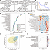 Figure 1
Metabolomics analysis of patients with ALS and healthy individuals. (A) Flow chart of metabolome analysis. For discovery cohort, we included patients with definite, probable, or possible ALS that met the revised El Escorial criteria and had disease duration less than 2 years. Patients with ALS were divided into rapidly progressive (Rapid ALS, n = 12) and slowly progressive (Slow ALS, n = 14) groups according to the median longitudinal change in ALSFRS-R (slope ALSFRS-R) from the first evaluation to 6 months. Rapid ALS was defined as a decline of ≥ 5 points in ALSFRS-R over 6 months (slope ALSFRS-R ≤ –0.83/month), and the others were classified as Slow ALS. Ten age- and sex-matched healthy controls were also included. In the replication cohort, 55 patients with ALS and 25 healthy controls were included. Patients with ALS were similarly classified using the same cut-off value (Rapid ALS, n = 30; Slow ALS, n = 25). (B) Kaplan-Meier curves of Rapid ALS and Slow ALS groups (median survival: 12 months vs 53 months, P = 0.0004, log-rank test). The primary endpoint was defined as the introduction of tracheostomy positive pressure ventilation (TPPV) or death of the patient. (C) Top 30 metabolites that discriminate among Rapid ALS, Slow ALS, and healthy controls by random forest classification. The metabolites were ranked according to their mean decrease accuracy. (D) Heatmap analysis of the top 20 metabolites identified by 1-way ANOVA. (E) Sparse partial least squares discriminant analysis (sPLS-DA) plots by MetaboAnalyst 6.0 to discriminate Rapid ALS, Slow ALS, and healthy controls (HC). (F) Loading plots of the top 10 metabolites by sPLS-DA shown in E.
Serum metabolomics detected 867 metabolites in total. Thirteen metabolites were significantly different among the 3 groups: Rapid ALS, Slow ALS, and healthy controls (Supplemental Figure 1 and Supplemental Table 3). Random forest analysis, whose predictive accuracy for discrimination of 3 groups was 61%, which is above random chance, showed that N-linoleoyl taurine, a metabolite belonging to NAT, was the most effective metabolite for distinguishing the groups (Figure 1C). NATs are taurine molecules conjugated to fatty acyl chains, and N-linoleoyl taurine is one representative species within this class. The top-ranked metabolites in the random forest analysis were mainly related to the expanded ECS including NATs, nucleic sugar, acylcarnitine, and polyamine metabolism. Similarly, sparse partial least squares discriminant analysis (sPLS-DA) and heatmap analysis also demonstrated that NATs, nucleic sugar, acylcarnitine, and polyamine metabolites were the discriminant metabolic factors for the 3 groups (Figure 1, D–F).
To identify metabolic factors relevant to the disease progression of ALS, we further performed a comparison between the Rapid ALS and Slow ALS groups. Volcano plot and heatmap analyses showed an increase in NATs and polyamine metabolites in the Rapid ALS group, and pathway enrichment analyses also showed alterations in those metabolic pathways (Figure 2A and Supplemental Figures 2–4). In addition, altered levels of bile acid metabolites were also found in the Rapid ALS group with pathway analyses (Supplemental Figure 3 and Supplemental Table 4). According to these clinical metabolome analyses, we selected 5 metabolic pathways, NATs, polyamine/arginine, xanthine, bile acid, and acylcarnitine metabolism, as candidate metabolic biomarkers associated with the progression of ALS. Detailed alterations in identified metabolic pathways among the 3 groups are presented in Supplemental Table 5.
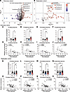 Figure 2
Alteration in N-acyl taurine metabolism among patients with rapidly progressive ALS, patients with slowly progressive ALS, and healthy individuals and their correlation with disease progression in the discovery cohort and the replication cohort. (A) Volcano plot of serum metabolomics analysis comparing between patients with rapid ALS and patients with slow ALS in the discovery cohort. (B) Volcano plot of serum lipid mediator analysis comparing between patients with rapid ALS and patients with slow ALS in the replication cohort. (C–F) Serum levels of N-acyl taurine metabolism in the discovery cohort (C, N-oleoyl taurine; D, N-stearoyl taurine; E, N-linoleoyl taurine; F, N-palmitoyl taurine). (G–J) Correlation between the serum levels of N-acyl taurines and the prospective, longitudinal change in ALSFRS-R between the first and second evaluations (slope ALSFRS-R) in the discovery cohort (G, N-oleoyl taurine; H, N-stearoyl taurine; I, N- linoleoyl taurine; J, N-palmitoyl taurine). (K–N) Serum levels of N-acyl taurine metabolism in the replication cohort (K, N-oleoyl taurine; L, N-stearoyl taurine; M, N- linoleoyl taurine; N, N-palmitoyl taurine). (O–R) Correlation between the serum levels of N-acyl taurines and slope ALSFRS-R in the replication cohort (O, N-oleoyl taurine; P, N-stearoyl taurine; Q, N-linoleoyl taurine; R, N-palmitoyl taurine). The discovery cohort was analyzed by broad untargeted metabolomics, whereas the replication cohort was analyzed using a targeted lipid mediator panel; therefore, the total number of detected analytes differed between the 2 cohorts. ALSFRS-R slope values were transformed using the Yeo-Johnson transformation before correlation analysis. One-way ANOVA and Tukey’s post hoc analysis were performed (*P < 0.05, **P < 0.01, and ***P < 0.001). HC, healthy controls; R, Rapid ALS; S, Slow ALS. A coefficient value (r) of 0.40–0.59 is considered moderate, while 0.60–0.79 is considered strong in Spearman’s rank correlation coefficient. Data are shown as mean ± SEM.
Among the identified metabolic pathways, the alterations of NATs were prominent, which belong to the expanded endocannabinoid system and act as a lipid mediators. To validate the changes in NATs, we performed a comprehensive lipid mediator analysis in the replication cohort (ALS, n = 55; healthy controls, n = 25) (Figure 1A). The smaller number of analytes detected in the replication cohort likely resulted from the use of a targeted lipid mediator validation assay, in contrast to the broad untargeted metabolomics platform used for the discovery cohort. Among the 75 lipid mediators analyzed, NAT levels were higher in the Rapid ALS group compared with the Slow ALS group, consistent with the discovery cohort, demonstrating reproducible results (Figure 2, A and B). A detailed analysis of the 4 detected NAT species revealed that, in both cohorts, their concentrations were elevated in the Rapid ALS group compared with healthy controls and the Slow ALS group. Furthermore, their levels showed a correlation with the rate of disease progression, as indicated by the slope of ALSFRS-R (Figure 2, C–R). When analyzed separately by sex, similar trends were observed in both male and female patients in both cohorts (Supplemental Figures 5 and 6). In addition, survival analyses were conducted by stratifying patients in both cohorts into 2 groups based on the median serum concentrations of each NAT. These analyses revealed that patients with higher NAT levels exhibited shorter survival durations, and this trend was consistently observed across both cohorts (Figure 3). These findings are also consistent with the observed correlations between NAT levels and disease progression rates. The other metabolites in the ECS such as N-acyl ethanolamines (NAE) were not altered among the 3 groups (Supplemental Figure 7).
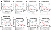 Figure 3
Kaplan-Meier survival curves based on serum N-acyl taurine levels in the discovery and replication cohorts. (A–D) Kaplan-Meier survival curves of the discovery cohort stratified by serum levels of N-acyl taurines: (A) N-oleoyl taurine, (B) N-stearoyl taurine, (C) N-linoleoyl taurine, and (D) N-palmitoyl taurine. (E–H) Kaplan-Meier survival curves of the replication cohort for the corresponding metabolites: (E) N-oleoyl taurine, (F) N-stearoyl taurine, (G) N-linoleoyl taurine, and (H) N-palmitoyl taurine. In both cohorts, patients were divided into high and low groups based on the median serum concentration of each N-acyl taurine. The primary endpoint was defined as the introduction of tracheostomy positive pressure ventilation (TPPV) or death of the patient. Log-rank test was used to compare survival between groups.
In vitro drug screening. The results of the clinical study on the relationship between the metabolome and disease progression of sporadic patients with ALS led us to drug screening using cellular models of ALS. A total of 29 chemical compounds that potentially modulate the focused metabolic pathways (the expanded ECS including NATs, polyamine/arginine, xanthine, bile acid, and acylcarnitine), together with riluzole and edaravone as controls, were administered to 2 cellular models of ALS: NSC-34 cells transiently expressing mutant TDP-43A315T and those expressing mutant SOD1G93A (Figure 4, A–M, and Supplemental Table 6). The 29 compounds were selected as a representative and experimentally feasible set of modulators for the progression-related metabolic pathways identified in the patient metabolomics data, with emphasis on translational relevance and suitability for human use whenever possible. Cell viability was assessed using a water-soluble tetrazolium (WST) assay. Among the tested compounds, intervention in the endocannabinoid system demonstrated the most pronounced effect. PF-04457845 and N-stearoyl taurine improved cell viability in both models in a concentration-dependent manner (Figure 4, B and D). Additionally, N-palmitoyl taurine improved cell viability in both models, although efficacy was observed at a single concentration (Figure 4E). While arachidonoyl ethanolamide (AEA, anandamide) improved the viability of NSC-34 cells expressing mutant TDP-43A315T, such efficacy was not verified in the cells expressing mutant SOD1G93A (Figure 4C). The efficacies of PF-04457845 and N-stearoyl taurine were further investigated by assessing lactate dehydrogenase (LDH) release. The results demonstrated a significant reduction in LDH release (Figure 4, F and G), corroborating their potential as therapeutic agents. To evaluate its effect in a human ALS disease model, we further administered PF-04457845 to motor neurons differentiated from iPS cells (iPS-MN) derived from 3 patients with ALS (Figure 4H). Concentration-dependent neuroprotective effects were demonstrated by IncuCyte SX5 (Figure 4, I–L) and LDH assay (Figure 4M), as evidenced by preserved neurite length, number of cell clusters, and reduced LDH release. No significant line-dependent difference in treatment effect was detected among the iPSC lines. To assess disease specificity in vitro, PF-04457845 was also evaluated in mock-transfected NSC-34 cells and control iPSC-derived motor neurons; however, no significant effect of treatment was observed on WST-8 assay or LDH release in mock-transfected NSC-34 cells — nor was there a significant effect of treatment observed on neuronal morphology or LDH release in control iPSC-derived motor neurons (Supplemental Figures 8 and 9). These data suggest that PF-04457845 has potential as a therapeutic candidate for ALS.
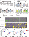 Figure 4
Cellular assays of compounds targeting the expanded endocannabinoid system and efficacy of PF-04457845 in NSC-34 cells and iPSC-derived motor neurons from sporadic patients with ALS. We tested a total of 29 compounds that target the identified metabolic pathways. We used NSC-34 cells transiently overexpressing TDP-43A315T or SOD1G93A and assessed cell viability using the WST-8 assay and cytotoxicity using the lactate dehydrogenase (LDH) assay. Results for the representative agents that modulate the expanded endocannabinoid system including N-acyl taurines are shown in this figure, and the other results are summarized in Supplemental Table 6. (A) Protocol of the cell viability assay. (B–E) Effects of compounds targeting expanded endocannabinoid system (B, PF-04457845; C, arachidonoyl ethanolamide [AEA]), and N-acyl taurine in expanded endocannabinoid system (D, N-stearoyl taurine; E, N-palmitoyl taurine), on cell viability. (F and G) Neuroprotective effects of PF-04457845 and N-stearoyl taurine were examined by LDH assays (F, PF-04457845; G, N-stearoyl taurine). (H) Protocol of administration of PF-04457845 on iPSC-derived motor neurons (iPS-MN) derived from 3 sporadic patients with ALS. (I) Analysis of cell morphology by IncuCyte SX5. iPS-MN were captured by phase separation and neurites (purple) and cell clusters (yellow) were automatically recognized by NeuroTrack algorithm. (J) The result of neurite length (N = 3 each from 3 iPS-MN). (K) The result of number of cell clusters (N = 3 each from 3 iPS-MN). (L) The result of area of cell clusters (N = 3 each from 3 iPS-MN). (M) The result of LDH assay (N = 3 each from 3 iPS-MN). For NSC-34 cells, 1-way ANOVA followed by Dunnett’s multiple-comparisons test was performed, with each concentration compared with the baseline condition. For iPSC-derived motor neurons, data were analyzed using a mixed-effects model, with treatment as a fixed effect and iPSC line as a random effect, followed by Dunnett’s multiple-comparisons test. (*P < 0.05, **P < 0.01, ****P < 0.0001). Scale bars: 200 μm. Data are shown as mean ± SEM.
PF-04457845 is a FAAH inhibitor. FAAH degrades NATs and NAEs, and knockdown of FAAH partially attenuates motor dysfunction in SOD1G93A transgenic mice (28). Based on the results of cellular screening and the putative mechanism of the hit compounds, we further investigated PF-04457845 in vivo.
In vivo analysis of hit compounds in SOD1G93A transgenic mice. The treatment of SOD1G93A transgenic mice with PF-04457845 was started at the age of 8 weeks and continued until the ethical endpoint. Body weight measurements, grip strength, and rotarod tests were repeated weekly from the age of 8 weeks to 19 weeks, which was the end stage of this model. PF-04457845 was mixed with chow at a dose of 0.001%, which corresponded to ~1 mg/kg/day and was administered to SOD1G93A transgenic mice. The survival of SOD1G93A transgenic mice administered PF-04457845 was extended by 8.5 days compared with that of untreated mice (Figure 5A). The administration of PF-04457845 significantly improved grip strength and rotarod test, whereas body weight was not significantly different (Figure 5, B–D). Survival and motor functional tests were also analyzed separately by the sex of the mice, yielding similar results (male mice, Supplemental Figure 10, A–D; female mice, Supplemental Figure 10, E–H). To assess possible nonspecific effects, PF-04457845 was also administered to WT C57BL/6 and B6/SJL mice. However, no significant effect of treatment was observed on body weight, grip strength, or rotarod performance, supporting the idea that its beneficial effects are disease-specific (Supplemental Figures 11 and 12).
 Figure 5
In vivo analysis of hit compounds in SOD1G93A transgenic mice. (A) Survival of SOD1G93A transgenic mice treated with PF-04457845 (~1 mg/kg/day, PF-04457845) or unmodified chow (Untreated) (n = 28, 34; median survival, 138, 129.5 days, respectively; P = 0.0001, log-rank test). PF-04457845 extended the survival of SOD1G93A transgenic mice compared with untreated mice. (B–D) Body weight, grip strength, and rotarod test in SOD1G93A transgenic mice treated with PF-04457845 or unmodified chow. Body weight was not significantly different, whereas grip strength (P < 0.0001) and rotarod test (P = 0.0004) were significantly improved by PF-04457845 (2-way ANOVA). To impute values after the endpoint, the final body weights were carried forward and the values of grip strength and rotarod were imputed to zero. (E–L) IHC images and quantitative analysis of immunoreactivity for choline acetyltransferase (ChAT) (E and F), glial fibrillary acidic protein (GFAP) (G and H), ionized calcium-binding adapter molecule 1 (IBA-1) (I and J), and Sry-related HMG-BOX gene 10 (SOX10) (K and L) in the ventral horn of the spinal cords of 16-week-old mice treated with PF-04457845 (n = 4) or unmodified chow (n = 4). Scale bars: 100 μm. Data are shown as mean ± SEM. Survival was analyzed using the Kaplan-Meier method and compared by the log-rank test. Body weight and motor functions were analyzed by 2-way ANOVA, and P values for the treatment effect are shown in the figure. Quantified pathology measures were compared using 2-sided unpaired t tests. *P < 0.05.
To examine the effect of PF-04457845 on the motor neurons of SOD1G93A transgenic mice, paraffin-embedded sections of lumbar spinal cord (L5) samples from 16-week-old mice were analyzed by immunohistochemistry. The number of motor neurons with immunoreactivity for choline acetyltransferase within the lumbar spinal cord was significantly preserved in the SOD1G93A mice treated with PF-04457845 compared with untreated mice (Figure 5, E and F). The proliferation of astrocytes, microglia and oligodendrocytes was evaluated with anti–glial fibrillary acidic protein (anti-GFAP) antibody (Figure 5, G and H), anti–ionized calcium-binding adapter molecule 1 (anti–IBA-1) antibody (Figure 5, I and J), and anti–Sry-related HMG-BOX gene 10 (anti-SOX10) antibody (Figure 5, K and L), respectively. The proportion of astrocytes was significantly decreased by PF-04457845 treatment, although the proportions of microglia and oligodendrocytes were unchanged.
Lipidomics analysis of the murine spinal cords. To clarify the influence of PF-04457845 on lipid metabolism, a comprehensive lipidomics analysis was conducted on the spinal cords of 3 distinct groups: wild-type mice, untreated SOD1G93A transgenic mice, and SOD1G93A transgenic mice treated with PF-04457845. A total of 799 lipid metabolites were identified in the process (Supplemental Table 7). Subsequent sPLS-DA analysis effectively segregated these 3 groups, underscoring that the differentiation was primarily attributed to the concentrations of NATs and NAEs, both of which are known substrates of FAAH (Figure 6, A and B). By examining the levels of specific metabolites belonging to NATs and NAEs, we found that both lipids experienced an increase after treatment with PF-04457845, demonstrating that orally administered PF-04457845 successfully upregulates NATs and NAEs in the central nervous system of the SOD1G93A transgenic mice. (Figure 6, C and D). Furthermore, 3 of the 4 NAT species identified in the sera of patients with ALS — N-palmitoyl taurine(16:0), N-stearoyl taurine(18:0), and N-oleoyl taurine(18:1) — were detected by lipidomics. Notably, the levels of all these NATs were elevated in the spinal cord of untreated SOD1G93A transgenic mice compared with WT mice, being consistent with the increase observed in patients with rapid ALS. Such changes were not observed for NAEs (Figure 6, C–E, and Supplemental Figure 13). These results indicate that elevation of neural NATs levels represents a common pathogenic mechanism shared by SOD1G93A transgenic mice and patients with Rapid ALS.
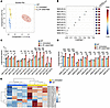 Figure 6
Lipidomics analysis of spinal cords of WT and SOD1G93A transgenic mice with or without treatment with PF-04457845. (A) Sparse partial least squares discriminant analysis (sPLS-DA) plots by MetaboAnalyst 6.0 to discriminate WT mice (WT, n = 4), untreated SOD1G93A transgenic mice (S_Untreated, n = 4), and SOD1G93A transgenic mice treated with PF-04457845 (S_PF-04457845, n = 4). Loading plots of the top 10 lipids by sPLS-DA are shown in B. (C) Log transformed concentrations of N-acyl taurines (NAT). (D) Log transformed concentrations of N-acyl ethanolamine (NAE). (E) Heatmap analysis of N-acyl taurines comparing untreated SOD1G93A transgenic mice and WT mice. Data are shown as mean ± SEM. *P < 0.05, **P < 0.01, and ***P < 0.001, ****P < 0.0001. One-way ANOVA and Tukey’s post hoc analysis were performed.
RNA and protein analysis of the spinal cord of SOD1G93A transgenic mice treated with PF-04457845. To explore the mechanism of phenotypic amelioration in SOD1G93A transgenic mice by PF-04457845, we performed RNA-Seq on their spinal cord. Enrichment analysis of the RNA-Seq data revealed significant involvement of pathways related to macrophage and microglial differentiation and lipoprotein metabolism in PF-04457845–treated mice (n = 4) compared with untreated mice (n = 4) (Figure 7, A and B, and Supplemental Table 8). Ingenuity Pathway Analysis (IPA) of the RNA-Seq data revealed activation of the IL-13 pathway in SOD1G93A transgenic mice treated with PF-04457845 (Figure 7C). In line with this finding, Cd36, a downstream molecule of IL-13, was upregulated in the spinal cord of PF-04457845–treated mice (Supplemental Figure 14). CD36+ microglia regulate lipid metabolism, promote myelin debris clearance, and support antioxidant responses (29, 30). Lipoprotein lipase (LPL) is also known to be expressed in microglial subtypes such as disease-associated microglia (DAM) and proliferative region–associated microglia (PAM) (31, 32). Stimulation of CB2, an endocannabinoid receptor expressed on microglia, is reported to suppress inflammatory signaling and promote an antiinflammatory, metabolically supportive microglial state (33, 34). Therefore, we examined microglial markers and molecules downstream of CB2 receptor signaling in the mouse spinal cord by Western blotting. ERK1/2 are key kinases in the MAPK pathway and function as downstream effectors of cannabinoid receptor associated signaling. The ratio of phosphorylated ERK1/2 to total ERK1/2 was increased in PF-04457845–treated mice compared with untreated mice, suggesting activation of the CB1/CB2 signaling pathway (Figure 7, D and E) (35). The protein levels of IBA-1 were not significantly changed by PF-04457845 treatment, consistent with the histopathological findings (Figure 7, D and F). The protein levels of CD86, a proinflammatory microglial marker, were decreased, whereas those of CD36 and LPL were increased in PF-04457845–treated mice, supporting a shift toward a neuroprotective microglial phenotype (Figure 7, D and G–I). The expression of SOD1 was not affected by PF-04457845 (Figure 7, D and J).
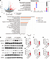 Figure 7
RNA-Seq analysis of the spinal cords from SOD1G93A transgenic mice treated with PF-04457845. (A) Volcano plot of the differentially expressed genes of the bulk spinal cords between the mice treated with PF-04457845 (n = 4) and the untreated mice (n = 4). (B and C) Gene Ontology enrichment analysis for up-regulated genes (B) and Ingenuity Pathway Analysis (IPA) (C) analysis of genes with significant expression changes (fold change < 1/1.5 or > 1., P < 0.05) in the spinal cords of 16-week-old SOD1G93A transgenic mice treated with PF-04457845 (n = 4) compared with untreated mice (n = 4). (D) Western blot analysis of proteins downstream of cannabinoid receptor type 2 (CB2), microglial markers, and superoxide dismutase 1 (SOD1) in the spinal cords of 16-week-old SOD1G93A transgenic mice treated with PF-04457845 (S_PF-04457845, n = 5) and untreated SOD1G93A transgenic mice (S_untreated, n = 4). (E–J) Quantitative analysis of phosphorylated ERK/ERK (E), ionized calcium-binding adapter molecule 1 (IBA-1) (F), CD86 (G), CD36 (H), lipoprotein lipase (LPL) (I), and SOD1 (J). The levels of IBA-1, CD86, CD36, LPL, and SOD1 were normalized to GAPDH levels. Data are shown as mean ± SEM. *P < 0.05, unpaired 2-sided t test.
Transcriptome alteration in neurons by PF-04457845. The bulk RNA-Seq of the spinal cord primarily revealed altered signaling related to the CB2 receptor, which is mainly expressed in microglia and oligodendrocytes. Given that neurons, as well as astrocytes, express CB1 receptors, the treatment-related changes in neurons did not appear to be reflected in the RNA-Seq data. To examine the effects of treatment with PF-04457845 on neurons, we performed single-nucleus RNA-Seq (snRNA-Seq) on the spinal cords from SOD1G93A transgenic mice treated with PF-04457845 or unmodified chow (Figure 8A and Supplemental Table 9). Sequencing quality was high, with > 90% of bases at Q30 in the RNA reads, a mean of 50,000 reads per cell, and > 76% of reads mapped to the genome. Unsupervised clustering identified 6 major cell types, and each cluster expressed cell type–specific cell markers (Figure 8, B and C). The cluster of neurons expressed Cnr1, the gene encoding the CB1 receptor; however, Cnr2, the CB2 receptor, was barely expressed in the neuronal population (Figure 8, D and E). The administration of PF-04457845 upregulated the expression of genes including Tshz3, Ppp3r1, and Cyfip1 in neurons (Figure 8F). Enrichment analysis revealed that PF-04457845 activates synapse plasticity and neuronal development, although no specific pathway was downregulated with the treatment (Figure 8, G and H). In addition, although the abundance of Cnr1+ neurons was not significantly altered, pathway analysis revealed treatment-associated transcriptional changes in this neuronal subset (Supplemental Figure 15). In oligodendrocytes, pathway analysis revealed treatment-associated changes related to myelination, oligodendrocyte differentiation, and cytoskeletal organization, suggesting transcriptional remodeling without a major change in oligodendrocyte abundance (Figure 5, K and L, and Supplemental Figure 16). In microglia, PF-04457845 treatment was associated with downregulation of pathways related to cytokine production, and cytoskeletal/cell migration programs, together with upregulation of pathways related to synapse organization and trans-synaptic signaling (Supplemental Figure 17).
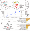 Figure 8
Single-nucleus RNA-Seq analysis of the spinal cords from SOD1G93A transgenic mice treated with PF-04457845. We performed single-nucleus RNA-seq on the spinal cords of 16-week-old SOD1G93A transgenic mice treated with PF-04457845 (S_PF-04457845) or unmodified chow (S_Untreated). (A) t-SNE plot color-coded by sample, with red for untreated mice and blue for PF-04457845–treated mice. (B) t-SNE plots of all cells sequenced showing 6 cell types. (C) Dot plot depicting the expression of specific markers for each cell type. (D and E) Expression levels of CB1 (D) and CB2 (E) in the neuron cluster of each sample. (F) Volcano plot of the differentially expressed genes in the neuron cluster from the mice treated with PF-04457845 compared with that from the untreated mice. (G and H) Gene Ontology enrichment analysis for upregulated (G) and downregulated (H) genes between the neuron clusters from the mice treated with PF-04457845 and the untreated mice.


