
Research ArticleImmunologyInflammation
Open Access |  10.1172/jci.insight.200002
Truncating RELA variants drive autoinflammation and autoimmunity by impairing the negative feedback control of NF-κB
Nadja Lucas,1 Sophia Weidler,1 Antonia A. Eicher,2 Baerbel Keller,3,4 Özlem Satirer,5 Adam Desrochers,1 Timothy J.S. Ramnarine,2 Mohammad Mokhtari,2 Sophie Elstner,1 Timmy Strauss,1 Simon W. Mages,2 Arek Kendirli,6 Oana Cristina Buzoianu,5 Tobias B. Haack,7 Lina Igel,8 Tim Niehues,8 Sandra von Hardenberg,9 Maria Fasshauer,10 Rami Abou Jamra,11 Hagen Ott,12 Ulrike Hüffmeier,13 Catharina Schuetz,1,14 Marisa Bijwaard,1 Susan Wagner,1 Paulina Switala,1 Sarah Koss,1 Jurek Schultz,15 Stefanie Kretschmer,1 Jasmin Kümmerle-Deschner,5 Christine Wolf,1 Johanna Klughammer,2 and Min Ae Lee-Kirsch1,14,16
1Department of Pediatrics, Medizinische Fakultät Carl Gustav Carus, Technische Universität Dresden, Dresden, Germany.
2Gene Center and Department of Biochemistry, Ludwig-Maximilians-Universität München, Munich, Germany.
3Department of Rheumatology and Clinical Immunology, and
4Center for Chronic Immunodeficiency (CCI), Medical Center - University of Freiburg, Faculty of Medicine, University of Freiburg, Freiburg, Germany.
5Division of Paediatric Rheumatology and Autoinflammation Reference Centre Tübingen, Department of Paediatrics, University Hospital Tübingen, Tübingen, Germany.
6Institute of Clinical Neuroimmunology, University Hospital, Ludwig-Maximilians-Universität München, Munich, Germany.
7Institute of Medical Genetics and Applied Genomics, University Hospital Tübingen, Tübingen, Germany.
8Department of Pediatrics, Helios Klinik Krefeld, Krefeld, Germany.
9Department of Human Genetics, Hannover Medical School, Hannover, Germany.
10Hospital for Children and Adolescents, Hospital St. Georg, Academic Teaching Hospital of the University of Leipzig, Leipzig, Germany.
11Institute of Human Genetics, University of Leipzig Medical Center, Leipzig, Germany.
12Section of Pediatric Dermatology, Munich University Center for Children with Medical and Developmental Complexity, München, Germany.
13Institute of Human Genetics, Universitätsklinikum Erlangen, Friedrich-Alexander-Universität Erlangen-Nürnberg, Erlangen, Germany.
14German Center for Child and Adolescent Health (DZKJ), partner site Leipzig/Dresden, Dresden, Germany.
15Department of Pediatric Surgery, Medizinische Fakultät Carl Gustav Carus, and
16University Center for Rare Diseases, Medizinische Fakultät Carl Gustav Carus, Technische Universität Dresden, Dresden, Germany.
Address correspondence to: Min Ae Lee-Kirsch, Department of Pediatrics, Medizinische Fakultät Carl Gustav Carus, Technische Universität Dresden, Fetscherstraße 74, 01307 Dresden, Germany. Phone: 0049.351.45816878; Email: minae.lee-kirsch@uniklinikum-dresden.de. Or to: Johanna Klughammer, Gene Center and Department of Biochemistry, Ludwig-Maximilians-Universität München, Feodor-Lynen-Straße 25, 81377 Munich, Germany. Phone: 0049.89.2180.71050; Email: klughammer@genzentrum.lmu.de.
Authorship note: NL, S. Weidler, and AE contributed equally to this work. JK and MALK are co–corresponding authors.
Find articles by Lucas, N. in: PubMed | Google Scholar
1Department of Pediatrics, Medizinische Fakultät Carl Gustav Carus, Technische Universität Dresden, Dresden, Germany.
2Gene Center and Department of Biochemistry, Ludwig-Maximilians-Universität München, Munich, Germany.
3Department of Rheumatology and Clinical Immunology, and
4Center for Chronic Immunodeficiency (CCI), Medical Center - University of Freiburg, Faculty of Medicine, University of Freiburg, Freiburg, Germany.
5Division of Paediatric Rheumatology and Autoinflammation Reference Centre Tübingen, Department of Paediatrics, University Hospital Tübingen, Tübingen, Germany.
6Institute of Clinical Neuroimmunology, University Hospital, Ludwig-Maximilians-Universität München, Munich, Germany.
7Institute of Medical Genetics and Applied Genomics, University Hospital Tübingen, Tübingen, Germany.
8Department of Pediatrics, Helios Klinik Krefeld, Krefeld, Germany.
9Department of Human Genetics, Hannover Medical School, Hannover, Germany.
10Hospital for Children and Adolescents, Hospital St. Georg, Academic Teaching Hospital of the University of Leipzig, Leipzig, Germany.
11Institute of Human Genetics, University of Leipzig Medical Center, Leipzig, Germany.
12Section of Pediatric Dermatology, Munich University Center for Children with Medical and Developmental Complexity, München, Germany.
13Institute of Human Genetics, Universitätsklinikum Erlangen, Friedrich-Alexander-Universität Erlangen-Nürnberg, Erlangen, Germany.
14German Center for Child and Adolescent Health (DZKJ), partner site Leipzig/Dresden, Dresden, Germany.
15Department of Pediatric Surgery, Medizinische Fakultät Carl Gustav Carus, and
16University Center for Rare Diseases, Medizinische Fakultät Carl Gustav Carus, Technische Universität Dresden, Dresden, Germany.
Address correspondence to: Min Ae Lee-Kirsch, Department of Pediatrics, Medizinische Fakultät Carl Gustav Carus, Technische Universität Dresden, Fetscherstraße 74, 01307 Dresden, Germany. Phone: 0049.351.45816878; Email: minae.lee-kirsch@uniklinikum-dresden.de. Or to: Johanna Klughammer, Gene Center and Department of Biochemistry, Ludwig-Maximilians-Universität München, Feodor-Lynen-Straße 25, 81377 Munich, Germany. Phone: 0049.89.2180.71050; Email: klughammer@genzentrum.lmu.de.
Authorship note: NL, S. Weidler, and AE contributed equally to this work. JK and MALK are co–corresponding authors.
Find articles by Weidler, S. in: PubMed | Google Scholar
1Department of Pediatrics, Medizinische Fakultät Carl Gustav Carus, Technische Universität Dresden, Dresden, Germany.
2Gene Center and Department of Biochemistry, Ludwig-Maximilians-Universität München, Munich, Germany.
3Department of Rheumatology and Clinical Immunology, and
4Center for Chronic Immunodeficiency (CCI), Medical Center - University of Freiburg, Faculty of Medicine, University of Freiburg, Freiburg, Germany.
5Division of Paediatric Rheumatology and Autoinflammation Reference Centre Tübingen, Department of Paediatrics, University Hospital Tübingen, Tübingen, Germany.
6Institute of Clinical Neuroimmunology, University Hospital, Ludwig-Maximilians-Universität München, Munich, Germany.
7Institute of Medical Genetics and Applied Genomics, University Hospital Tübingen, Tübingen, Germany.
8Department of Pediatrics, Helios Klinik Krefeld, Krefeld, Germany.
9Department of Human Genetics, Hannover Medical School, Hannover, Germany.
10Hospital for Children and Adolescents, Hospital St. Georg, Academic Teaching Hospital of the University of Leipzig, Leipzig, Germany.
11Institute of Human Genetics, University of Leipzig Medical Center, Leipzig, Germany.
12Section of Pediatric Dermatology, Munich University Center for Children with Medical and Developmental Complexity, München, Germany.
13Institute of Human Genetics, Universitätsklinikum Erlangen, Friedrich-Alexander-Universität Erlangen-Nürnberg, Erlangen, Germany.
14German Center for Child and Adolescent Health (DZKJ), partner site Leipzig/Dresden, Dresden, Germany.
15Department of Pediatric Surgery, Medizinische Fakultät Carl Gustav Carus, and
16University Center for Rare Diseases, Medizinische Fakultät Carl Gustav Carus, Technische Universität Dresden, Dresden, Germany.
Address correspondence to: Min Ae Lee-Kirsch, Department of Pediatrics, Medizinische Fakultät Carl Gustav Carus, Technische Universität Dresden, Fetscherstraße 74, 01307 Dresden, Germany. Phone: 0049.351.45816878; Email: minae.lee-kirsch@uniklinikum-dresden.de. Or to: Johanna Klughammer, Gene Center and Department of Biochemistry, Ludwig-Maximilians-Universität München, Feodor-Lynen-Straße 25, 81377 Munich, Germany. Phone: 0049.89.2180.71050; Email: klughammer@genzentrum.lmu.de.
Authorship note: NL, S. Weidler, and AE contributed equally to this work. JK and MALK are co–corresponding authors.
Find articles by Eicher, A. in: PubMed | Google Scholar
1Department of Pediatrics, Medizinische Fakultät Carl Gustav Carus, Technische Universität Dresden, Dresden, Germany.
2Gene Center and Department of Biochemistry, Ludwig-Maximilians-Universität München, Munich, Germany.
3Department of Rheumatology and Clinical Immunology, and
4Center for Chronic Immunodeficiency (CCI), Medical Center - University of Freiburg, Faculty of Medicine, University of Freiburg, Freiburg, Germany.
5Division of Paediatric Rheumatology and Autoinflammation Reference Centre Tübingen, Department of Paediatrics, University Hospital Tübingen, Tübingen, Germany.
6Institute of Clinical Neuroimmunology, University Hospital, Ludwig-Maximilians-Universität München, Munich, Germany.
7Institute of Medical Genetics and Applied Genomics, University Hospital Tübingen, Tübingen, Germany.
8Department of Pediatrics, Helios Klinik Krefeld, Krefeld, Germany.
9Department of Human Genetics, Hannover Medical School, Hannover, Germany.
10Hospital for Children and Adolescents, Hospital St. Georg, Academic Teaching Hospital of the University of Leipzig, Leipzig, Germany.
11Institute of Human Genetics, University of Leipzig Medical Center, Leipzig, Germany.
12Section of Pediatric Dermatology, Munich University Center for Children with Medical and Developmental Complexity, München, Germany.
13Institute of Human Genetics, Universitätsklinikum Erlangen, Friedrich-Alexander-Universität Erlangen-Nürnberg, Erlangen, Germany.
14German Center for Child and Adolescent Health (DZKJ), partner site Leipzig/Dresden, Dresden, Germany.
15Department of Pediatric Surgery, Medizinische Fakultät Carl Gustav Carus, and
16University Center for Rare Diseases, Medizinische Fakultät Carl Gustav Carus, Technische Universität Dresden, Dresden, Germany.
Address correspondence to: Min Ae Lee-Kirsch, Department of Pediatrics, Medizinische Fakultät Carl Gustav Carus, Technische Universität Dresden, Fetscherstraße 74, 01307 Dresden, Germany. Phone: 0049.351.45816878; Email: minae.lee-kirsch@uniklinikum-dresden.de. Or to: Johanna Klughammer, Gene Center and Department of Biochemistry, Ludwig-Maximilians-Universität München, Feodor-Lynen-Straße 25, 81377 Munich, Germany. Phone: 0049.89.2180.71050; Email: klughammer@genzentrum.lmu.de.
Authorship note: NL, S. Weidler, and AE contributed equally to this work. JK and MALK are co–corresponding authors.
Find articles by Keller, B. in: PubMed | Google Scholar
1Department of Pediatrics, Medizinische Fakultät Carl Gustav Carus, Technische Universität Dresden, Dresden, Germany.
2Gene Center and Department of Biochemistry, Ludwig-Maximilians-Universität München, Munich, Germany.
3Department of Rheumatology and Clinical Immunology, and
4Center for Chronic Immunodeficiency (CCI), Medical Center - University of Freiburg, Faculty of Medicine, University of Freiburg, Freiburg, Germany.
5Division of Paediatric Rheumatology and Autoinflammation Reference Centre Tübingen, Department of Paediatrics, University Hospital Tübingen, Tübingen, Germany.
6Institute of Clinical Neuroimmunology, University Hospital, Ludwig-Maximilians-Universität München, Munich, Germany.
7Institute of Medical Genetics and Applied Genomics, University Hospital Tübingen, Tübingen, Germany.
8Department of Pediatrics, Helios Klinik Krefeld, Krefeld, Germany.
9Department of Human Genetics, Hannover Medical School, Hannover, Germany.
10Hospital for Children and Adolescents, Hospital St. Georg, Academic Teaching Hospital of the University of Leipzig, Leipzig, Germany.
11Institute of Human Genetics, University of Leipzig Medical Center, Leipzig, Germany.
12Section of Pediatric Dermatology, Munich University Center for Children with Medical and Developmental Complexity, München, Germany.
13Institute of Human Genetics, Universitätsklinikum Erlangen, Friedrich-Alexander-Universität Erlangen-Nürnberg, Erlangen, Germany.
14German Center for Child and Adolescent Health (DZKJ), partner site Leipzig/Dresden, Dresden, Germany.
15Department of Pediatric Surgery, Medizinische Fakultät Carl Gustav Carus, and
16University Center for Rare Diseases, Medizinische Fakultät Carl Gustav Carus, Technische Universität Dresden, Dresden, Germany.
Address correspondence to: Min Ae Lee-Kirsch, Department of Pediatrics, Medizinische Fakultät Carl Gustav Carus, Technische Universität Dresden, Fetscherstraße 74, 01307 Dresden, Germany. Phone: 0049.351.45816878; Email: minae.lee-kirsch@uniklinikum-dresden.de. Or to: Johanna Klughammer, Gene Center and Department of Biochemistry, Ludwig-Maximilians-Universität München, Feodor-Lynen-Straße 25, 81377 Munich, Germany. Phone: 0049.89.2180.71050; Email: klughammer@genzentrum.lmu.de.
Authorship note: NL, S. Weidler, and AE contributed equally to this work. JK and MALK are co–corresponding authors.
Find articles by Satirer, Ö. in: PubMed | Google Scholar
1Department of Pediatrics, Medizinische Fakultät Carl Gustav Carus, Technische Universität Dresden, Dresden, Germany.
2Gene Center and Department of Biochemistry, Ludwig-Maximilians-Universität München, Munich, Germany.
3Department of Rheumatology and Clinical Immunology, and
4Center for Chronic Immunodeficiency (CCI), Medical Center - University of Freiburg, Faculty of Medicine, University of Freiburg, Freiburg, Germany.
5Division of Paediatric Rheumatology and Autoinflammation Reference Centre Tübingen, Department of Paediatrics, University Hospital Tübingen, Tübingen, Germany.
6Institute of Clinical Neuroimmunology, University Hospital, Ludwig-Maximilians-Universität München, Munich, Germany.
7Institute of Medical Genetics and Applied Genomics, University Hospital Tübingen, Tübingen, Germany.
8Department of Pediatrics, Helios Klinik Krefeld, Krefeld, Germany.
9Department of Human Genetics, Hannover Medical School, Hannover, Germany.
10Hospital for Children and Adolescents, Hospital St. Georg, Academic Teaching Hospital of the University of Leipzig, Leipzig, Germany.
11Institute of Human Genetics, University of Leipzig Medical Center, Leipzig, Germany.
12Section of Pediatric Dermatology, Munich University Center for Children with Medical and Developmental Complexity, München, Germany.
13Institute of Human Genetics, Universitätsklinikum Erlangen, Friedrich-Alexander-Universität Erlangen-Nürnberg, Erlangen, Germany.
14German Center for Child and Adolescent Health (DZKJ), partner site Leipzig/Dresden, Dresden, Germany.
15Department of Pediatric Surgery, Medizinische Fakultät Carl Gustav Carus, and
16University Center for Rare Diseases, Medizinische Fakultät Carl Gustav Carus, Technische Universität Dresden, Dresden, Germany.
Address correspondence to: Min Ae Lee-Kirsch, Department of Pediatrics, Medizinische Fakultät Carl Gustav Carus, Technische Universität Dresden, Fetscherstraße 74, 01307 Dresden, Germany. Phone: 0049.351.45816878; Email: minae.lee-kirsch@uniklinikum-dresden.de. Or to: Johanna Klughammer, Gene Center and Department of Biochemistry, Ludwig-Maximilians-Universität München, Feodor-Lynen-Straße 25, 81377 Munich, Germany. Phone: 0049.89.2180.71050; Email: klughammer@genzentrum.lmu.de.
Authorship note: NL, S. Weidler, and AE contributed equally to this work. JK and MALK are co–corresponding authors.
Find articles by Desrochers, A. in: PubMed | Google Scholar
1Department of Pediatrics, Medizinische Fakultät Carl Gustav Carus, Technische Universität Dresden, Dresden, Germany.
2Gene Center and Department of Biochemistry, Ludwig-Maximilians-Universität München, Munich, Germany.
3Department of Rheumatology and Clinical Immunology, and
4Center for Chronic Immunodeficiency (CCI), Medical Center - University of Freiburg, Faculty of Medicine, University of Freiburg, Freiburg, Germany.
5Division of Paediatric Rheumatology and Autoinflammation Reference Centre Tübingen, Department of Paediatrics, University Hospital Tübingen, Tübingen, Germany.
6Institute of Clinical Neuroimmunology, University Hospital, Ludwig-Maximilians-Universität München, Munich, Germany.
7Institute of Medical Genetics and Applied Genomics, University Hospital Tübingen, Tübingen, Germany.
8Department of Pediatrics, Helios Klinik Krefeld, Krefeld, Germany.
9Department of Human Genetics, Hannover Medical School, Hannover, Germany.
10Hospital for Children and Adolescents, Hospital St. Georg, Academic Teaching Hospital of the University of Leipzig, Leipzig, Germany.
11Institute of Human Genetics, University of Leipzig Medical Center, Leipzig, Germany.
12Section of Pediatric Dermatology, Munich University Center for Children with Medical and Developmental Complexity, München, Germany.
13Institute of Human Genetics, Universitätsklinikum Erlangen, Friedrich-Alexander-Universität Erlangen-Nürnberg, Erlangen, Germany.
14German Center for Child and Adolescent Health (DZKJ), partner site Leipzig/Dresden, Dresden, Germany.
15Department of Pediatric Surgery, Medizinische Fakultät Carl Gustav Carus, and
16University Center for Rare Diseases, Medizinische Fakultät Carl Gustav Carus, Technische Universität Dresden, Dresden, Germany.
Address correspondence to: Min Ae Lee-Kirsch, Department of Pediatrics, Medizinische Fakultät Carl Gustav Carus, Technische Universität Dresden, Fetscherstraße 74, 01307 Dresden, Germany. Phone: 0049.351.45816878; Email: minae.lee-kirsch@uniklinikum-dresden.de. Or to: Johanna Klughammer, Gene Center and Department of Biochemistry, Ludwig-Maximilians-Universität München, Feodor-Lynen-Straße 25, 81377 Munich, Germany. Phone: 0049.89.2180.71050; Email: klughammer@genzentrum.lmu.de.
Authorship note: NL, S. Weidler, and AE contributed equally to this work. JK and MALK are co–corresponding authors.
Find articles by Ramnarine, T. in: PubMed | Google Scholar
1Department of Pediatrics, Medizinische Fakultät Carl Gustav Carus, Technische Universität Dresden, Dresden, Germany.
2Gene Center and Department of Biochemistry, Ludwig-Maximilians-Universität München, Munich, Germany.
3Department of Rheumatology and Clinical Immunology, and
4Center for Chronic Immunodeficiency (CCI), Medical Center - University of Freiburg, Faculty of Medicine, University of Freiburg, Freiburg, Germany.
5Division of Paediatric Rheumatology and Autoinflammation Reference Centre Tübingen, Department of Paediatrics, University Hospital Tübingen, Tübingen, Germany.
6Institute of Clinical Neuroimmunology, University Hospital, Ludwig-Maximilians-Universität München, Munich, Germany.
7Institute of Medical Genetics and Applied Genomics, University Hospital Tübingen, Tübingen, Germany.
8Department of Pediatrics, Helios Klinik Krefeld, Krefeld, Germany.
9Department of Human Genetics, Hannover Medical School, Hannover, Germany.
10Hospital for Children and Adolescents, Hospital St. Georg, Academic Teaching Hospital of the University of Leipzig, Leipzig, Germany.
11Institute of Human Genetics, University of Leipzig Medical Center, Leipzig, Germany.
12Section of Pediatric Dermatology, Munich University Center for Children with Medical and Developmental Complexity, München, Germany.
13Institute of Human Genetics, Universitätsklinikum Erlangen, Friedrich-Alexander-Universität Erlangen-Nürnberg, Erlangen, Germany.
14German Center for Child and Adolescent Health (DZKJ), partner site Leipzig/Dresden, Dresden, Germany.
15Department of Pediatric Surgery, Medizinische Fakultät Carl Gustav Carus, and
16University Center for Rare Diseases, Medizinische Fakultät Carl Gustav Carus, Technische Universität Dresden, Dresden, Germany.
Address correspondence to: Min Ae Lee-Kirsch, Department of Pediatrics, Medizinische Fakultät Carl Gustav Carus, Technische Universität Dresden, Fetscherstraße 74, 01307 Dresden, Germany. Phone: 0049.351.45816878; Email: minae.lee-kirsch@uniklinikum-dresden.de. Or to: Johanna Klughammer, Gene Center and Department of Biochemistry, Ludwig-Maximilians-Universität München, Feodor-Lynen-Straße 25, 81377 Munich, Germany. Phone: 0049.89.2180.71050; Email: klughammer@genzentrum.lmu.de.
Authorship note: NL, S. Weidler, and AE contributed equally to this work. JK and MALK are co–corresponding authors.
Find articles by Mokhtari, M. in: PubMed | Google Scholar
1Department of Pediatrics, Medizinische Fakultät Carl Gustav Carus, Technische Universität Dresden, Dresden, Germany.
2Gene Center and Department of Biochemistry, Ludwig-Maximilians-Universität München, Munich, Germany.
3Department of Rheumatology and Clinical Immunology, and
4Center for Chronic Immunodeficiency (CCI), Medical Center - University of Freiburg, Faculty of Medicine, University of Freiburg, Freiburg, Germany.
5Division of Paediatric Rheumatology and Autoinflammation Reference Centre Tübingen, Department of Paediatrics, University Hospital Tübingen, Tübingen, Germany.
6Institute of Clinical Neuroimmunology, University Hospital, Ludwig-Maximilians-Universität München, Munich, Germany.
7Institute of Medical Genetics and Applied Genomics, University Hospital Tübingen, Tübingen, Germany.
8Department of Pediatrics, Helios Klinik Krefeld, Krefeld, Germany.
9Department of Human Genetics, Hannover Medical School, Hannover, Germany.
10Hospital for Children and Adolescents, Hospital St. Georg, Academic Teaching Hospital of the University of Leipzig, Leipzig, Germany.
11Institute of Human Genetics, University of Leipzig Medical Center, Leipzig, Germany.
12Section of Pediatric Dermatology, Munich University Center for Children with Medical and Developmental Complexity, München, Germany.
13Institute of Human Genetics, Universitätsklinikum Erlangen, Friedrich-Alexander-Universität Erlangen-Nürnberg, Erlangen, Germany.
14German Center for Child and Adolescent Health (DZKJ), partner site Leipzig/Dresden, Dresden, Germany.
15Department of Pediatric Surgery, Medizinische Fakultät Carl Gustav Carus, and
16University Center for Rare Diseases, Medizinische Fakultät Carl Gustav Carus, Technische Universität Dresden, Dresden, Germany.
Address correspondence to: Min Ae Lee-Kirsch, Department of Pediatrics, Medizinische Fakultät Carl Gustav Carus, Technische Universität Dresden, Fetscherstraße 74, 01307 Dresden, Germany. Phone: 0049.351.45816878; Email: minae.lee-kirsch@uniklinikum-dresden.de. Or to: Johanna Klughammer, Gene Center and Department of Biochemistry, Ludwig-Maximilians-Universität München, Feodor-Lynen-Straße 25, 81377 Munich, Germany. Phone: 0049.89.2180.71050; Email: klughammer@genzentrum.lmu.de.
Authorship note: NL, S. Weidler, and AE contributed equally to this work. JK and MALK are co–corresponding authors.
Find articles by Elstner, S. in: PubMed | Google Scholar
1Department of Pediatrics, Medizinische Fakultät Carl Gustav Carus, Technische Universität Dresden, Dresden, Germany.
2Gene Center and Department of Biochemistry, Ludwig-Maximilians-Universität München, Munich, Germany.
3Department of Rheumatology and Clinical Immunology, and
4Center for Chronic Immunodeficiency (CCI), Medical Center - University of Freiburg, Faculty of Medicine, University of Freiburg, Freiburg, Germany.
5Division of Paediatric Rheumatology and Autoinflammation Reference Centre Tübingen, Department of Paediatrics, University Hospital Tübingen, Tübingen, Germany.
6Institute of Clinical Neuroimmunology, University Hospital, Ludwig-Maximilians-Universität München, Munich, Germany.
7Institute of Medical Genetics and Applied Genomics, University Hospital Tübingen, Tübingen, Germany.
8Department of Pediatrics, Helios Klinik Krefeld, Krefeld, Germany.
9Department of Human Genetics, Hannover Medical School, Hannover, Germany.
10Hospital for Children and Adolescents, Hospital St. Georg, Academic Teaching Hospital of the University of Leipzig, Leipzig, Germany.
11Institute of Human Genetics, University of Leipzig Medical Center, Leipzig, Germany.
12Section of Pediatric Dermatology, Munich University Center for Children with Medical and Developmental Complexity, München, Germany.
13Institute of Human Genetics, Universitätsklinikum Erlangen, Friedrich-Alexander-Universität Erlangen-Nürnberg, Erlangen, Germany.
14German Center for Child and Adolescent Health (DZKJ), partner site Leipzig/Dresden, Dresden, Germany.
15Department of Pediatric Surgery, Medizinische Fakultät Carl Gustav Carus, and
16University Center for Rare Diseases, Medizinische Fakultät Carl Gustav Carus, Technische Universität Dresden, Dresden, Germany.
Address correspondence to: Min Ae Lee-Kirsch, Department of Pediatrics, Medizinische Fakultät Carl Gustav Carus, Technische Universität Dresden, Fetscherstraße 74, 01307 Dresden, Germany. Phone: 0049.351.45816878; Email: minae.lee-kirsch@uniklinikum-dresden.de. Or to: Johanna Klughammer, Gene Center and Department of Biochemistry, Ludwig-Maximilians-Universität München, Feodor-Lynen-Straße 25, 81377 Munich, Germany. Phone: 0049.89.2180.71050; Email: klughammer@genzentrum.lmu.de.
Authorship note: NL, S. Weidler, and AE contributed equally to this work. JK and MALK are co–corresponding authors.
Find articles by Strauss, T. in: PubMed | Google Scholar
1Department of Pediatrics, Medizinische Fakultät Carl Gustav Carus, Technische Universität Dresden, Dresden, Germany.
2Gene Center and Department of Biochemistry, Ludwig-Maximilians-Universität München, Munich, Germany.
3Department of Rheumatology and Clinical Immunology, and
4Center for Chronic Immunodeficiency (CCI), Medical Center - University of Freiburg, Faculty of Medicine, University of Freiburg, Freiburg, Germany.
5Division of Paediatric Rheumatology and Autoinflammation Reference Centre Tübingen, Department of Paediatrics, University Hospital Tübingen, Tübingen, Germany.
6Institute of Clinical Neuroimmunology, University Hospital, Ludwig-Maximilians-Universität München, Munich, Germany.
7Institute of Medical Genetics and Applied Genomics, University Hospital Tübingen, Tübingen, Germany.
8Department of Pediatrics, Helios Klinik Krefeld, Krefeld, Germany.
9Department of Human Genetics, Hannover Medical School, Hannover, Germany.
10Hospital for Children and Adolescents, Hospital St. Georg, Academic Teaching Hospital of the University of Leipzig, Leipzig, Germany.
11Institute of Human Genetics, University of Leipzig Medical Center, Leipzig, Germany.
12Section of Pediatric Dermatology, Munich University Center for Children with Medical and Developmental Complexity, München, Germany.
13Institute of Human Genetics, Universitätsklinikum Erlangen, Friedrich-Alexander-Universität Erlangen-Nürnberg, Erlangen, Germany.
14German Center for Child and Adolescent Health (DZKJ), partner site Leipzig/Dresden, Dresden, Germany.
15Department of Pediatric Surgery, Medizinische Fakultät Carl Gustav Carus, and
16University Center for Rare Diseases, Medizinische Fakultät Carl Gustav Carus, Technische Universität Dresden, Dresden, Germany.
Address correspondence to: Min Ae Lee-Kirsch, Department of Pediatrics, Medizinische Fakultät Carl Gustav Carus, Technische Universität Dresden, Fetscherstraße 74, 01307 Dresden, Germany. Phone: 0049.351.45816878; Email: minae.lee-kirsch@uniklinikum-dresden.de. Or to: Johanna Klughammer, Gene Center and Department of Biochemistry, Ludwig-Maximilians-Universität München, Feodor-Lynen-Straße 25, 81377 Munich, Germany. Phone: 0049.89.2180.71050; Email: klughammer@genzentrum.lmu.de.
Authorship note: NL, S. Weidler, and AE contributed equally to this work. JK and MALK are co–corresponding authors.
Find articles by Mages, S. in: PubMed | Google Scholar
1Department of Pediatrics, Medizinische Fakultät Carl Gustav Carus, Technische Universität Dresden, Dresden, Germany.
2Gene Center and Department of Biochemistry, Ludwig-Maximilians-Universität München, Munich, Germany.
3Department of Rheumatology and Clinical Immunology, and
4Center for Chronic Immunodeficiency (CCI), Medical Center - University of Freiburg, Faculty of Medicine, University of Freiburg, Freiburg, Germany.
5Division of Paediatric Rheumatology and Autoinflammation Reference Centre Tübingen, Department of Paediatrics, University Hospital Tübingen, Tübingen, Germany.
6Institute of Clinical Neuroimmunology, University Hospital, Ludwig-Maximilians-Universität München, Munich, Germany.
7Institute of Medical Genetics and Applied Genomics, University Hospital Tübingen, Tübingen, Germany.
8Department of Pediatrics, Helios Klinik Krefeld, Krefeld, Germany.
9Department of Human Genetics, Hannover Medical School, Hannover, Germany.
10Hospital for Children and Adolescents, Hospital St. Georg, Academic Teaching Hospital of the University of Leipzig, Leipzig, Germany.
11Institute of Human Genetics, University of Leipzig Medical Center, Leipzig, Germany.
12Section of Pediatric Dermatology, Munich University Center for Children with Medical and Developmental Complexity, München, Germany.
13Institute of Human Genetics, Universitätsklinikum Erlangen, Friedrich-Alexander-Universität Erlangen-Nürnberg, Erlangen, Germany.
14German Center for Child and Adolescent Health (DZKJ), partner site Leipzig/Dresden, Dresden, Germany.
15Department of Pediatric Surgery, Medizinische Fakultät Carl Gustav Carus, and
16University Center for Rare Diseases, Medizinische Fakultät Carl Gustav Carus, Technische Universität Dresden, Dresden, Germany.
Address correspondence to: Min Ae Lee-Kirsch, Department of Pediatrics, Medizinische Fakultät Carl Gustav Carus, Technische Universität Dresden, Fetscherstraße 74, 01307 Dresden, Germany. Phone: 0049.351.45816878; Email: minae.lee-kirsch@uniklinikum-dresden.de. Or to: Johanna Klughammer, Gene Center and Department of Biochemistry, Ludwig-Maximilians-Universität München, Feodor-Lynen-Straße 25, 81377 Munich, Germany. Phone: 0049.89.2180.71050; Email: klughammer@genzentrum.lmu.de.
Authorship note: NL, S. Weidler, and AE contributed equally to this work. JK and MALK are co–corresponding authors.
Find articles by Kendirli, A. in: PubMed | Google Scholar
1Department of Pediatrics, Medizinische Fakultät Carl Gustav Carus, Technische Universität Dresden, Dresden, Germany.
2Gene Center and Department of Biochemistry, Ludwig-Maximilians-Universität München, Munich, Germany.
3Department of Rheumatology and Clinical Immunology, and
4Center for Chronic Immunodeficiency (CCI), Medical Center - University of Freiburg, Faculty of Medicine, University of Freiburg, Freiburg, Germany.
5Division of Paediatric Rheumatology and Autoinflammation Reference Centre Tübingen, Department of Paediatrics, University Hospital Tübingen, Tübingen, Germany.
6Institute of Clinical Neuroimmunology, University Hospital, Ludwig-Maximilians-Universität München, Munich, Germany.
7Institute of Medical Genetics and Applied Genomics, University Hospital Tübingen, Tübingen, Germany.
8Department of Pediatrics, Helios Klinik Krefeld, Krefeld, Germany.
9Department of Human Genetics, Hannover Medical School, Hannover, Germany.
10Hospital for Children and Adolescents, Hospital St. Georg, Academic Teaching Hospital of the University of Leipzig, Leipzig, Germany.
11Institute of Human Genetics, University of Leipzig Medical Center, Leipzig, Germany.
12Section of Pediatric Dermatology, Munich University Center for Children with Medical and Developmental Complexity, München, Germany.
13Institute of Human Genetics, Universitätsklinikum Erlangen, Friedrich-Alexander-Universität Erlangen-Nürnberg, Erlangen, Germany.
14German Center for Child and Adolescent Health (DZKJ), partner site Leipzig/Dresden, Dresden, Germany.
15Department of Pediatric Surgery, Medizinische Fakultät Carl Gustav Carus, and
16University Center for Rare Diseases, Medizinische Fakultät Carl Gustav Carus, Technische Universität Dresden, Dresden, Germany.
Address correspondence to: Min Ae Lee-Kirsch, Department of Pediatrics, Medizinische Fakultät Carl Gustav Carus, Technische Universität Dresden, Fetscherstraße 74, 01307 Dresden, Germany. Phone: 0049.351.45816878; Email: minae.lee-kirsch@uniklinikum-dresden.de. Or to: Johanna Klughammer, Gene Center and Department of Biochemistry, Ludwig-Maximilians-Universität München, Feodor-Lynen-Straße 25, 81377 Munich, Germany. Phone: 0049.89.2180.71050; Email: klughammer@genzentrum.lmu.de.
Authorship note: NL, S. Weidler, and AE contributed equally to this work. JK and MALK are co–corresponding authors.
Find articles by Buzoianu, O. in: PubMed | Google Scholar
1Department of Pediatrics, Medizinische Fakultät Carl Gustav Carus, Technische Universität Dresden, Dresden, Germany.
2Gene Center and Department of Biochemistry, Ludwig-Maximilians-Universität München, Munich, Germany.
3Department of Rheumatology and Clinical Immunology, and
4Center for Chronic Immunodeficiency (CCI), Medical Center - University of Freiburg, Faculty of Medicine, University of Freiburg, Freiburg, Germany.
5Division of Paediatric Rheumatology and Autoinflammation Reference Centre Tübingen, Department of Paediatrics, University Hospital Tübingen, Tübingen, Germany.
6Institute of Clinical Neuroimmunology, University Hospital, Ludwig-Maximilians-Universität München, Munich, Germany.
7Institute of Medical Genetics and Applied Genomics, University Hospital Tübingen, Tübingen, Germany.
8Department of Pediatrics, Helios Klinik Krefeld, Krefeld, Germany.
9Department of Human Genetics, Hannover Medical School, Hannover, Germany.
10Hospital for Children and Adolescents, Hospital St. Georg, Academic Teaching Hospital of the University of Leipzig, Leipzig, Germany.
11Institute of Human Genetics, University of Leipzig Medical Center, Leipzig, Germany.
12Section of Pediatric Dermatology, Munich University Center for Children with Medical and Developmental Complexity, München, Germany.
13Institute of Human Genetics, Universitätsklinikum Erlangen, Friedrich-Alexander-Universität Erlangen-Nürnberg, Erlangen, Germany.
14German Center for Child and Adolescent Health (DZKJ), partner site Leipzig/Dresden, Dresden, Germany.
15Department of Pediatric Surgery, Medizinische Fakultät Carl Gustav Carus, and
16University Center for Rare Diseases, Medizinische Fakultät Carl Gustav Carus, Technische Universität Dresden, Dresden, Germany.
Address correspondence to: Min Ae Lee-Kirsch, Department of Pediatrics, Medizinische Fakultät Carl Gustav Carus, Technische Universität Dresden, Fetscherstraße 74, 01307 Dresden, Germany. Phone: 0049.351.45816878; Email: minae.lee-kirsch@uniklinikum-dresden.de. Or to: Johanna Klughammer, Gene Center and Department of Biochemistry, Ludwig-Maximilians-Universität München, Feodor-Lynen-Straße 25, 81377 Munich, Germany. Phone: 0049.89.2180.71050; Email: klughammer@genzentrum.lmu.de.
Authorship note: NL, S. Weidler, and AE contributed equally to this work. JK and MALK are co–corresponding authors.
Find articles by Haack, T. in: PubMed | Google Scholar
1Department of Pediatrics, Medizinische Fakultät Carl Gustav Carus, Technische Universität Dresden, Dresden, Germany.
2Gene Center and Department of Biochemistry, Ludwig-Maximilians-Universität München, Munich, Germany.
3Department of Rheumatology and Clinical Immunology, and
4Center for Chronic Immunodeficiency (CCI), Medical Center - University of Freiburg, Faculty of Medicine, University of Freiburg, Freiburg, Germany.
5Division of Paediatric Rheumatology and Autoinflammation Reference Centre Tübingen, Department of Paediatrics, University Hospital Tübingen, Tübingen, Germany.
6Institute of Clinical Neuroimmunology, University Hospital, Ludwig-Maximilians-Universität München, Munich, Germany.
7Institute of Medical Genetics and Applied Genomics, University Hospital Tübingen, Tübingen, Germany.
8Department of Pediatrics, Helios Klinik Krefeld, Krefeld, Germany.
9Department of Human Genetics, Hannover Medical School, Hannover, Germany.
10Hospital for Children and Adolescents, Hospital St. Georg, Academic Teaching Hospital of the University of Leipzig, Leipzig, Germany.
11Institute of Human Genetics, University of Leipzig Medical Center, Leipzig, Germany.
12Section of Pediatric Dermatology, Munich University Center for Children with Medical and Developmental Complexity, München, Germany.
13Institute of Human Genetics, Universitätsklinikum Erlangen, Friedrich-Alexander-Universität Erlangen-Nürnberg, Erlangen, Germany.
14German Center for Child and Adolescent Health (DZKJ), partner site Leipzig/Dresden, Dresden, Germany.
15Department of Pediatric Surgery, Medizinische Fakultät Carl Gustav Carus, and
16University Center for Rare Diseases, Medizinische Fakultät Carl Gustav Carus, Technische Universität Dresden, Dresden, Germany.
Address correspondence to: Min Ae Lee-Kirsch, Department of Pediatrics, Medizinische Fakultät Carl Gustav Carus, Technische Universität Dresden, Fetscherstraße 74, 01307 Dresden, Germany. Phone: 0049.351.45816878; Email: minae.lee-kirsch@uniklinikum-dresden.de. Or to: Johanna Klughammer, Gene Center and Department of Biochemistry, Ludwig-Maximilians-Universität München, Feodor-Lynen-Straße 25, 81377 Munich, Germany. Phone: 0049.89.2180.71050; Email: klughammer@genzentrum.lmu.de.
Authorship note: NL, S. Weidler, and AE contributed equally to this work. JK and MALK are co–corresponding authors.
Find articles by Igel, L. in: PubMed | Google Scholar
1Department of Pediatrics, Medizinische Fakultät Carl Gustav Carus, Technische Universität Dresden, Dresden, Germany.
2Gene Center and Department of Biochemistry, Ludwig-Maximilians-Universität München, Munich, Germany.
3Department of Rheumatology and Clinical Immunology, and
4Center for Chronic Immunodeficiency (CCI), Medical Center - University of Freiburg, Faculty of Medicine, University of Freiburg, Freiburg, Germany.
5Division of Paediatric Rheumatology and Autoinflammation Reference Centre Tübingen, Department of Paediatrics, University Hospital Tübingen, Tübingen, Germany.
6Institute of Clinical Neuroimmunology, University Hospital, Ludwig-Maximilians-Universität München, Munich, Germany.
7Institute of Medical Genetics and Applied Genomics, University Hospital Tübingen, Tübingen, Germany.
8Department of Pediatrics, Helios Klinik Krefeld, Krefeld, Germany.
9Department of Human Genetics, Hannover Medical School, Hannover, Germany.
10Hospital for Children and Adolescents, Hospital St. Georg, Academic Teaching Hospital of the University of Leipzig, Leipzig, Germany.
11Institute of Human Genetics, University of Leipzig Medical Center, Leipzig, Germany.
12Section of Pediatric Dermatology, Munich University Center for Children with Medical and Developmental Complexity, München, Germany.
13Institute of Human Genetics, Universitätsklinikum Erlangen, Friedrich-Alexander-Universität Erlangen-Nürnberg, Erlangen, Germany.
14German Center for Child and Adolescent Health (DZKJ), partner site Leipzig/Dresden, Dresden, Germany.
15Department of Pediatric Surgery, Medizinische Fakultät Carl Gustav Carus, and
16University Center for Rare Diseases, Medizinische Fakultät Carl Gustav Carus, Technische Universität Dresden, Dresden, Germany.
Address correspondence to: Min Ae Lee-Kirsch, Department of Pediatrics, Medizinische Fakultät Carl Gustav Carus, Technische Universität Dresden, Fetscherstraße 74, 01307 Dresden, Germany. Phone: 0049.351.45816878; Email: minae.lee-kirsch@uniklinikum-dresden.de. Or to: Johanna Klughammer, Gene Center and Department of Biochemistry, Ludwig-Maximilians-Universität München, Feodor-Lynen-Straße 25, 81377 Munich, Germany. Phone: 0049.89.2180.71050; Email: klughammer@genzentrum.lmu.de.
Authorship note: NL, S. Weidler, and AE contributed equally to this work. JK and MALK are co–corresponding authors.
Find articles by Niehues, T. in: PubMed | Google Scholar
1Department of Pediatrics, Medizinische Fakultät Carl Gustav Carus, Technische Universität Dresden, Dresden, Germany.
2Gene Center and Department of Biochemistry, Ludwig-Maximilians-Universität München, Munich, Germany.
3Department of Rheumatology and Clinical Immunology, and
4Center for Chronic Immunodeficiency (CCI), Medical Center - University of Freiburg, Faculty of Medicine, University of Freiburg, Freiburg, Germany.
5Division of Paediatric Rheumatology and Autoinflammation Reference Centre Tübingen, Department of Paediatrics, University Hospital Tübingen, Tübingen, Germany.
6Institute of Clinical Neuroimmunology, University Hospital, Ludwig-Maximilians-Universität München, Munich, Germany.
7Institute of Medical Genetics and Applied Genomics, University Hospital Tübingen, Tübingen, Germany.
8Department of Pediatrics, Helios Klinik Krefeld, Krefeld, Germany.
9Department of Human Genetics, Hannover Medical School, Hannover, Germany.
10Hospital for Children and Adolescents, Hospital St. Georg, Academic Teaching Hospital of the University of Leipzig, Leipzig, Germany.
11Institute of Human Genetics, University of Leipzig Medical Center, Leipzig, Germany.
12Section of Pediatric Dermatology, Munich University Center for Children with Medical and Developmental Complexity, München, Germany.
13Institute of Human Genetics, Universitätsklinikum Erlangen, Friedrich-Alexander-Universität Erlangen-Nürnberg, Erlangen, Germany.
14German Center for Child and Adolescent Health (DZKJ), partner site Leipzig/Dresden, Dresden, Germany.
15Department of Pediatric Surgery, Medizinische Fakultät Carl Gustav Carus, and
16University Center for Rare Diseases, Medizinische Fakultät Carl Gustav Carus, Technische Universität Dresden, Dresden, Germany.
Address correspondence to: Min Ae Lee-Kirsch, Department of Pediatrics, Medizinische Fakultät Carl Gustav Carus, Technische Universität Dresden, Fetscherstraße 74, 01307 Dresden, Germany. Phone: 0049.351.45816878; Email: minae.lee-kirsch@uniklinikum-dresden.de. Or to: Johanna Klughammer, Gene Center and Department of Biochemistry, Ludwig-Maximilians-Universität München, Feodor-Lynen-Straße 25, 81377 Munich, Germany. Phone: 0049.89.2180.71050; Email: klughammer@genzentrum.lmu.de.
Authorship note: NL, S. Weidler, and AE contributed equally to this work. JK and MALK are co–corresponding authors.
Find articles by von Hardenberg, S. in: PubMed | Google Scholar
1Department of Pediatrics, Medizinische Fakultät Carl Gustav Carus, Technische Universität Dresden, Dresden, Germany.
2Gene Center and Department of Biochemistry, Ludwig-Maximilians-Universität München, Munich, Germany.
3Department of Rheumatology and Clinical Immunology, and
4Center for Chronic Immunodeficiency (CCI), Medical Center - University of Freiburg, Faculty of Medicine, University of Freiburg, Freiburg, Germany.
5Division of Paediatric Rheumatology and Autoinflammation Reference Centre Tübingen, Department of Paediatrics, University Hospital Tübingen, Tübingen, Germany.
6Institute of Clinical Neuroimmunology, University Hospital, Ludwig-Maximilians-Universität München, Munich, Germany.
7Institute of Medical Genetics and Applied Genomics, University Hospital Tübingen, Tübingen, Germany.
8Department of Pediatrics, Helios Klinik Krefeld, Krefeld, Germany.
9Department of Human Genetics, Hannover Medical School, Hannover, Germany.
10Hospital for Children and Adolescents, Hospital St. Georg, Academic Teaching Hospital of the University of Leipzig, Leipzig, Germany.
11Institute of Human Genetics, University of Leipzig Medical Center, Leipzig, Germany.
12Section of Pediatric Dermatology, Munich University Center for Children with Medical and Developmental Complexity, München, Germany.
13Institute of Human Genetics, Universitätsklinikum Erlangen, Friedrich-Alexander-Universität Erlangen-Nürnberg, Erlangen, Germany.
14German Center for Child and Adolescent Health (DZKJ), partner site Leipzig/Dresden, Dresden, Germany.
15Department of Pediatric Surgery, Medizinische Fakultät Carl Gustav Carus, and
16University Center for Rare Diseases, Medizinische Fakultät Carl Gustav Carus, Technische Universität Dresden, Dresden, Germany.
Address correspondence to: Min Ae Lee-Kirsch, Department of Pediatrics, Medizinische Fakultät Carl Gustav Carus, Technische Universität Dresden, Fetscherstraße 74, 01307 Dresden, Germany. Phone: 0049.351.45816878; Email: minae.lee-kirsch@uniklinikum-dresden.de. Or to: Johanna Klughammer, Gene Center and Department of Biochemistry, Ludwig-Maximilians-Universität München, Feodor-Lynen-Straße 25, 81377 Munich, Germany. Phone: 0049.89.2180.71050; Email: klughammer@genzentrum.lmu.de.
Authorship note: NL, S. Weidler, and AE contributed equally to this work. JK and MALK are co–corresponding authors.
Find articles by Fasshauer, M. in: PubMed | Google Scholar
1Department of Pediatrics, Medizinische Fakultät Carl Gustav Carus, Technische Universität Dresden, Dresden, Germany.
2Gene Center and Department of Biochemistry, Ludwig-Maximilians-Universität München, Munich, Germany.
3Department of Rheumatology and Clinical Immunology, and
4Center for Chronic Immunodeficiency (CCI), Medical Center - University of Freiburg, Faculty of Medicine, University of Freiburg, Freiburg, Germany.
5Division of Paediatric Rheumatology and Autoinflammation Reference Centre Tübingen, Department of Paediatrics, University Hospital Tübingen, Tübingen, Germany.
6Institute of Clinical Neuroimmunology, University Hospital, Ludwig-Maximilians-Universität München, Munich, Germany.
7Institute of Medical Genetics and Applied Genomics, University Hospital Tübingen, Tübingen, Germany.
8Department of Pediatrics, Helios Klinik Krefeld, Krefeld, Germany.
9Department of Human Genetics, Hannover Medical School, Hannover, Germany.
10Hospital for Children and Adolescents, Hospital St. Georg, Academic Teaching Hospital of the University of Leipzig, Leipzig, Germany.
11Institute of Human Genetics, University of Leipzig Medical Center, Leipzig, Germany.
12Section of Pediatric Dermatology, Munich University Center for Children with Medical and Developmental Complexity, München, Germany.
13Institute of Human Genetics, Universitätsklinikum Erlangen, Friedrich-Alexander-Universität Erlangen-Nürnberg, Erlangen, Germany.
14German Center for Child and Adolescent Health (DZKJ), partner site Leipzig/Dresden, Dresden, Germany.
15Department of Pediatric Surgery, Medizinische Fakultät Carl Gustav Carus, and
16University Center for Rare Diseases, Medizinische Fakultät Carl Gustav Carus, Technische Universität Dresden, Dresden, Germany.
Address correspondence to: Min Ae Lee-Kirsch, Department of Pediatrics, Medizinische Fakultät Carl Gustav Carus, Technische Universität Dresden, Fetscherstraße 74, 01307 Dresden, Germany. Phone: 0049.351.45816878; Email: minae.lee-kirsch@uniklinikum-dresden.de. Or to: Johanna Klughammer, Gene Center and Department of Biochemistry, Ludwig-Maximilians-Universität München, Feodor-Lynen-Straße 25, 81377 Munich, Germany. Phone: 0049.89.2180.71050; Email: klughammer@genzentrum.lmu.de.
Authorship note: NL, S. Weidler, and AE contributed equally to this work. JK and MALK are co–corresponding authors.
Find articles by Abou Jamra, R. in: PubMed | Google Scholar
1Department of Pediatrics, Medizinische Fakultät Carl Gustav Carus, Technische Universität Dresden, Dresden, Germany.
2Gene Center and Department of Biochemistry, Ludwig-Maximilians-Universität München, Munich, Germany.
3Department of Rheumatology and Clinical Immunology, and
4Center for Chronic Immunodeficiency (CCI), Medical Center - University of Freiburg, Faculty of Medicine, University of Freiburg, Freiburg, Germany.
5Division of Paediatric Rheumatology and Autoinflammation Reference Centre Tübingen, Department of Paediatrics, University Hospital Tübingen, Tübingen, Germany.
6Institute of Clinical Neuroimmunology, University Hospital, Ludwig-Maximilians-Universität München, Munich, Germany.
7Institute of Medical Genetics and Applied Genomics, University Hospital Tübingen, Tübingen, Germany.
8Department of Pediatrics, Helios Klinik Krefeld, Krefeld, Germany.
9Department of Human Genetics, Hannover Medical School, Hannover, Germany.
10Hospital for Children and Adolescents, Hospital St. Georg, Academic Teaching Hospital of the University of Leipzig, Leipzig, Germany.
11Institute of Human Genetics, University of Leipzig Medical Center, Leipzig, Germany.
12Section of Pediatric Dermatology, Munich University Center for Children with Medical and Developmental Complexity, München, Germany.
13Institute of Human Genetics, Universitätsklinikum Erlangen, Friedrich-Alexander-Universität Erlangen-Nürnberg, Erlangen, Germany.
14German Center for Child and Adolescent Health (DZKJ), partner site Leipzig/Dresden, Dresden, Germany.
15Department of Pediatric Surgery, Medizinische Fakultät Carl Gustav Carus, and
16University Center for Rare Diseases, Medizinische Fakultät Carl Gustav Carus, Technische Universität Dresden, Dresden, Germany.
Address correspondence to: Min Ae Lee-Kirsch, Department of Pediatrics, Medizinische Fakultät Carl Gustav Carus, Technische Universität Dresden, Fetscherstraße 74, 01307 Dresden, Germany. Phone: 0049.351.45816878; Email: minae.lee-kirsch@uniklinikum-dresden.de. Or to: Johanna Klughammer, Gene Center and Department of Biochemistry, Ludwig-Maximilians-Universität München, Feodor-Lynen-Straße 25, 81377 Munich, Germany. Phone: 0049.89.2180.71050; Email: klughammer@genzentrum.lmu.de.
Authorship note: NL, S. Weidler, and AE contributed equally to this work. JK and MALK are co–corresponding authors.
Find articles by Ott, H. in: PubMed | Google Scholar
1Department of Pediatrics, Medizinische Fakultät Carl Gustav Carus, Technische Universität Dresden, Dresden, Germany.
2Gene Center and Department of Biochemistry, Ludwig-Maximilians-Universität München, Munich, Germany.
3Department of Rheumatology and Clinical Immunology, and
4Center for Chronic Immunodeficiency (CCI), Medical Center - University of Freiburg, Faculty of Medicine, University of Freiburg, Freiburg, Germany.
5Division of Paediatric Rheumatology and Autoinflammation Reference Centre Tübingen, Department of Paediatrics, University Hospital Tübingen, Tübingen, Germany.
6Institute of Clinical Neuroimmunology, University Hospital, Ludwig-Maximilians-Universität München, Munich, Germany.
7Institute of Medical Genetics and Applied Genomics, University Hospital Tübingen, Tübingen, Germany.
8Department of Pediatrics, Helios Klinik Krefeld, Krefeld, Germany.
9Department of Human Genetics, Hannover Medical School, Hannover, Germany.
10Hospital for Children and Adolescents, Hospital St. Georg, Academic Teaching Hospital of the University of Leipzig, Leipzig, Germany.
11Institute of Human Genetics, University of Leipzig Medical Center, Leipzig, Germany.
12Section of Pediatric Dermatology, Munich University Center for Children with Medical and Developmental Complexity, München, Germany.
13Institute of Human Genetics, Universitätsklinikum Erlangen, Friedrich-Alexander-Universität Erlangen-Nürnberg, Erlangen, Germany.
14German Center for Child and Adolescent Health (DZKJ), partner site Leipzig/Dresden, Dresden, Germany.
15Department of Pediatric Surgery, Medizinische Fakultät Carl Gustav Carus, and
16University Center for Rare Diseases, Medizinische Fakultät Carl Gustav Carus, Technische Universität Dresden, Dresden, Germany.
Address correspondence to: Min Ae Lee-Kirsch, Department of Pediatrics, Medizinische Fakultät Carl Gustav Carus, Technische Universität Dresden, Fetscherstraße 74, 01307 Dresden, Germany. Phone: 0049.351.45816878; Email: minae.lee-kirsch@uniklinikum-dresden.de. Or to: Johanna Klughammer, Gene Center and Department of Biochemistry, Ludwig-Maximilians-Universität München, Feodor-Lynen-Straße 25, 81377 Munich, Germany. Phone: 0049.89.2180.71050; Email: klughammer@genzentrum.lmu.de.
Authorship note: NL, S. Weidler, and AE contributed equally to this work. JK and MALK are co–corresponding authors.
Find articles by Hüffmeier, U. in: PubMed | Google Scholar
1Department of Pediatrics, Medizinische Fakultät Carl Gustav Carus, Technische Universität Dresden, Dresden, Germany.
2Gene Center and Department of Biochemistry, Ludwig-Maximilians-Universität München, Munich, Germany.
3Department of Rheumatology and Clinical Immunology, and
4Center for Chronic Immunodeficiency (CCI), Medical Center - University of Freiburg, Faculty of Medicine, University of Freiburg, Freiburg, Germany.
5Division of Paediatric Rheumatology and Autoinflammation Reference Centre Tübingen, Department of Paediatrics, University Hospital Tübingen, Tübingen, Germany.
6Institute of Clinical Neuroimmunology, University Hospital, Ludwig-Maximilians-Universität München, Munich, Germany.
7Institute of Medical Genetics and Applied Genomics, University Hospital Tübingen, Tübingen, Germany.
8Department of Pediatrics, Helios Klinik Krefeld, Krefeld, Germany.
9Department of Human Genetics, Hannover Medical School, Hannover, Germany.
10Hospital for Children and Adolescents, Hospital St. Georg, Academic Teaching Hospital of the University of Leipzig, Leipzig, Germany.
11Institute of Human Genetics, University of Leipzig Medical Center, Leipzig, Germany.
12Section of Pediatric Dermatology, Munich University Center for Children with Medical and Developmental Complexity, München, Germany.
13Institute of Human Genetics, Universitätsklinikum Erlangen, Friedrich-Alexander-Universität Erlangen-Nürnberg, Erlangen, Germany.
14German Center for Child and Adolescent Health (DZKJ), partner site Leipzig/Dresden, Dresden, Germany.
15Department of Pediatric Surgery, Medizinische Fakultät Carl Gustav Carus, and
16University Center for Rare Diseases, Medizinische Fakultät Carl Gustav Carus, Technische Universität Dresden, Dresden, Germany.
Address correspondence to: Min Ae Lee-Kirsch, Department of Pediatrics, Medizinische Fakultät Carl Gustav Carus, Technische Universität Dresden, Fetscherstraße 74, 01307 Dresden, Germany. Phone: 0049.351.45816878; Email: minae.lee-kirsch@uniklinikum-dresden.de. Or to: Johanna Klughammer, Gene Center and Department of Biochemistry, Ludwig-Maximilians-Universität München, Feodor-Lynen-Straße 25, 81377 Munich, Germany. Phone: 0049.89.2180.71050; Email: klughammer@genzentrum.lmu.de.
Authorship note: NL, S. Weidler, and AE contributed equally to this work. JK and MALK are co–corresponding authors.
Find articles by Schuetz, C. in: PubMed | Google Scholar
1Department of Pediatrics, Medizinische Fakultät Carl Gustav Carus, Technische Universität Dresden, Dresden, Germany.
2Gene Center and Department of Biochemistry, Ludwig-Maximilians-Universität München, Munich, Germany.
3Department of Rheumatology and Clinical Immunology, and
4Center for Chronic Immunodeficiency (CCI), Medical Center - University of Freiburg, Faculty of Medicine, University of Freiburg, Freiburg, Germany.
5Division of Paediatric Rheumatology and Autoinflammation Reference Centre Tübingen, Department of Paediatrics, University Hospital Tübingen, Tübingen, Germany.
6Institute of Clinical Neuroimmunology, University Hospital, Ludwig-Maximilians-Universität München, Munich, Germany.
7Institute of Medical Genetics and Applied Genomics, University Hospital Tübingen, Tübingen, Germany.
8Department of Pediatrics, Helios Klinik Krefeld, Krefeld, Germany.
9Department of Human Genetics, Hannover Medical School, Hannover, Germany.
10Hospital for Children and Adolescents, Hospital St. Georg, Academic Teaching Hospital of the University of Leipzig, Leipzig, Germany.
11Institute of Human Genetics, University of Leipzig Medical Center, Leipzig, Germany.
12Section of Pediatric Dermatology, Munich University Center for Children with Medical and Developmental Complexity, München, Germany.
13Institute of Human Genetics, Universitätsklinikum Erlangen, Friedrich-Alexander-Universität Erlangen-Nürnberg, Erlangen, Germany.
14German Center for Child and Adolescent Health (DZKJ), partner site Leipzig/Dresden, Dresden, Germany.
15Department of Pediatric Surgery, Medizinische Fakultät Carl Gustav Carus, and
16University Center for Rare Diseases, Medizinische Fakultät Carl Gustav Carus, Technische Universität Dresden, Dresden, Germany.
Address correspondence to: Min Ae Lee-Kirsch, Department of Pediatrics, Medizinische Fakultät Carl Gustav Carus, Technische Universität Dresden, Fetscherstraße 74, 01307 Dresden, Germany. Phone: 0049.351.45816878; Email: minae.lee-kirsch@uniklinikum-dresden.de. Or to: Johanna Klughammer, Gene Center and Department of Biochemistry, Ludwig-Maximilians-Universität München, Feodor-Lynen-Straße 25, 81377 Munich, Germany. Phone: 0049.89.2180.71050; Email: klughammer@genzentrum.lmu.de.
Authorship note: NL, S. Weidler, and AE contributed equally to this work. JK and MALK are co–corresponding authors.
Find articles by Bijwaard, M. in: PubMed | Google Scholar
1Department of Pediatrics, Medizinische Fakultät Carl Gustav Carus, Technische Universität Dresden, Dresden, Germany.
2Gene Center and Department of Biochemistry, Ludwig-Maximilians-Universität München, Munich, Germany.
3Department of Rheumatology and Clinical Immunology, and
4Center for Chronic Immunodeficiency (CCI), Medical Center - University of Freiburg, Faculty of Medicine, University of Freiburg, Freiburg, Germany.
5Division of Paediatric Rheumatology and Autoinflammation Reference Centre Tübingen, Department of Paediatrics, University Hospital Tübingen, Tübingen, Germany.
6Institute of Clinical Neuroimmunology, University Hospital, Ludwig-Maximilians-Universität München, Munich, Germany.
7Institute of Medical Genetics and Applied Genomics, University Hospital Tübingen, Tübingen, Germany.
8Department of Pediatrics, Helios Klinik Krefeld, Krefeld, Germany.
9Department of Human Genetics, Hannover Medical School, Hannover, Germany.
10Hospital for Children and Adolescents, Hospital St. Georg, Academic Teaching Hospital of the University of Leipzig, Leipzig, Germany.
11Institute of Human Genetics, University of Leipzig Medical Center, Leipzig, Germany.
12Section of Pediatric Dermatology, Munich University Center for Children with Medical and Developmental Complexity, München, Germany.
13Institute of Human Genetics, Universitätsklinikum Erlangen, Friedrich-Alexander-Universität Erlangen-Nürnberg, Erlangen, Germany.
14German Center for Child and Adolescent Health (DZKJ), partner site Leipzig/Dresden, Dresden, Germany.
15Department of Pediatric Surgery, Medizinische Fakultät Carl Gustav Carus, and
16University Center for Rare Diseases, Medizinische Fakultät Carl Gustav Carus, Technische Universität Dresden, Dresden, Germany.
Address correspondence to: Min Ae Lee-Kirsch, Department of Pediatrics, Medizinische Fakultät Carl Gustav Carus, Technische Universität Dresden, Fetscherstraße 74, 01307 Dresden, Germany. Phone: 0049.351.45816878; Email: minae.lee-kirsch@uniklinikum-dresden.de. Or to: Johanna Klughammer, Gene Center and Department of Biochemistry, Ludwig-Maximilians-Universität München, Feodor-Lynen-Straße 25, 81377 Munich, Germany. Phone: 0049.89.2180.71050; Email: klughammer@genzentrum.lmu.de.
Authorship note: NL, S. Weidler, and AE contributed equally to this work. JK and MALK are co–corresponding authors.
Find articles by Wagner, S. in: PubMed | Google Scholar
1Department of Pediatrics, Medizinische Fakultät Carl Gustav Carus, Technische Universität Dresden, Dresden, Germany.
2Gene Center and Department of Biochemistry, Ludwig-Maximilians-Universität München, Munich, Germany.
3Department of Rheumatology and Clinical Immunology, and
4Center for Chronic Immunodeficiency (CCI), Medical Center - University of Freiburg, Faculty of Medicine, University of Freiburg, Freiburg, Germany.
5Division of Paediatric Rheumatology and Autoinflammation Reference Centre Tübingen, Department of Paediatrics, University Hospital Tübingen, Tübingen, Germany.
6Institute of Clinical Neuroimmunology, University Hospital, Ludwig-Maximilians-Universität München, Munich, Germany.
7Institute of Medical Genetics and Applied Genomics, University Hospital Tübingen, Tübingen, Germany.
8Department of Pediatrics, Helios Klinik Krefeld, Krefeld, Germany.
9Department of Human Genetics, Hannover Medical School, Hannover, Germany.
10Hospital for Children and Adolescents, Hospital St. Georg, Academic Teaching Hospital of the University of Leipzig, Leipzig, Germany.
11Institute of Human Genetics, University of Leipzig Medical Center, Leipzig, Germany.
12Section of Pediatric Dermatology, Munich University Center for Children with Medical and Developmental Complexity, München, Germany.
13Institute of Human Genetics, Universitätsklinikum Erlangen, Friedrich-Alexander-Universität Erlangen-Nürnberg, Erlangen, Germany.
14German Center for Child and Adolescent Health (DZKJ), partner site Leipzig/Dresden, Dresden, Germany.
15Department of Pediatric Surgery, Medizinische Fakultät Carl Gustav Carus, and
16University Center for Rare Diseases, Medizinische Fakultät Carl Gustav Carus, Technische Universität Dresden, Dresden, Germany.
Address correspondence to: Min Ae Lee-Kirsch, Department of Pediatrics, Medizinische Fakultät Carl Gustav Carus, Technische Universität Dresden, Fetscherstraße 74, 01307 Dresden, Germany. Phone: 0049.351.45816878; Email: minae.lee-kirsch@uniklinikum-dresden.de. Or to: Johanna Klughammer, Gene Center and Department of Biochemistry, Ludwig-Maximilians-Universität München, Feodor-Lynen-Straße 25, 81377 Munich, Germany. Phone: 0049.89.2180.71050; Email: klughammer@genzentrum.lmu.de.
Authorship note: NL, S. Weidler, and AE contributed equally to this work. JK and MALK are co–corresponding authors.
Find articles by Switala, P. in: PubMed | Google Scholar
1Department of Pediatrics, Medizinische Fakultät Carl Gustav Carus, Technische Universität Dresden, Dresden, Germany.
2Gene Center and Department of Biochemistry, Ludwig-Maximilians-Universität München, Munich, Germany.
3Department of Rheumatology and Clinical Immunology, and
4Center for Chronic Immunodeficiency (CCI), Medical Center - University of Freiburg, Faculty of Medicine, University of Freiburg, Freiburg, Germany.
5Division of Paediatric Rheumatology and Autoinflammation Reference Centre Tübingen, Department of Paediatrics, University Hospital Tübingen, Tübingen, Germany.
6Institute of Clinical Neuroimmunology, University Hospital, Ludwig-Maximilians-Universität München, Munich, Germany.
7Institute of Medical Genetics and Applied Genomics, University Hospital Tübingen, Tübingen, Germany.
8Department of Pediatrics, Helios Klinik Krefeld, Krefeld, Germany.
9Department of Human Genetics, Hannover Medical School, Hannover, Germany.
10Hospital for Children and Adolescents, Hospital St. Georg, Academic Teaching Hospital of the University of Leipzig, Leipzig, Germany.
11Institute of Human Genetics, University of Leipzig Medical Center, Leipzig, Germany.
12Section of Pediatric Dermatology, Munich University Center for Children with Medical and Developmental Complexity, München, Germany.
13Institute of Human Genetics, Universitätsklinikum Erlangen, Friedrich-Alexander-Universität Erlangen-Nürnberg, Erlangen, Germany.
14German Center for Child and Adolescent Health (DZKJ), partner site Leipzig/Dresden, Dresden, Germany.
15Department of Pediatric Surgery, Medizinische Fakultät Carl Gustav Carus, and
16University Center for Rare Diseases, Medizinische Fakultät Carl Gustav Carus, Technische Universität Dresden, Dresden, Germany.
Address correspondence to: Min Ae Lee-Kirsch, Department of Pediatrics, Medizinische Fakultät Carl Gustav Carus, Technische Universität Dresden, Fetscherstraße 74, 01307 Dresden, Germany. Phone: 0049.351.45816878; Email: minae.lee-kirsch@uniklinikum-dresden.de. Or to: Johanna Klughammer, Gene Center and Department of Biochemistry, Ludwig-Maximilians-Universität München, Feodor-Lynen-Straße 25, 81377 Munich, Germany. Phone: 0049.89.2180.71050; Email: klughammer@genzentrum.lmu.de.
Authorship note: NL, S. Weidler, and AE contributed equally to this work. JK and MALK are co–corresponding authors.
Find articles by Koss, S. in: PubMed | Google Scholar
1Department of Pediatrics, Medizinische Fakultät Carl Gustav Carus, Technische Universität Dresden, Dresden, Germany.
2Gene Center and Department of Biochemistry, Ludwig-Maximilians-Universität München, Munich, Germany.
3Department of Rheumatology and Clinical Immunology, and
4Center for Chronic Immunodeficiency (CCI), Medical Center - University of Freiburg, Faculty of Medicine, University of Freiburg, Freiburg, Germany.
5Division of Paediatric Rheumatology and Autoinflammation Reference Centre Tübingen, Department of Paediatrics, University Hospital Tübingen, Tübingen, Germany.
6Institute of Clinical Neuroimmunology, University Hospital, Ludwig-Maximilians-Universität München, Munich, Germany.
7Institute of Medical Genetics and Applied Genomics, University Hospital Tübingen, Tübingen, Germany.
8Department of Pediatrics, Helios Klinik Krefeld, Krefeld, Germany.
9Department of Human Genetics, Hannover Medical School, Hannover, Germany.
10Hospital for Children and Adolescents, Hospital St. Georg, Academic Teaching Hospital of the University of Leipzig, Leipzig, Germany.
11Institute of Human Genetics, University of Leipzig Medical Center, Leipzig, Germany.
12Section of Pediatric Dermatology, Munich University Center for Children with Medical and Developmental Complexity, München, Germany.
13Institute of Human Genetics, Universitätsklinikum Erlangen, Friedrich-Alexander-Universität Erlangen-Nürnberg, Erlangen, Germany.
14German Center for Child and Adolescent Health (DZKJ), partner site Leipzig/Dresden, Dresden, Germany.
15Department of Pediatric Surgery, Medizinische Fakultät Carl Gustav Carus, and
16University Center for Rare Diseases, Medizinische Fakultät Carl Gustav Carus, Technische Universität Dresden, Dresden, Germany.
Address correspondence to: Min Ae Lee-Kirsch, Department of Pediatrics, Medizinische Fakultät Carl Gustav Carus, Technische Universität Dresden, Fetscherstraße 74, 01307 Dresden, Germany. Phone: 0049.351.45816878; Email: minae.lee-kirsch@uniklinikum-dresden.de. Or to: Johanna Klughammer, Gene Center and Department of Biochemistry, Ludwig-Maximilians-Universität München, Feodor-Lynen-Straße 25, 81377 Munich, Germany. Phone: 0049.89.2180.71050; Email: klughammer@genzentrum.lmu.de.
Authorship note: NL, S. Weidler, and AE contributed equally to this work. JK and MALK are co–corresponding authors.
Find articles by Schultz, J. in: PubMed | Google Scholar
1Department of Pediatrics, Medizinische Fakultät Carl Gustav Carus, Technische Universität Dresden, Dresden, Germany.
2Gene Center and Department of Biochemistry, Ludwig-Maximilians-Universität München, Munich, Germany.
3Department of Rheumatology and Clinical Immunology, and
4Center for Chronic Immunodeficiency (CCI), Medical Center - University of Freiburg, Faculty of Medicine, University of Freiburg, Freiburg, Germany.
5Division of Paediatric Rheumatology and Autoinflammation Reference Centre Tübingen, Department of Paediatrics, University Hospital Tübingen, Tübingen, Germany.
6Institute of Clinical Neuroimmunology, University Hospital, Ludwig-Maximilians-Universität München, Munich, Germany.
7Institute of Medical Genetics and Applied Genomics, University Hospital Tübingen, Tübingen, Germany.
8Department of Pediatrics, Helios Klinik Krefeld, Krefeld, Germany.
9Department of Human Genetics, Hannover Medical School, Hannover, Germany.
10Hospital for Children and Adolescents, Hospital St. Georg, Academic Teaching Hospital of the University of Leipzig, Leipzig, Germany.
11Institute of Human Genetics, University of Leipzig Medical Center, Leipzig, Germany.
12Section of Pediatric Dermatology, Munich University Center for Children with Medical and Developmental Complexity, München, Germany.
13Institute of Human Genetics, Universitätsklinikum Erlangen, Friedrich-Alexander-Universität Erlangen-Nürnberg, Erlangen, Germany.
14German Center for Child and Adolescent Health (DZKJ), partner site Leipzig/Dresden, Dresden, Germany.
15Department of Pediatric Surgery, Medizinische Fakultät Carl Gustav Carus, and
16University Center for Rare Diseases, Medizinische Fakultät Carl Gustav Carus, Technische Universität Dresden, Dresden, Germany.
Address correspondence to: Min Ae Lee-Kirsch, Department of Pediatrics, Medizinische Fakultät Carl Gustav Carus, Technische Universität Dresden, Fetscherstraße 74, 01307 Dresden, Germany. Phone: 0049.351.45816878; Email: minae.lee-kirsch@uniklinikum-dresden.de. Or to: Johanna Klughammer, Gene Center and Department of Biochemistry, Ludwig-Maximilians-Universität München, Feodor-Lynen-Straße 25, 81377 Munich, Germany. Phone: 0049.89.2180.71050; Email: klughammer@genzentrum.lmu.de.
Authorship note: NL, S. Weidler, and AE contributed equally to this work. JK and MALK are co–corresponding authors.
Find articles by Kretschmer, S. in: PubMed | Google Scholar
1Department of Pediatrics, Medizinische Fakultät Carl Gustav Carus, Technische Universität Dresden, Dresden, Germany.
2Gene Center and Department of Biochemistry, Ludwig-Maximilians-Universität München, Munich, Germany.
3Department of Rheumatology and Clinical Immunology, and
4Center for Chronic Immunodeficiency (CCI), Medical Center - University of Freiburg, Faculty of Medicine, University of Freiburg, Freiburg, Germany.
5Division of Paediatric Rheumatology and Autoinflammation Reference Centre Tübingen, Department of Paediatrics, University Hospital Tübingen, Tübingen, Germany.
6Institute of Clinical Neuroimmunology, University Hospital, Ludwig-Maximilians-Universität München, Munich, Germany.
7Institute of Medical Genetics and Applied Genomics, University Hospital Tübingen, Tübingen, Germany.
8Department of Pediatrics, Helios Klinik Krefeld, Krefeld, Germany.
9Department of Human Genetics, Hannover Medical School, Hannover, Germany.
10Hospital for Children and Adolescents, Hospital St. Georg, Academic Teaching Hospital of the University of Leipzig, Leipzig, Germany.
11Institute of Human Genetics, University of Leipzig Medical Center, Leipzig, Germany.
12Section of Pediatric Dermatology, Munich University Center for Children with Medical and Developmental Complexity, München, Germany.
13Institute of Human Genetics, Universitätsklinikum Erlangen, Friedrich-Alexander-Universität Erlangen-Nürnberg, Erlangen, Germany.
14German Center for Child and Adolescent Health (DZKJ), partner site Leipzig/Dresden, Dresden, Germany.
15Department of Pediatric Surgery, Medizinische Fakultät Carl Gustav Carus, and
16University Center for Rare Diseases, Medizinische Fakultät Carl Gustav Carus, Technische Universität Dresden, Dresden, Germany.
Address correspondence to: Min Ae Lee-Kirsch, Department of Pediatrics, Medizinische Fakultät Carl Gustav Carus, Technische Universität Dresden, Fetscherstraße 74, 01307 Dresden, Germany. Phone: 0049.351.45816878; Email: minae.lee-kirsch@uniklinikum-dresden.de. Or to: Johanna Klughammer, Gene Center and Department of Biochemistry, Ludwig-Maximilians-Universität München, Feodor-Lynen-Straße 25, 81377 Munich, Germany. Phone: 0049.89.2180.71050; Email: klughammer@genzentrum.lmu.de.
Authorship note: NL, S. Weidler, and AE contributed equally to this work. JK and MALK are co–corresponding authors.
Find articles by Kümmerle-Deschner, J. in: PubMed | Google Scholar
1Department of Pediatrics, Medizinische Fakultät Carl Gustav Carus, Technische Universität Dresden, Dresden, Germany.
2Gene Center and Department of Biochemistry, Ludwig-Maximilians-Universität München, Munich, Germany.
3Department of Rheumatology and Clinical Immunology, and
4Center for Chronic Immunodeficiency (CCI), Medical Center - University of Freiburg, Faculty of Medicine, University of Freiburg, Freiburg, Germany.
5Division of Paediatric Rheumatology and Autoinflammation Reference Centre Tübingen, Department of Paediatrics, University Hospital Tübingen, Tübingen, Germany.
6Institute of Clinical Neuroimmunology, University Hospital, Ludwig-Maximilians-Universität München, Munich, Germany.
7Institute of Medical Genetics and Applied Genomics, University Hospital Tübingen, Tübingen, Germany.
8Department of Pediatrics, Helios Klinik Krefeld, Krefeld, Germany.
9Department of Human Genetics, Hannover Medical School, Hannover, Germany.
10Hospital for Children and Adolescents, Hospital St. Georg, Academic Teaching Hospital of the University of Leipzig, Leipzig, Germany.
11Institute of Human Genetics, University of Leipzig Medical Center, Leipzig, Germany.
12Section of Pediatric Dermatology, Munich University Center for Children with Medical and Developmental Complexity, München, Germany.
13Institute of Human Genetics, Universitätsklinikum Erlangen, Friedrich-Alexander-Universität Erlangen-Nürnberg, Erlangen, Germany.
14German Center for Child and Adolescent Health (DZKJ), partner site Leipzig/Dresden, Dresden, Germany.
15Department of Pediatric Surgery, Medizinische Fakultät Carl Gustav Carus, and
16University Center for Rare Diseases, Medizinische Fakultät Carl Gustav Carus, Technische Universität Dresden, Dresden, Germany.
Address correspondence to: Min Ae Lee-Kirsch, Department of Pediatrics, Medizinische Fakultät Carl Gustav Carus, Technische Universität Dresden, Fetscherstraße 74, 01307 Dresden, Germany. Phone: 0049.351.45816878; Email: minae.lee-kirsch@uniklinikum-dresden.de. Or to: Johanna Klughammer, Gene Center and Department of Biochemistry, Ludwig-Maximilians-Universität München, Feodor-Lynen-Straße 25, 81377 Munich, Germany. Phone: 0049.89.2180.71050; Email: klughammer@genzentrum.lmu.de.
Authorship note: NL, S. Weidler, and AE contributed equally to this work. JK and MALK are co–corresponding authors.
Find articles by Wolf, C. in: PubMed | Google Scholar
1Department of Pediatrics, Medizinische Fakultät Carl Gustav Carus, Technische Universität Dresden, Dresden, Germany.
2Gene Center and Department of Biochemistry, Ludwig-Maximilians-Universität München, Munich, Germany.
3Department of Rheumatology and Clinical Immunology, and
4Center for Chronic Immunodeficiency (CCI), Medical Center - University of Freiburg, Faculty of Medicine, University of Freiburg, Freiburg, Germany.
5Division of Paediatric Rheumatology and Autoinflammation Reference Centre Tübingen, Department of Paediatrics, University Hospital Tübingen, Tübingen, Germany.
6Institute of Clinical Neuroimmunology, University Hospital, Ludwig-Maximilians-Universität München, Munich, Germany.
7Institute of Medical Genetics and Applied Genomics, University Hospital Tübingen, Tübingen, Germany.
8Department of Pediatrics, Helios Klinik Krefeld, Krefeld, Germany.
9Department of Human Genetics, Hannover Medical School, Hannover, Germany.
10Hospital for Children and Adolescents, Hospital St. Georg, Academic Teaching Hospital of the University of Leipzig, Leipzig, Germany.
11Institute of Human Genetics, University of Leipzig Medical Center, Leipzig, Germany.
12Section of Pediatric Dermatology, Munich University Center for Children with Medical and Developmental Complexity, München, Germany.
13Institute of Human Genetics, Universitätsklinikum Erlangen, Friedrich-Alexander-Universität Erlangen-Nürnberg, Erlangen, Germany.
14German Center for Child and Adolescent Health (DZKJ), partner site Leipzig/Dresden, Dresden, Germany.
15Department of Pediatric Surgery, Medizinische Fakultät Carl Gustav Carus, and
16University Center for Rare Diseases, Medizinische Fakultät Carl Gustav Carus, Technische Universität Dresden, Dresden, Germany.
Address correspondence to: Min Ae Lee-Kirsch, Department of Pediatrics, Medizinische Fakultät Carl Gustav Carus, Technische Universität Dresden, Fetscherstraße 74, 01307 Dresden, Germany. Phone: 0049.351.45816878; Email: minae.lee-kirsch@uniklinikum-dresden.de. Or to: Johanna Klughammer, Gene Center and Department of Biochemistry, Ludwig-Maximilians-Universität München, Feodor-Lynen-Straße 25, 81377 Munich, Germany. Phone: 0049.89.2180.71050; Email: klughammer@genzentrum.lmu.de.
Authorship note: NL, S. Weidler, and AE contributed equally to this work. JK and MALK are co–corresponding authors.
Find articles by Klughammer, J. in: PubMed | Google Scholar
1Department of Pediatrics, Medizinische Fakultät Carl Gustav Carus, Technische Universität Dresden, Dresden, Germany.
2Gene Center and Department of Biochemistry, Ludwig-Maximilians-Universität München, Munich, Germany.
3Department of Rheumatology and Clinical Immunology, and
4Center for Chronic Immunodeficiency (CCI), Medical Center - University of Freiburg, Faculty of Medicine, University of Freiburg, Freiburg, Germany.
5Division of Paediatric Rheumatology and Autoinflammation Reference Centre Tübingen, Department of Paediatrics, University Hospital Tübingen, Tübingen, Germany.
6Institute of Clinical Neuroimmunology, University Hospital, Ludwig-Maximilians-Universität München, Munich, Germany.
7Institute of Medical Genetics and Applied Genomics, University Hospital Tübingen, Tübingen, Germany.
8Department of Pediatrics, Helios Klinik Krefeld, Krefeld, Germany.
9Department of Human Genetics, Hannover Medical School, Hannover, Germany.
10Hospital for Children and Adolescents, Hospital St. Georg, Academic Teaching Hospital of the University of Leipzig, Leipzig, Germany.
11Institute of Human Genetics, University of Leipzig Medical Center, Leipzig, Germany.
12Section of Pediatric Dermatology, Munich University Center for Children with Medical and Developmental Complexity, München, Germany.
13Institute of Human Genetics, Universitätsklinikum Erlangen, Friedrich-Alexander-Universität Erlangen-Nürnberg, Erlangen, Germany.
14German Center for Child and Adolescent Health (DZKJ), partner site Leipzig/Dresden, Dresden, Germany.
15Department of Pediatric Surgery, Medizinische Fakultät Carl Gustav Carus, and
16University Center for Rare Diseases, Medizinische Fakultät Carl Gustav Carus, Technische Universität Dresden, Dresden, Germany.
Address correspondence to: Min Ae Lee-Kirsch, Department of Pediatrics, Medizinische Fakultät Carl Gustav Carus, Technische Universität Dresden, Fetscherstraße 74, 01307 Dresden, Germany. Phone: 0049.351.45816878; Email: minae.lee-kirsch@uniklinikum-dresden.de. Or to: Johanna Klughammer, Gene Center and Department of Biochemistry, Ludwig-Maximilians-Universität München, Feodor-Lynen-Straße 25, 81377 Munich, Germany. Phone: 0049.89.2180.71050; Email: klughammer@genzentrum.lmu.de.
Authorship note: NL, S. Weidler, and AE contributed equally to this work. JK and MALK are co–corresponding authors.
Find articles by Lee-Kirsch, M. in: PubMed | Google Scholar
Published August 11, 2026 - More info
JCI Insight. 2026;11(18):e200002. https://doi.org/10.1172/jci.insight.200002.
© 2026 Lucas et al. This work is licensed under the Creative Commons Attribution 4.0 International License. To view a copy of this license, visit http://creativecommons.org/licenses/by/4.0/.
Received: September 11, 2025; Accepted: August 5, 2026


-
Results
Autoinflammation and autoimmunity in patients with truncating RELA mutations. We studied 6 patients from 5 unrelated families (Figure 1, A and B) who presented with signs of autoinflammation and autoimmunity of varying degrees of severity (Table 1). Patient 1 (P1) presented at the age of 3 years with recurrent fevers; arthralgias; and painful erythematous nodules on the head (Figure 1C), face, and back. Histology of a nodule biopsy was consistent with panniculitis. At the age of 5, she developed lichenoid ulcerating skin lesions in the genital area and osteitis of the head, neck, and mandibular joints. Laboratory tests showed elevated levels of erythrocyte sedimentation rate (ESR), C-reactive protein (CRP), IL-6, soluble IL-2 receptor (sIL-2R), serum amyloid a (SAA), and calprotectin. P1 had hypergammaglobulinemia, elevated antinuclear antibodies (ANA; 1:160 to 1:1,100) and an elevated IFN score. Immunophenotyping showed an expansion of IgM++CD38++ transitional B cells and normal T cells. Genetic testing revealed a heterozygous nonsense variant in the RELA gene (NM_021975.4; c.592C>T, p.Arg198*) (Figure 1A). The variant was also identified in the patient’s father (P2), who developed autoimmunity at the age of 26 years with vitiligo on the legs, trunk, and face. Like his daughter, he also had an elevated IFN signature. P3 presented with inflammatory symptoms at the age of 15 years, with recurrent oral (Figure 1D) and genital ulcers; malar rash; arthralgia of the wrists, interphalangeal joints, knees, and back; and intermittent abdominal pain with diarrhea and elevated calprotectin. A lesional skin biopsy showed inflammatory infiltrates, vasculitis, and microabscesses compatible with Behçet′s disease. Laboratory findings included intermittent lymphopenia with unremarkable lymphocyte differentiation, hypergammaglobulinemia, elevated ANA (1:1,280 to 1:20,480) and SS-B/La antibodies (strongly positive), and an increased IFN score. Genetic testing revealed a heterozygous de novo RELA variant (c.1515_1516del; p.Ala507Profs*25) resulting in a frameshift and premature stop codon (Figure 1A). P4 developed recurrent fevers from the age of 18 months associated with fatigue, intermittent abdominal pain and diarrhea, and oral aphthae. Laboratory tests repeatedly showed increased CRP, sIL-2R, SAA, calprotectin, and ANA (1:160). Immunophenotyping showed increased γδ T cells, CD38++CD138+ plasma cells, and CD19+CD38++CD138– plasmablasts, indicative of autoimmunity. Exome analysis of P4 revealed a heterozygous de novo RELA variant (c.1114C>T, p.Gln372*). P5 presented with severe protein-losing enteropathy at 3 months of age. He also developed refractory atopic eczema from the age of 5 months, which progressed to generalized erythroderma (Figure 1E). Laboratory investigations revealed iron deficiency anemia, hypalbuminemia, hypogammaglobulinemia with normal B cell counts, and IgE-mediated sensitization to several foods. P5 also had hypothyroidism requiring hormone replacement. Treatment with dupilumab (IL-4/IL-13 antibody) led to a marked improvement of the skin (Figure 1E), which still required intensive topical treatment with pimecrolimus and prednicarbate. On a hypoallergenic amino acid–based formula, enteropathy and weight gain improved. Genetic testing identified a heterozygous RELA nonsense variant (c.506C>G, p.Ser169*). P6 developed fever episodes and skin rashes in the genital area at 10 months of age (Figure 1F), along with elevated CRP. Whole-exome sequencing revealed a heterozygous de novo RELA variant (c.592C>T, p.Arg198*), which had also been detected in the unrelated patients P1 and P2. None of the patients showed an increased susceptibility to infections.
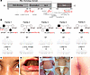 Figure 1
Genetic and clinical findings in patients with truncating RELA variants. (A) Schematic p65 domain architecture. NLS, nuclear localization signal; TAD, transactivation domain. Newly identified (red) and previously described (gray) pathogenic RELA variants. (B) Pedigrees and sequence pherograms depicting RELA variants. (C) Erythema nodosum in periorbital region of P1. (D) Oral aphthous ulcer in P3. (E) Generalized refractory eczema in P5 and marked clinical improvement under systemic dupilumab. There was no clinical data available for the father of P5. (F) Perianal ulcer in P6.
All identified RELA variants had not previously been described in the context of an inflammatory disease phenotype and were absent from the gnomAD v4.1.0 database, except for A507Pfs*25 with a very low allele frequency of 0.000006 (Table 1). Given a loss of function intolerance (pLI) score of 1 for the RELA gene (https://gnomad.broadinstitute.org/gene/ENSG00000173039?dataset=gnomad_r4), all identified variants were predicted to be deleterious. Consistent with previous reports (13), lymphoblastoid cells from P1–P4 carrying truncating RELA mutations showed expression of mutant p65, and WT p65 expression was reduced compared with cells from healthy controls (Supplemental Figure 1A; supplemental material available online with this article; https://doi.org/10.1172/jci.insight.200002DS1). As expected, the p65R198* mutant, which lacks both the Rel homology domain required for dimerization and the transactivation domain, was unable to form homodimers with WT p65 or heterodimers with p50 and could not transactivate an NF-κB reporter gene. Likewise, the truncating mutants S169*, Q372*, and A507Pfs*25 also showed significantly reduced reporter activity (Supplemental Figure 1, B and C).
Increased levels of proinflammatory cytokines at baseline and in response to immune stimulation. Consistent with clinical signs of autoinflammation and autoimmunity, cytokine profiling of patient sera revealed elevated levels of a broad range of proinflammatory cytokines, including IL-8, IL-10, TNF, MCP-1 (CCL2), and IFN-γ compared with healthy controls (Figure 2, A–E). Notably, the inflammasome-dependent cytokines IL-1β and IL-18 (P1–P3) and the Th2-dependent cytokine IL-33 (P2) were also markedly elevated at baseline (Figure 2, F–H). In addition, all analyzed patients showed evidence of constitutive type I IFN activation, as shown by increased serum levels of IFN-λ (P1, P3, and P4), elevation of the type I IFN–dependent chemokine CXCL10 (P1, P2, P5) (Figure 2, I and J), or an increased blood IFN signature (P1–P3) (Figure 2K), in line with previous reports (11).
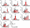 Figure 2
Increased secretion of proinflammatory cytokines in RELA-perturbed patient cells. (A–J) Serum levels of IL-8, IL-10, TNF, CCL2, IFN-γ, IL-1β, IL-18, IL-33, IFN-λ1, and CXCL10, collected at different time points, from patients (P1–P5) compared with 4 healthy controls (Co). Data are plotted as mean ± SD, *P < 0.05; **P < 0.01; ***P < 0.001; ****P < 0.0001 versus mean of WT controls, Kruskal-Wallis test (Dunn’s multiple-comparison test) for IL-8, CCL2, and IL-18 and 1-way ANOVA (Dunnett’s multiple-comparison test) for IL-10, TNF, IFN-γ, IL-1β, IL-33, IFN-λ1, and CXCL10. (K) IFN scores based on expression of IFN-stimulated genes in PBMCs. An IFN score of 12.49 (dashed line) indicates the median IFN score of 10 healthy controls + 2 SD.
To further assess induced cytokine responses, we conducted whole-blood stimulation assays using different immunostimulatory ligands. After stimulation with increasing doses of the TLR4 agonist LPS, all examined patients (P1–P5) secreted significantly higher amounts of IL-6 at the lowest dose of 1 ng/mL, consistent with hypersensitivity to LPS, whereas at higher doses of 2.5 and 5 ng/mL, patients P3–P5 showed increased IL-6 secretion (Figure 3A). Similarly, TNF secretion was significantly increased by LPS stimulation in a dose-dependent manner in P1–P4 (Figure 3B). Notably, all patients also secreted higher amounts of IL-1β in response to LPS compared with controls, indicating hypersensitivity to inflammasome activation (Figure 3C). In addition, all patients showed increased IL-10 secretion upon LPS stimulation (Figure 3D). Similar to blood cells, primary fibroblasts from P1 were hyperresponsive to TNF stimulation compared with a WT control, as evidenced by a greater induction of the IL6 gene (Supplemental Figure 2A). We next examined the patients’ response to stimulation of the type I IFN–inducing nucleic acid sensors TLR7 and TLR9, which have recently been shown to be hyperreactive in patients with truncating RELA mutations (11). Upon stimulation of whole blood with increasing doses of the TLR7 agonist R837, 2 (P4, P5) out of 5 patients responded with increased secretion of CXCL10, whereas CXCL10 secretion from patients P1, P2, and P3 did not differ from WT controls (Figure 3E). Similarly, after stimulation with the TLR9 agonist ODN2006, only P4 showed an increased CXCL10 response compared with WT controls (Figure 3F). Thus, in our cohort, constitutive type I IFN activation observed in all patients was not consistently associated with TLR7 and TLR9 hypersensitivity. Collectively, these findings demonstrate a global activation of multiple inflammatory pathways in patients with truncating RELA mutations that is accompanied by hyperresponsiveness to stimulation with LPS and TNF, and, to a lesser extent, to stimulation of TLR7 and TLR9.
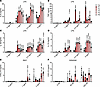 Figure 3
Enhanced cytokine responses after stimulation. (A–D) Secretion of IL-6, TNF, IL-1β, and IL-10 by whole blood from patients (P1–P5) after 24-hour stimulation with increasing concentrations of the TLR4 agonist LPS compared with 4 healthy controls (Co). Mean ± SD of 2 to 3 independent experiments. *P < 0.05; **P < 0.01; ****P < 0.0001 versus mean of WT controls, 2-way ANOVA (Dunnett’s multiple-comparison test). (E and F) Secretion of CXCL10 by whole blood from patients after 24-hour stimulation with increasing doses of the TLR7 agonist R837 or the TLR9 agonist ODN2006 compared with 4 healthy controls. Mean ± SD of 2 to 3 independent experiments. **P < 0.01; ***P < 0.001; ****P < 0.0001 versus mean of WT controls, 2-way ANOVA (Dunnett’s multiple-comparison test).
Altered canonical NF-κB signaling in patient cells. The finding of upregulated proinflammatory cytokines in patient blood, both at steady state and in response to stimuli utilizing the canonical NF-κB pathway, is surprising given the inability of mutant p65 to perform its core function of dimer formation and gene transcription activation (Supplemental Figure 1, B and C). To investigate this further, we first examined the subcellular levels of p65 in patient cells. To distinguish WT from mutant p65, we used an antibody that recognizes the C-terminus and therefore only WT p65. Confocal imaging revealed reduced levels of cytosolic and nuclear WT p65 in P1 compared with the control (Figure 4, A and B), both in unstimulated cells and after TNF stimulation, consistent with reduced WT p65 dosage. An approximately 50% decrease of WT p65 in mutant cells was confirmed by Western blot analysis of cytosolic and nuclear fractions (Figure 4, C and D).
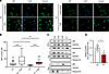 Figure 4
Altered nuclear translocation of p65 in patient fibroblasts. (A) Representative images of fibroblasts from patient and control immunostained for p65 (green) without and after TNF stimulation (10 ng/mL, 1 hour). Nuclei were stained with DAPI. Scale bars: 20 μm. (B) Quantitative analysis of nuclear p65 intensity. MFI in patient cells (n = 30–35) versus WT control cells (n = 28–32). Data presented as Tukey’s box plots with the box indicating the IQR, the center line indicating the median, and whiskers extending to 1.5 × IQR. *P < 0.05; **P < 0.01; ****P < 0.0001 versus mean of WT controls. One-way ANOVA (Šídák’s multiple-comparison test) was used for statistical analyses. (C) Representative immunoblot of p65 expression in whole cell lysate (WCL) and cytosolic (Cyt.) and nuclear fractions (Nuc.) of fibroblasts from P1 and 3 WT controls (Co1–3) after subcellular fractionation. GAPDH (cytoplasmic) and Histone H3 (nuclear) were used as loading controls. (D) Quantification of relative nuclear p65 in patient fibroblasts (P1) compared with WT controls, normalized to Histone H3. Mean ± SEM of at least 3 independent experiments. *P < 0.05 versus controls, Mann-Whitney U test.
We next analyzed canonical NF-κB signaling in lymphocytes from patients P1 and P2 by flow cytometry. We first examined the degradation of the inhibitory IκBα protein as the initial step in the signaling cascade leading to the release of the activated NF-κB dimer into the nucleus. Remarkably, in the unstimulated condition, IκBα protein, which is tightly regulated by the canonical NF-κB pathway, was lower in naive B, CD4+, and CD8+ T cells from both patients compared with healthy controls (Figure 5A). After stimulation of naive B cells with anti-IgM and PMA and of naive CD4+ and CD8+ T cells with PMA, respectively, the degradation of IκBα measured at 20 minutes after stimulation did not differ between patients and healthy controls (Figure 5A and Supplemental Figure 3A), suggesting normal stimulus-induced initiation of canonical NF-κB signaling. We then measured the activation-induced phosphorylation of p65. Consistent with the observed lower levels of p65 in patient cells at basal state (Figure 4, A and B), the levels of phosphorylated p65 (pSer529) were also lower compared with healthy controls in both naive B cells stimulated with anti-IgM or PMA and naive CD4+ and CD8+ T cells stimulated with PMA (Figure 5B and Supplemental Figure 3B). Thus, although basal expression of p65 was reduced, its phosphorylation in response to stimulation occurred normally. The normal IκBα degradation kinetics were consistent with the reduced but intact TNF-induced nuclear translocation of WT p65 observed by confocal microscopy at 30 minutes after stimulation (Figure 4, A and B).
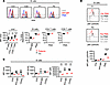 Figure 5
Altered NF-κB signaling in patient lymphocytes. (A) IκBα degradation in primary naive B cells stimulated with anti-IgM and PMA, and in T cells stimulated with PMA, of P1 and P2. IκBα protein levels at baseline and after stimulation in naive B, CD4+, and CD8+ T cells compared with healthy controls (Co). One-way ANOVA (Šídák’s multiple-comparison test); *P < 0.05; **P < 0.01; ***P < 0.001 versus mean of WT controls. (B) MFI of phosphorylated p65 upon activation with PMA in naive B cells of P1 and P2 compared with healthy controls. One-way ANOVA (Šídák’s multiple-comparison test); ****P < 0.0001 versus mean of WT controls. (C) Protein levels of p65 and its targets IκBα (NFKBIA) and BclXL (BCL2L1) after stimulation of PBMCs of P1 and P2 with anti-IgM and CD40L for 36 hours. One-way ANOVA (Šídák′s multiple-comparison test); **P < 0.01; ***P < 0.001 versus mean of WT controls.
To assess the effects of canonical NF-κB pathway stimulation on downstream targets, we stimulated PBMCs with anti-IgM and CD40L and then measured the protein levels of IκBα (NFKBIA) and Bcl-XL (BCL2L1) in naive B cells after 36 hours. Notably, although IκBα protein levels were adequately induced in B cells from healthy controls, they remained low in patient cells (Figure 5C and Supplemental Figure 3C). In contrast, poststimulation induction of Bcl-XL, which acts as an antiapoptotic protein, did not differ between patient cells and controls (Figure 5C and Supplemental Figure 3C). Consistent with the RELA haploinsufficiency, p65 protein levels in both unstimulated and stimulated cells were greatly reduced in patient B cells (Figure 5C and Supplemental Figure 3C). Taken together, these results indicate that activation of canonical NF-κB signaling upstream of p65 occurs normally in patient cells, albeit at lower levels. However, p65 activation was not accompanied by adequate poststimulation induction of IκBα, which could impair NF-κB inhibition at steady state and result in imbalanced NF-κB signaling.
Impaired negative feedback control of NF-κB in patient cells and RELA gene-edited HEK293T cells. The transcriptional activity of NF-κB is tightly repressed by IκB proteins, including IκBα, IκBβ, and IκBε, through the formation of stable IκB/NF-κB complexes (2). After stimulus-induced IκB degradation, one of the earliest NF-κB transcriptional targets to be re-expressed is IκBα, providing negative feedback control (14). Consistent with this IκBα-dependent negative feedback mechanism, transfection of WT p65 into HEK293T cells resulted in a strong induction of IκBα (Figure 6A). In contrast, transfection of mutant p65R198* had no effect on IκBα expression (Figure 6A), in agreement with its inability to transactivate a reporter gene (Supplemental Figure 1C).
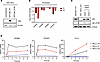 Figure 6
Impaired negative feedback control of NF-κB in patient cells and RELAR198* KI cells. (A) IκBα protein expression in HEK293T cells transfected with either GFP-tagged WT p65 (GFP-p65_WT) or mutant p65R198* (GFP-p65_R198*); β-actin was probed as loading control. (B) Differential gene expression of NF-κB inhibitors NFKBIA (IκBα), NFKBIB (IκBβ), NFKBIE (IκBε), and TNFAIP3 (A20) in patient fibroblasts (P1, P2), shown as log2 fold-change compared with 3 WT controls. (C) Representative immunoblot of p65 expression in WT HEK293T cells (HEK293T WT) compared with homozygous RELA KO (RELA_KO) and 2 independent heterozygous RELAR198* KI (RELA_KI) clones (clones 1 and 2). (D) Relative expression of NFKBIA, TNFAIP3, and CCL2 in WT HEK293T cells (RELA_WT), RELA KO (RELA_KO), and RELAR198* KI cells (RELA_KI) after stimulation with 100 ng/mL TNF for indicated time periods. Representative data from 1 out of 3 independent experiments run in triplicates. Data are plotted as mean ± SEM.
Notably, at steady state, IκBα levels also strongly influence basal NF-κB activity by controlling the levels of nuclear NF-κB, which actively shuttles between the nucleus and cytosol. Although free IκBα is intrinsically unstable and rapidly degraded in an IKK-independent manner, NF-κB–bound IκBα is more stable (15). Thus, besides regulating its transcription, NF-κB also determines the fate of its own inhibitor IκBα through stabilization. Given the inability of mutant p65 to induce IκBα in HEK293T cells (Figure 6A), we reasoned that reduced steady-state levels of p65 might affect basal transcription of IκB protein-coding genes. We therefore examined the consequences of reduced WT p65 dosage on the expression of the NFKBIA (IκBα), NFKBIB (IκBβ), and NFKBIE (IκBε) genes in fibroblasts from P1 and P2. RNA-Seq revealed a marked decrease in the expression of all 3 NF-κB inhibitors in patient fibroblasts compared with 3 WT controls (Figure 6B), mirroring the reduced basal levels of IκBα observed in unstimulated patient lymphocytes (Figure 5A). Moreover, gene expression of the NF-κB inhibitor TNFAIP3 was also reduced in patient fibroblasts (Figure 6B). Thus, at steady state, loss of a functional RELA allele in patient cells causes a shift in the balance between NF-κB activating and inhibitory factors toward activation.
To further investigate the functional consequences of the reduction of NF-κB inhibitory factors at the cellular level, we turned to a reductionist approach. We generated HEK293T cells (RELA_WT) with either a complete homozygous KO of the RELA gene (RELA_KO) or with a heterozygous KI of the R198* mutation at the RELA locus (RELA_KI) by CRISPR/Cas or prime editing, respectively (Figure 6C). Introduction of the R198* RELA mutation into HEK293T cells had no effect on cytokine gene expression in unstimulated cells (Supplemental Figure 4A). After stimulation of cells with 100 ng/mL of TNF, expression of the NF-κB inhibitory gene NFKBIA was strongly induced within 60 minutes in a RELA gene dose-dependent manner. Thus, the highest transcriptional activation of NFKBIA was seen in RELA_WT cells and the lowest in RELA_KO cells; RELA_KI cells showed an intermediate transcriptional response (Figure 6D), confirming the observation made in patient lymphocytes (Figure 5A) that a lack of RELA gene dose results in a correspondingly reduced induction of its inhibitor IκBα. Consistent with complete absence of functional p65 in RELA_KO cells, TNF stimulation had essentially no effect on the expression of TNFAIP3 and CCL2, 2 bona fide NF-κB target genes (Figure 6D). Normally, TNF-induced expression of TNFAIP3, which functions as a key negative regulator of NF-κB signaling, peaks within 1 hour with a kinetic similar to NFKBIA (16). However, in contrast to RELA_WT cells, the induction of TNFAIP3 was substantially reduced in RELA_KI cells, reflecting a weaker transactivation potential exerted by a single RELA allele (Figure 6D). Interestingly, in RELA_KI cells, in which the induction of the inhibitory genes NFKBIA and TNFAIP3 was only half that of RELA_WT cells, the proinflammatory CCL2 gene was strongly induced by more than 2.5-fold at 3 hours and more than 2.2-fold at 6 hours compared with RELA_WT cells (Figure 6D). Collectively, these findings suggest that the heightened proinflammatory response of RELA_KI cells to TNF stimulation may be due to a lack of inhibitory control of the NF-κB pathway.
Upregulation of cell type–specific proinflammatory and cell death pathways in patient cells. To further investigate the transcriptional changes related to truncating RELA mutations in a cell type–resolved, untargeted manner, we performed single-cell RNA-Seq (scRNA-Seq) in PBMCs from P1, P2, and P3 along with 5 sex- and age-matched healthy controls. We identified and annotated all major cell types (naive and memory B cells, naive and central memory CD4+ and CD8+ T cells, γδ T cells, NK cells, classical and nonclassical monocytes, DCs, and plasmacytoid DCs [pDCs]) using annotation transfer and marker-based curation (Figure 7A and Supplemental Figure 5A). In line with clinical and laboratory data (Figure 2K), cells from all 3 patients expressed IFN-stimulated genes more highly than control cells — a signal mainly driven by myeloid cells but also present to a lesser degree in lymphoid cells (Supplemental Figure 5B). Cell type composition varied strongly, with no consistent differences between patient and control samples (Supplemental Figure 5C).
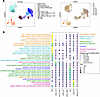 Figure 7
Cell type–specific transcriptomic alterations in RELA-perturbed patient cells. (A) Batch-corrected UMAP embedding of all cells profiled with scRNA-Seq from patient (P1–P3) and control (sc Co1–sc Co5) PBMCs. (B) GSEA enrichment of selected GO Biological Processes terms in differentially expressed genes between patient and control cells of the indicated cell types. For monocytes, only P3 and 2 controls (sc Co1, sc Co2) could be compared due to lack of monocytes in the other samples (see Methods). NES, normalized enrichment score.
Consistent with the hyperinflammatory phenotype observed in patients, gene set enrichment analysis (GSEA) revealed strong activation of multiple pathways related to proinflammatory cytokine production and signaling, including type I IFN, IFN-γ, and IL-1, particularly in monocytes (Figure 7B). Among the top-ranked gene sets enriched in monocytes were type I IFN signaling pathway (Gene Ontology [GO] 0060337) and response to IFN-γ (GO 0034341) (Figure 7B and Figure 8A). The IFN-stimulated genes ISG15 and APOBEC3G, as well as the antiviral factor BST2, which can amplify proinflammatory responses via NF-κB signaling, showed increased and relatively homogeneous expression in both classical and nonclassical monocytes (Figure 8B and Supplemental Figure 5D). In contrast, the proinflammatory cytokine CCL2 and NFKBIA, both NF-κB target genes, were more prominently upregulated in classical monocytes (Figure 8B), potentially indicating more WT-like NF-κB activity in classical monocytes.
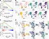 Figure 8
Activation of inflammatory and cell death pathways in RELA-perturbed patient cells. (A) GSEA leading-edge plot for the GO term type I IFN signaling pathway enriched in monocytes from P3 and control (sc Co1 and sc Co2) PBMCs. (B) UMAP embedding of monocytes colored by the sample assignment or the expression of the indicated genes in individual cells. (C) GSEA leading-edge plot for the GO term “regulation of necroptotic process” enriched in NK cells from patient (P1–P3) and control (sc Co1–sc Co5) PBMCs. (D) UMAP embedding of NK cells colored by the sample assignment or the expression of the indicated genes in individual cells.
In naive B cells from patients, NIK/NF-κB signaling (GO 0038061) emerged as the most highly enriched pathway (Figure 7B), suggesting a negative regulatory crosstalk of p65 on the alternative NF-κB signaling pathway in circulating B cells. Transcription factor activity analysis, considering the expression of all known target genes for a given transcription factor, further revealed elevated activity scores for STAT1, NFKB, and RELA in patient B cells (Supplemental Figure 6A). Patient B cells also showed elevated expression of the NF-κB inhibitors and target genes (NFKBIA, NFKBID, and NFKBIE), unlike patient T cells (Supplemental Figure 6B), providing evidence of constitutive NF-κB pathway activation in B cells.
In contrast, patient T and NK cells, in which IκBα protein levels were more strongly reduced than in B cells (Figure 5A), showed upregulation of cell death pathways, including both apoptosis and necroptosis (Figure 7B, Figure 8C, and Supplemental Figure 6C), the latter being a lytic, proinflammatory form of programmed cell death. This signature was most pronounced in NK cells (Figure 7B and Figure 8C). Apoptosis as an enriched term was also evident in naive CD4+ and CD8+ T cells but not in monocytes or B cells (Supplemental Figure 6C). Interestingly, the genes driving the apoptosis signal in T cells and NK cells showed substantial overlap with each other, but were largely distinct from those in monocytes (Supplemental Figure 6D). Notably, CASP8 and ZBP1 emerged among the most upregulated differentially expressed genes in NK cells (Supplemental Figure 6E) but still showed heterogeneous expression among patient and control cells (Figure 8D). CASP8 encodes caspase-8, a critical initiator of apoptosis downstream of death receptor signaling; ZBP1 (Z-DNA binding protein 1) functions as a cytosolic nucleic acid sensor that can activate both necroptosis and apoptosis. Through these mechanisms, ZBP1 contributes to inflammation via the release of danger-associated molecular patterns (DAMPs) (17, 18). Accordingly, the highest ranked gene set enriched in patient NK cells was regulation of necroptotic process (GO 0060544) (Figure 7B). This enrichment was among others driven by increased expression of CASP8, ZBP1, and CYLD, and JUN was also among the top differentially expressed genes (Figure 8D, Supplemental Figure 5D, and Supplemental Figure 6E), consistent with chronic activation of both apoptotic and necroptotic cell death pathways. In addition, transcription factor activity analysis revealed the highest activity scores for JUN, ATF4, and CREB1 in CD4+ naive T cells from patients (Supplemental Figure 6A), reflecting a state of activation and stress-associated dysfunction. Jun is associated with T cell activation, promotes Th1 and Th17 differentiation, and has been linked to T cell exhaustion (19). ATF4 acts as a central mediator of the integrated stress response and regulates the expression of proinflammatory cytokines under cellular stress conditions (20). CREB1 contributes to the transcription of key cytokines such as IL-2 (essential for T cell proliferation); IFN-γ (critical for Th1 responses); and under certain conditions, IL-17 (involved in Th17 differentiation) (21). Together, these transcriptional signatures support a bias toward proinflammatory effector T cells, primarily Th1 and Th17, which are known to play key roles in initiating and sustaining chronic inflammation. Overall, the cell type–resolved transcriptional comparison of patient versus control immune cells uncovered a differential signature marked by reduced NF-κB inhibition and heightened apoptotic/necroptotic signaling, particularly in T and NK cells.
To verify that these findings are unique to RELA-mutated patients and not a broader feature of inflammatory processes, we analyzed publicly available scRNA-Seq data from SLE and COVID-19 cohorts using the same pipeline (Supplemental Figure 7). Genes previously highlighted as differentially expressed in patient monocytes and NK cells, such as APOBEC3A, ISG15, and JUN (Figure 8D, Supplemental Figure 5D, and Supplemental Figure 6E), showed no significant upregulation in the corresponding cell types for either SLE or COVID-19 (Supplemental Figure 7, C and D). GSEA confirmed heightened inflammatory signatures in SLE and COVID-19, as expected. Similarly, pathways central to our interpretation of the RELA mutation were not perturbed in the same way in the SLE and COVID-19 cohorts. Notably, necroptosis-related pathways were not enriched in COVID-19 at all and were restricted to T cells in SLE, whereas in our data, these signatures were prominently enriched only in NK cells (Supplemental Figure 7E). These comparisons support a disease-specific transcriptional program of RELA mutations compared with other inflammatory diseases.
Enhanced sensitivity to TNF-induced apoptosis and necroptosis in patient cells and RELA gene-edited HEK293T cells. Upon TNF receptor engagement, adaptor proteins including RIPK1 and cIAP1/2 assemble complex I, a membrane-associated platform where ubiquitinated RIPK1 activates NF-κB and promotes survival (22). Deubiquitination of RIPK1 by CYLD, A20, or OTULIN shifts signaling toward cytosolic complex II, composed of FADD, procaspase-8, and RIPK1, where caspase-8 activation initiates apoptosis and simultaneously suppresses necroptosis by cleaving RIPK1 and RIPK3. In the absence or inhibition of caspase-8, RIPK1 and RIPK3 interact via RHIM domains to form the necrosome, which drives inflammatory necroptosis, particularly in response to TNF (23).
Given the increased apoptosis/necroptosis signaling (Figure 7B and Supplemental Figure 6C); upregulation of CASP8 and CYLD (Figure 8D and Supplemental Figure 6, D and E); and a reduction in negative regulation of NF-κB signaling (Figure 5A), observed predominantly in patient T and NK cells, we hypothesized that patient cells may be intrinsically more sensitive to cell death. To test this, we stimulated patient cells with TNF in the presence of the SMAC mimetic birinapant, which inhibits cIAPs and promotes apoptosis (24). In a parallel condition, cells were stimulated with TNF and cotreated with birinapant and the pan-caspase inhibitor emricasan to block apoptosis and enforce necroptosis (25). Upon treatment of fibroblasts from P1 and P2 with birinapant, TNF stimulation led to a stronger induction of cleaved caspase-3, indicating enhanced apoptosis compared with WT controls (Figure 9A). Similarly, cotreatment with birinapant and emricasan resulted in markedly increased phosphorylation of RIPK1 at Ser166, a hallmark of necroptosis activation, compared with WT controls, suggesting heightened necroptotic potential in patient cells (Figure 9A). Notably, RELA_KI cells, expressing only 1 functional RELA allele, also exhibited increased apoptosis and necroptosis in response to TNF stimulation compared with RELA_WT or RELA_KO cells (Figure 9B), confirming that RELA haploinsufficiency underlies the heightened sensitivity to TNF-induced cell death observed in patient cells. Densitometric quantification of all immunoblots in patient fibroblasts and RELA_WT/KO/KI HEK293T cells confirmed significantly increased levels of cleaved caspase-3 under apoptotic conditions (Figure 9A) and phosphorylated RIPK1 under necroptotic conditions (Figure 9B). Collectively, these findings demonstrate that the loss of a functional RELA allele compromises TNF tolerance and predisposes cells to inflammatory cell death.
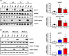 Figure 9
Increased sensitivity to apoptosis and necroptosis conferred by the RELAR198* mutation. (A) Left: representative immunoblots showing increased phosphorylated RIPK1 (pRIPK1; Ser166) and cleaved caspase-3 (CASP3) in patient fibroblasts (P1, P2) versus WT controls (Co1, Co2). GAPDH was stained as loading control. Right: quantification of 3 independent experiments (ImageJ [NIH], normalized to GAPDH). *P < 0.05 versus WT (Mann-Whitney U test; mean ± SEM). (B) Left: representative immunoblots showing increased pRIPK1 and cleaved CASP3 in RELA_WT, RELA_KO, RELA_KI HEK293T cells. GAPDH was probed as loading control. Right: quantification of 3 to 4 independent experiments (ImageJ [NIH], normalized to GAPDH). *P < 0.05 versus theoretical mean = 1 (1-sample t test; mean ± SEM). Apo: TNF + birinapant; Nec: TNF + birinapant + emricasan.


