
Research ArticleCardiologyMetabolism
Open Access |  10.1172/jci.insight.200381
A Slc5a6-deficient mouse model reveals metabolically driven cardiomyopathy with therapeutic potential for vitamin-based intervention
Millie O. Fullerton,1 Lauren C. Phillips,1,2 Rachael E. Redgrave,1 Luke Spray,3 Vincent Haufroid,4 George Merces,5 Scott T. Kerridge,1 Gavin D. Richardson,1 Nathalie Mercier,6 Dominique Roland,7 Rebecca Crossley,1 Andrew D.H. Morgan,1 Joseph P. Dewulf,4,8,9 John Burn,1,3 Simon D. Bamforth,1 and Helen M. Phillips1
1Biosciences Institute, Newcastle University, Newcastle upon Tyne, United Kingdom.
2Department of Pharmacology, University of Oxford, United Kingdom.
3Translational and Clinical Research Institute, Newcastle University, Newcastle upon Tyne, United Kingdom.
4Louvain Center for Toxicology and Applied Pharmacology (LTAP), Institut de Recherche Expérimentale et Clinique (IREC), UCLouvain, Brussels, Belgium.
5Image Analysis Unit, Newcastle University, Newcastle upon Tyne, United Kingdom.
6Department of Pediatrics, CHU HELORA Jo, La Louvière, Belgium.
7Department of Human Genetics, Center for Inherited Metabolic Disorders, Institute of Pathology and Genetics, Gosselies, Belgium.
8Metabolic Research Group, de Duve Institute, UCLouvain, Brussels, Belgium.
9Biochemical Genetics and Newborn Screening Laboratory, Department of laboratory medicine & Institut des Maladies Rares, Cliniques Universitaires Saint-Luc, UCLouvain, Brussels, Belgium.
Address correspondence to: Helen M. Phillips, Biosciences Institute, Faculty of Medical Sciences, Newcastle University, International Centre for Life, Central Parkway, Newcastle upon Tyne, NE1 3BZ, United Kingdom. Phone: 44.191.241.8698; Email: Helen.Phillips@newcastle.ac.uk.
Authorship note: MOF and LCP contributed equally to this work.
Find articles by Fullerton, M. in: PubMed | Google Scholar
1Biosciences Institute, Newcastle University, Newcastle upon Tyne, United Kingdom.
2Department of Pharmacology, University of Oxford, United Kingdom.
3Translational and Clinical Research Institute, Newcastle University, Newcastle upon Tyne, United Kingdom.
4Louvain Center for Toxicology and Applied Pharmacology (LTAP), Institut de Recherche Expérimentale et Clinique (IREC), UCLouvain, Brussels, Belgium.
5Image Analysis Unit, Newcastle University, Newcastle upon Tyne, United Kingdom.
6Department of Pediatrics, CHU HELORA Jo, La Louvière, Belgium.
7Department of Human Genetics, Center for Inherited Metabolic Disorders, Institute of Pathology and Genetics, Gosselies, Belgium.
8Metabolic Research Group, de Duve Institute, UCLouvain, Brussels, Belgium.
9Biochemical Genetics and Newborn Screening Laboratory, Department of laboratory medicine & Institut des Maladies Rares, Cliniques Universitaires Saint-Luc, UCLouvain, Brussels, Belgium.
Address correspondence to: Helen M. Phillips, Biosciences Institute, Faculty of Medical Sciences, Newcastle University, International Centre for Life, Central Parkway, Newcastle upon Tyne, NE1 3BZ, United Kingdom. Phone: 44.191.241.8698; Email: Helen.Phillips@newcastle.ac.uk.
Authorship note: MOF and LCP contributed equally to this work.
Find articles by Phillips, L. in: PubMed | Google Scholar
1Biosciences Institute, Newcastle University, Newcastle upon Tyne, United Kingdom.
2Department of Pharmacology, University of Oxford, United Kingdom.
3Translational and Clinical Research Institute, Newcastle University, Newcastle upon Tyne, United Kingdom.
4Louvain Center for Toxicology and Applied Pharmacology (LTAP), Institut de Recherche Expérimentale et Clinique (IREC), UCLouvain, Brussels, Belgium.
5Image Analysis Unit, Newcastle University, Newcastle upon Tyne, United Kingdom.
6Department of Pediatrics, CHU HELORA Jo, La Louvière, Belgium.
7Department of Human Genetics, Center for Inherited Metabolic Disorders, Institute of Pathology and Genetics, Gosselies, Belgium.
8Metabolic Research Group, de Duve Institute, UCLouvain, Brussels, Belgium.
9Biochemical Genetics and Newborn Screening Laboratory, Department of laboratory medicine & Institut des Maladies Rares, Cliniques Universitaires Saint-Luc, UCLouvain, Brussels, Belgium.
Address correspondence to: Helen M. Phillips, Biosciences Institute, Faculty of Medical Sciences, Newcastle University, International Centre for Life, Central Parkway, Newcastle upon Tyne, NE1 3BZ, United Kingdom. Phone: 44.191.241.8698; Email: Helen.Phillips@newcastle.ac.uk.
Authorship note: MOF and LCP contributed equally to this work.
Find articles by Redgrave, R. in: PubMed | Google Scholar
1Biosciences Institute, Newcastle University, Newcastle upon Tyne, United Kingdom.
2Department of Pharmacology, University of Oxford, United Kingdom.
3Translational and Clinical Research Institute, Newcastle University, Newcastle upon Tyne, United Kingdom.
4Louvain Center for Toxicology and Applied Pharmacology (LTAP), Institut de Recherche Expérimentale et Clinique (IREC), UCLouvain, Brussels, Belgium.
5Image Analysis Unit, Newcastle University, Newcastle upon Tyne, United Kingdom.
6Department of Pediatrics, CHU HELORA Jo, La Louvière, Belgium.
7Department of Human Genetics, Center for Inherited Metabolic Disorders, Institute of Pathology and Genetics, Gosselies, Belgium.
8Metabolic Research Group, de Duve Institute, UCLouvain, Brussels, Belgium.
9Biochemical Genetics and Newborn Screening Laboratory, Department of laboratory medicine & Institut des Maladies Rares, Cliniques Universitaires Saint-Luc, UCLouvain, Brussels, Belgium.
Address correspondence to: Helen M. Phillips, Biosciences Institute, Faculty of Medical Sciences, Newcastle University, International Centre for Life, Central Parkway, Newcastle upon Tyne, NE1 3BZ, United Kingdom. Phone: 44.191.241.8698; Email: Helen.Phillips@newcastle.ac.uk.
Authorship note: MOF and LCP contributed equally to this work.
Find articles by Spray, L. in: PubMed | Google Scholar
1Biosciences Institute, Newcastle University, Newcastle upon Tyne, United Kingdom.
2Department of Pharmacology, University of Oxford, United Kingdom.
3Translational and Clinical Research Institute, Newcastle University, Newcastle upon Tyne, United Kingdom.
4Louvain Center for Toxicology and Applied Pharmacology (LTAP), Institut de Recherche Expérimentale et Clinique (IREC), UCLouvain, Brussels, Belgium.
5Image Analysis Unit, Newcastle University, Newcastle upon Tyne, United Kingdom.
6Department of Pediatrics, CHU HELORA Jo, La Louvière, Belgium.
7Department of Human Genetics, Center for Inherited Metabolic Disorders, Institute of Pathology and Genetics, Gosselies, Belgium.
8Metabolic Research Group, de Duve Institute, UCLouvain, Brussels, Belgium.
9Biochemical Genetics and Newborn Screening Laboratory, Department of laboratory medicine & Institut des Maladies Rares, Cliniques Universitaires Saint-Luc, UCLouvain, Brussels, Belgium.
Address correspondence to: Helen M. Phillips, Biosciences Institute, Faculty of Medical Sciences, Newcastle University, International Centre for Life, Central Parkway, Newcastle upon Tyne, NE1 3BZ, United Kingdom. Phone: 44.191.241.8698; Email: Helen.Phillips@newcastle.ac.uk.
Authorship note: MOF and LCP contributed equally to this work.
Find articles by Haufroid, V. in: PubMed | Google Scholar
1Biosciences Institute, Newcastle University, Newcastle upon Tyne, United Kingdom.
2Department of Pharmacology, University of Oxford, United Kingdom.
3Translational and Clinical Research Institute, Newcastle University, Newcastle upon Tyne, United Kingdom.
4Louvain Center for Toxicology and Applied Pharmacology (LTAP), Institut de Recherche Expérimentale et Clinique (IREC), UCLouvain, Brussels, Belgium.
5Image Analysis Unit, Newcastle University, Newcastle upon Tyne, United Kingdom.
6Department of Pediatrics, CHU HELORA Jo, La Louvière, Belgium.
7Department of Human Genetics, Center for Inherited Metabolic Disorders, Institute of Pathology and Genetics, Gosselies, Belgium.
8Metabolic Research Group, de Duve Institute, UCLouvain, Brussels, Belgium.
9Biochemical Genetics and Newborn Screening Laboratory, Department of laboratory medicine & Institut des Maladies Rares, Cliniques Universitaires Saint-Luc, UCLouvain, Brussels, Belgium.
Address correspondence to: Helen M. Phillips, Biosciences Institute, Faculty of Medical Sciences, Newcastle University, International Centre for Life, Central Parkway, Newcastle upon Tyne, NE1 3BZ, United Kingdom. Phone: 44.191.241.8698; Email: Helen.Phillips@newcastle.ac.uk.
Authorship note: MOF and LCP contributed equally to this work.
Find articles by Merces, G. in: PubMed | Google Scholar
1Biosciences Institute, Newcastle University, Newcastle upon Tyne, United Kingdom.
2Department of Pharmacology, University of Oxford, United Kingdom.
3Translational and Clinical Research Institute, Newcastle University, Newcastle upon Tyne, United Kingdom.
4Louvain Center for Toxicology and Applied Pharmacology (LTAP), Institut de Recherche Expérimentale et Clinique (IREC), UCLouvain, Brussels, Belgium.
5Image Analysis Unit, Newcastle University, Newcastle upon Tyne, United Kingdom.
6Department of Pediatrics, CHU HELORA Jo, La Louvière, Belgium.
7Department of Human Genetics, Center for Inherited Metabolic Disorders, Institute of Pathology and Genetics, Gosselies, Belgium.
8Metabolic Research Group, de Duve Institute, UCLouvain, Brussels, Belgium.
9Biochemical Genetics and Newborn Screening Laboratory, Department of laboratory medicine & Institut des Maladies Rares, Cliniques Universitaires Saint-Luc, UCLouvain, Brussels, Belgium.
Address correspondence to: Helen M. Phillips, Biosciences Institute, Faculty of Medical Sciences, Newcastle University, International Centre for Life, Central Parkway, Newcastle upon Tyne, NE1 3BZ, United Kingdom. Phone: 44.191.241.8698; Email: Helen.Phillips@newcastle.ac.uk.
Authorship note: MOF and LCP contributed equally to this work.
Find articles by Kerridge, S. in: PubMed | Google Scholar
1Biosciences Institute, Newcastle University, Newcastle upon Tyne, United Kingdom.
2Department of Pharmacology, University of Oxford, United Kingdom.
3Translational and Clinical Research Institute, Newcastle University, Newcastle upon Tyne, United Kingdom.
4Louvain Center for Toxicology and Applied Pharmacology (LTAP), Institut de Recherche Expérimentale et Clinique (IREC), UCLouvain, Brussels, Belgium.
5Image Analysis Unit, Newcastle University, Newcastle upon Tyne, United Kingdom.
6Department of Pediatrics, CHU HELORA Jo, La Louvière, Belgium.
7Department of Human Genetics, Center for Inherited Metabolic Disorders, Institute of Pathology and Genetics, Gosselies, Belgium.
8Metabolic Research Group, de Duve Institute, UCLouvain, Brussels, Belgium.
9Biochemical Genetics and Newborn Screening Laboratory, Department of laboratory medicine & Institut des Maladies Rares, Cliniques Universitaires Saint-Luc, UCLouvain, Brussels, Belgium.
Address correspondence to: Helen M. Phillips, Biosciences Institute, Faculty of Medical Sciences, Newcastle University, International Centre for Life, Central Parkway, Newcastle upon Tyne, NE1 3BZ, United Kingdom. Phone: 44.191.241.8698; Email: Helen.Phillips@newcastle.ac.uk.
Authorship note: MOF and LCP contributed equally to this work.
Find articles by
Richardson, G.
in:
PubMed
|
Google Scholar
|

1Biosciences Institute, Newcastle University, Newcastle upon Tyne, United Kingdom.
2Department of Pharmacology, University of Oxford, United Kingdom.
3Translational and Clinical Research Institute, Newcastle University, Newcastle upon Tyne, United Kingdom.
4Louvain Center for Toxicology and Applied Pharmacology (LTAP), Institut de Recherche Expérimentale et Clinique (IREC), UCLouvain, Brussels, Belgium.
5Image Analysis Unit, Newcastle University, Newcastle upon Tyne, United Kingdom.
6Department of Pediatrics, CHU HELORA Jo, La Louvière, Belgium.
7Department of Human Genetics, Center for Inherited Metabolic Disorders, Institute of Pathology and Genetics, Gosselies, Belgium.
8Metabolic Research Group, de Duve Institute, UCLouvain, Brussels, Belgium.
9Biochemical Genetics and Newborn Screening Laboratory, Department of laboratory medicine & Institut des Maladies Rares, Cliniques Universitaires Saint-Luc, UCLouvain, Brussels, Belgium.
Address correspondence to: Helen M. Phillips, Biosciences Institute, Faculty of Medical Sciences, Newcastle University, International Centre for Life, Central Parkway, Newcastle upon Tyne, NE1 3BZ, United Kingdom. Phone: 44.191.241.8698; Email: Helen.Phillips@newcastle.ac.uk.
Authorship note: MOF and LCP contributed equally to this work.
Find articles by Mercier, N. in: PubMed | Google Scholar
1Biosciences Institute, Newcastle University, Newcastle upon Tyne, United Kingdom.
2Department of Pharmacology, University of Oxford, United Kingdom.
3Translational and Clinical Research Institute, Newcastle University, Newcastle upon Tyne, United Kingdom.
4Louvain Center for Toxicology and Applied Pharmacology (LTAP), Institut de Recherche Expérimentale et Clinique (IREC), UCLouvain, Brussels, Belgium.
5Image Analysis Unit, Newcastle University, Newcastle upon Tyne, United Kingdom.
6Department of Pediatrics, CHU HELORA Jo, La Louvière, Belgium.
7Department of Human Genetics, Center for Inherited Metabolic Disorders, Institute of Pathology and Genetics, Gosselies, Belgium.
8Metabolic Research Group, de Duve Institute, UCLouvain, Brussels, Belgium.
9Biochemical Genetics and Newborn Screening Laboratory, Department of laboratory medicine & Institut des Maladies Rares, Cliniques Universitaires Saint-Luc, UCLouvain, Brussels, Belgium.
Address correspondence to: Helen M. Phillips, Biosciences Institute, Faculty of Medical Sciences, Newcastle University, International Centre for Life, Central Parkway, Newcastle upon Tyne, NE1 3BZ, United Kingdom. Phone: 44.191.241.8698; Email: Helen.Phillips@newcastle.ac.uk.
Authorship note: MOF and LCP contributed equally to this work.
Find articles by Roland, D. in: PubMed | Google Scholar
1Biosciences Institute, Newcastle University, Newcastle upon Tyne, United Kingdom.
2Department of Pharmacology, University of Oxford, United Kingdom.
3Translational and Clinical Research Institute, Newcastle University, Newcastle upon Tyne, United Kingdom.
4Louvain Center for Toxicology and Applied Pharmacology (LTAP), Institut de Recherche Expérimentale et Clinique (IREC), UCLouvain, Brussels, Belgium.
5Image Analysis Unit, Newcastle University, Newcastle upon Tyne, United Kingdom.
6Department of Pediatrics, CHU HELORA Jo, La Louvière, Belgium.
7Department of Human Genetics, Center for Inherited Metabolic Disorders, Institute of Pathology and Genetics, Gosselies, Belgium.
8Metabolic Research Group, de Duve Institute, UCLouvain, Brussels, Belgium.
9Biochemical Genetics and Newborn Screening Laboratory, Department of laboratory medicine & Institut des Maladies Rares, Cliniques Universitaires Saint-Luc, UCLouvain, Brussels, Belgium.
Address correspondence to: Helen M. Phillips, Biosciences Institute, Faculty of Medical Sciences, Newcastle University, International Centre for Life, Central Parkway, Newcastle upon Tyne, NE1 3BZ, United Kingdom. Phone: 44.191.241.8698; Email: Helen.Phillips@newcastle.ac.uk.
Authorship note: MOF and LCP contributed equally to this work.
Find articles by Crossley, R. in: PubMed | Google Scholar
1Biosciences Institute, Newcastle University, Newcastle upon Tyne, United Kingdom.
2Department of Pharmacology, University of Oxford, United Kingdom.
3Translational and Clinical Research Institute, Newcastle University, Newcastle upon Tyne, United Kingdom.
4Louvain Center for Toxicology and Applied Pharmacology (LTAP), Institut de Recherche Expérimentale et Clinique (IREC), UCLouvain, Brussels, Belgium.
5Image Analysis Unit, Newcastle University, Newcastle upon Tyne, United Kingdom.
6Department of Pediatrics, CHU HELORA Jo, La Louvière, Belgium.
7Department of Human Genetics, Center for Inherited Metabolic Disorders, Institute of Pathology and Genetics, Gosselies, Belgium.
8Metabolic Research Group, de Duve Institute, UCLouvain, Brussels, Belgium.
9Biochemical Genetics and Newborn Screening Laboratory, Department of laboratory medicine & Institut des Maladies Rares, Cliniques Universitaires Saint-Luc, UCLouvain, Brussels, Belgium.
Address correspondence to: Helen M. Phillips, Biosciences Institute, Faculty of Medical Sciences, Newcastle University, International Centre for Life, Central Parkway, Newcastle upon Tyne, NE1 3BZ, United Kingdom. Phone: 44.191.241.8698; Email: Helen.Phillips@newcastle.ac.uk.
Authorship note: MOF and LCP contributed equally to this work.
Find articles by Morgan, A. in: PubMed | Google Scholar
1Biosciences Institute, Newcastle University, Newcastle upon Tyne, United Kingdom.
2Department of Pharmacology, University of Oxford, United Kingdom.
3Translational and Clinical Research Institute, Newcastle University, Newcastle upon Tyne, United Kingdom.
4Louvain Center for Toxicology and Applied Pharmacology (LTAP), Institut de Recherche Expérimentale et Clinique (IREC), UCLouvain, Brussels, Belgium.
5Image Analysis Unit, Newcastle University, Newcastle upon Tyne, United Kingdom.
6Department of Pediatrics, CHU HELORA Jo, La Louvière, Belgium.
7Department of Human Genetics, Center for Inherited Metabolic Disorders, Institute of Pathology and Genetics, Gosselies, Belgium.
8Metabolic Research Group, de Duve Institute, UCLouvain, Brussels, Belgium.
9Biochemical Genetics and Newborn Screening Laboratory, Department of laboratory medicine & Institut des Maladies Rares, Cliniques Universitaires Saint-Luc, UCLouvain, Brussels, Belgium.
Address correspondence to: Helen M. Phillips, Biosciences Institute, Faculty of Medical Sciences, Newcastle University, International Centre for Life, Central Parkway, Newcastle upon Tyne, NE1 3BZ, United Kingdom. Phone: 44.191.241.8698; Email: Helen.Phillips@newcastle.ac.uk.
Authorship note: MOF and LCP contributed equally to this work.
Find articles by Dewulf, J. in: PubMed | Google Scholar
1Biosciences Institute, Newcastle University, Newcastle upon Tyne, United Kingdom.
2Department of Pharmacology, University of Oxford, United Kingdom.
3Translational and Clinical Research Institute, Newcastle University, Newcastle upon Tyne, United Kingdom.
4Louvain Center for Toxicology and Applied Pharmacology (LTAP), Institut de Recherche Expérimentale et Clinique (IREC), UCLouvain, Brussels, Belgium.
5Image Analysis Unit, Newcastle University, Newcastle upon Tyne, United Kingdom.
6Department of Pediatrics, CHU HELORA Jo, La Louvière, Belgium.
7Department of Human Genetics, Center for Inherited Metabolic Disorders, Institute of Pathology and Genetics, Gosselies, Belgium.
8Metabolic Research Group, de Duve Institute, UCLouvain, Brussels, Belgium.
9Biochemical Genetics and Newborn Screening Laboratory, Department of laboratory medicine & Institut des Maladies Rares, Cliniques Universitaires Saint-Luc, UCLouvain, Brussels, Belgium.
Address correspondence to: Helen M. Phillips, Biosciences Institute, Faculty of Medical Sciences, Newcastle University, International Centre for Life, Central Parkway, Newcastle upon Tyne, NE1 3BZ, United Kingdom. Phone: 44.191.241.8698; Email: Helen.Phillips@newcastle.ac.uk.
Authorship note: MOF and LCP contributed equally to this work.
Find articles by Burn, J. in: PubMed | Google Scholar
1Biosciences Institute, Newcastle University, Newcastle upon Tyne, United Kingdom.
2Department of Pharmacology, University of Oxford, United Kingdom.
3Translational and Clinical Research Institute, Newcastle University, Newcastle upon Tyne, United Kingdom.
4Louvain Center for Toxicology and Applied Pharmacology (LTAP), Institut de Recherche Expérimentale et Clinique (IREC), UCLouvain, Brussels, Belgium.
5Image Analysis Unit, Newcastle University, Newcastle upon Tyne, United Kingdom.
6Department of Pediatrics, CHU HELORA Jo, La Louvière, Belgium.
7Department of Human Genetics, Center for Inherited Metabolic Disorders, Institute of Pathology and Genetics, Gosselies, Belgium.
8Metabolic Research Group, de Duve Institute, UCLouvain, Brussels, Belgium.
9Biochemical Genetics and Newborn Screening Laboratory, Department of laboratory medicine & Institut des Maladies Rares, Cliniques Universitaires Saint-Luc, UCLouvain, Brussels, Belgium.
Address correspondence to: Helen M. Phillips, Biosciences Institute, Faculty of Medical Sciences, Newcastle University, International Centre for Life, Central Parkway, Newcastle upon Tyne, NE1 3BZ, United Kingdom. Phone: 44.191.241.8698; Email: Helen.Phillips@newcastle.ac.uk.
Authorship note: MOF and LCP contributed equally to this work.
Find articles by
Bamforth, S.
in:
PubMed
|
Google Scholar
|
1Biosciences Institute, Newcastle University, Newcastle upon Tyne, United Kingdom.
2Department of Pharmacology, University of Oxford, United Kingdom.
3Translational and Clinical Research Institute, Newcastle University, Newcastle upon Tyne, United Kingdom.
4Louvain Center for Toxicology and Applied Pharmacology (LTAP), Institut de Recherche Expérimentale et Clinique (IREC), UCLouvain, Brussels, Belgium.
5Image Analysis Unit, Newcastle University, Newcastle upon Tyne, United Kingdom.
6Department of Pediatrics, CHU HELORA Jo, La Louvière, Belgium.
7Department of Human Genetics, Center for Inherited Metabolic Disorders, Institute of Pathology and Genetics, Gosselies, Belgium.
8Metabolic Research Group, de Duve Institute, UCLouvain, Brussels, Belgium.
9Biochemical Genetics and Newborn Screening Laboratory, Department of laboratory medicine & Institut des Maladies Rares, Cliniques Universitaires Saint-Luc, UCLouvain, Brussels, Belgium.
Address correspondence to: Helen M. Phillips, Biosciences Institute, Faculty of Medical Sciences, Newcastle University, International Centre for Life, Central Parkway, Newcastle upon Tyne, NE1 3BZ, United Kingdom. Phone: 44.191.241.8698; Email: Helen.Phillips@newcastle.ac.uk.
Authorship note: MOF and LCP contributed equally to this work.
Find articles by Phillips, H. in: PubMed | Google Scholar
Published June 2, 2026 - More info
JCI Insight. 2026;11(14):e200381. https://doi.org/10.1172/jci.insight.200381.
© 2026 Fullerton et al. This work is licensed under the Creative Commons Attribution 4.0 International License. To view a copy of this license, visit http://creativecommons.org/licenses/by/4.0/.
Received: September 23, 2025; Accepted: May 21, 2026


-
Results
Clinical manifestations and progression of DCM in 2 consanguineous families. In Family 1, which is of Pakistani ethnicity, 3 siblings were born to healthy consanguineous parents (Figure 1A). All pregnancies carried to full term without any complications. Child II-1 died at 24 months due to DCM but no clinical details were available. Patient II-2 developed an acute illness at 9 months, characterized by hypoglycemia and lethargy, which rapidly progressed to respiratory failure, necessitating pediatric intensive care unit admission and mechanical ventilation for 2 weeks. During this time, total parenteral nutrition (TPN) was given, and a clinical diagnosis of DCM was entered in the patient’s clinical notes, although no objective measures of cardiac dilation were available. A subsequent admission occurred at 21 months due to cardiac dysfunction requiring inotropic support, and the patient developed pneumonia, left-sided paralysis, and seizures. Mild developmental delay was noted, and nutritional support was administered via nasogastric tube due to feeding difficulties. The patient then remained well for 2 years but relapsed at 4 years of age. Chest x-ray showed an enlarged dilated heart in Patient II-2, with a cardiothoracic ratio (CTR) of 0.57 (Figure 1B), consistent with cardiomegaly (21). Echocardiogram demonstrated globally impaired ventricular function with a left ventricular fractional shortening of only 5%, indicative of severe dysfunction. A left ventricular assist device (LVAD) was inserted and over the next 7 weeks repeated echocardiography demonstrated apparent improvement in left ventricular function. The patient was given vitamin supplementation of riboflavin, thiamine, ascorbic acid, and ubiquinone during this time. At 5 years old, another acute cardiac event triggered readmission with a second LVAD inserted for 2 weeks and TPN was administered. Cardiac function recovered and was stable for 2 years. At this point, the patient presented with lethargy and cardiorespiratory insufficiency and a third LVAD was inserted until a donor heart was transplanted 1 week later. Clinical follow-up showed no organ rejection and good cardiac function, and the stability of the transplant is maintained on tacrolimus 1 mg daily and mycophenolate mofetil 500 mg twice daily. Additional clinical complications included developmental delay, persistent left hemiplegia, complete areflexia, and recurrent seizures. Metabolic screening at 7 years demonstrated increased excretion of 3-hydroxyisovaleric acid (3-HIA) in urine, and increased 3-hydroxyisovaleryl-carnitine (C5OH) in both urine and a blood spot, consistent with 3-methylcrotonyl-Coenzyme A carboxylase (MCC) and biotinidase deficiency (22). At 14 years, based on the molecular findings (described below), supplementation was commenced with daily doses of pantothenic acid (550 mg), lipoic acid (400 mg), and biotin (10 mg). Subsequently daily ferrous sulphate (210 mg) was added, 4 years later, after diagnosis of iron deficiency associated with recurrent episodes of loose stools and hair loss. Epilepsy is controlled with twice daily zonesamide (200 mg), lamotrigine (225 mg), clonazepam (10 mg), and brivaracetam (50 mg). Infection is suppressed with 480 mg co-trimoxazole daily. Assessing the impact of the vitamin supplements has been challenging in an adolescent with multiple medical problems. The parents are excellent observers and consider growth, development, and epilepsy control to have all improved after 6 years on supplements. Puberty commenced normally. She continues to have occasional seizures coinciding with menstruation.
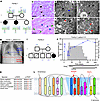 Figure 1
Clinical presentation and identification of the SLC5A6R253W mutation. (A) Pedigree of Family 1. The unaffected consanguineous parents and child II-3 were heterozygous for the SLC5A6 mutation (C/T) in R253, whereas children II-1 and II-2 both had DCM and were homozygous (T/T). (B) X-ray of child II-2 from Family 1, with a cardiothoracic ratio of 0.57, indicating an enlarged heart. (C–H) Imaging of a left ventricular heart biopsy from child II-2. (C and D) H&E-stained sections showed myocardial disarray with hypertrophied cardiomyocytes (black arrowheads), hyperchromatic nuclei (green arrowheads), and myofibrillar loss (blue arrowheads). (E–H) Sarcomeric wasting (black arrow in E), sarcomeric disarray (blue arrow in E), Z band disarray (black arrowheads in E), fragmented mitochondria (black arrowheads in F and G), and mitochondrial degradation (red arrowheads in F and H). (I) Pedigree of Family 2. The unaffected consanguineous parents were heterozygous for the SLC5A6 mutation (C/T) in P437, whereas Patient II-5 had DCM and was homozygous (T/T). Child II-2 died at 8 months from heart failure, but no genetic testing had been performed. (J) Timeline and treatment of Patient II-5 from Family 2. The ejection fraction (EF, blue line) and the administration of up to 5 cardiac drugs (gray histogram) at different time points are shown. The first day of admission to hospital is classified as day 0. The cardiac drugs were stopped on day 148 when the EF became stabilized. Vitamin treatment with biotin and pantothenic acid (PA) is ongoing, and the EF continues to be stabilized. (K) Amino acid residues R253 and P437 are both highly conserved across species. (L) Schematic of SMVT. The patient mutation, p.R253W (Family 1), is in the hinge region of the transporter and the p.P437L mutation (Family 2) is located within transmembrane domain 11. Scale bars: 20 μm (C and D), 2 μm (E), and 500 nm (F–H).
A myocardial biopsy obtained during LVAD insertion, stained by H&E, showed myocardial disarray, featuring hypertrophied cardiomyocytes (Figure 1C) with large, irregular, hyperchromatic nuclei. Pale stained cells correspond to myofibrillar loss within cardiomyocytes (Figure 1D). Transmission electron microscopy (TEM) images of the left ventricle revealed sarcomeric wasting and disarray. In some areas, the sarcomeres had fragmented, creating white patches, and the organized parallel alignment of the sarcomeres was lost. Z band disarray was evident, indicated by variations in the thickness of the lines. Additionally, large clusters of mitochondria were observed, with signs of fragmentation (Figure 1, E–H). The examining pathologist diagnosed cardiomyopathy with no morphological evidence as to the cause.
In Family 2 (15), consanguineous parents from Tunisia, had 5 children (Figure 1I). Child II-2 died of unexplained multisystemic decompensation and severe cardiac dysfunction at 8 months (genetic testing was not available at that time). Patient II-5 presented with DCM with severe cardiac decompensation at 5 months, with a left ventricular EF of 32% (normal > 60%; classed as day 0 in Figure 1J); however, further echocardiographic parameters or cardiac dimensional measurements could not be obtained. Treatment with cardiac drugs (adrenaline, milrinone, and furosemide) for 3 days resulted in EF improvement to 62% by day 10. However, by day 22, the patient had relapsed with an EF of 17%, necessitating intensive cardiac support with an increasing number of cardiac drugs (digoxin, furosemide, spironolactone, carvedilol, and lisinopril). Additionally, following an abnormal plasma acylcarnitine profile and a persistent increase in 3-HIA in the urine, biotin supplementation (10 mg on day 22, increased to 15 mg on day 33), and then pantothenic acid (100 mg from day 64) were initiated. Following the increase in biotin dose to 15 mg, the EF increased to 45% and the patient was maintained on vitamins and 4 cardiac drugs (digoxin, furosemide, carvedilol, and lisinopril) for a further 79 days. The number of cardiac drugs were slowly reduced as the EF rose to a healthy 66% on day 121 and 75% on day 150 (Figure 1J). Bivitamin supplementation remains ongoing, and the EF has remained within the normal range. The patient also presented with severe hypogammaglobulinemia that interestingly corrected itself after vitamin supplementation.
Pathogenic missense mutations in SLC5A6. Whole-exome sequencing of Family 1 revealed a homozygous missense variant in SLC5A6 ([GRCh37] Chr2:27427777 C>T) in Patients II-1 and II-2. The mutation results in a substitution of arginine at position 253 to a tryptophan (p.R253W). All unaffected family members were heterozygous for the variant (Figure 1A). The mutation, in exon 9 of SLC5A6, has an allele frequency of 0.00001983 (gnomADv4.1) and a high pathogenicity prediction (CADD score: 27.4).
Both the p.R253W and p.P437L mutation (C>T) in SLC5A6 previously reported in Family 2 (15) affect highly conserved amino acids (Figure 1K). Within the SMVT protein (, the p.R253W mutation is located on the extracellular loop between transmembrane domains 6 and 7, spanning the hinge region of the LeuT fold and p.P437L is within transmembrane domain 11 (Figure 1L). In silico protein modeling indicated that the substitution of arginine to tryptophan in p.R253W introduces a large bulky aromatic ring, which is predicted to induce steric hindrance and prevent the formation of a salt bridge with E91 (Supplemental Figure 1, A–E; supplemental material available online with this article; https://doi.org/10.1172/jci.insight.200381DS1). Similarly, P437L repositions the large intracellular loop and a polar bond is formed between L437 and V434 (Supplemental Figure 1, F–J). Therefore, structural modeling predicts that both of these pathogenic mutations in SLC5A6 may induce conformational changes in the tertiary structure of the SLC5A6 protein, but further experimental validation of the functional impact is required in future studies.
Slc5a6cKO mice develop cardiomyopathy. We confirmed that SLC5A6 was expressed in the mouse and human embryonic heart (Supplemental Figure 2, A–G). Initially, it was found that mice constitutively deficient for Slc5a6 (Supplemental Figure 2H) exhibited embryonic lethality, with underdeveloped embryos at embryonic day 9.5 (E9.5) and E10.5 (Supplemental Figure 2, I–L). The number of Slc5a6Tm1a/Tm1a mutant embryos was significantly underrepresented when collected between E9.5 and E15.5 (Supplemental Figure 2M).
A cardiac-specific Slc5a6-knockout (Slc5a6cKO) mouse line was then produced by crossing conditional Slc5a6Tm1c mice with the Tnt-Cre line (Supplemental Figure 2H). Reverse transcription PCR (RT-PCR) confirmed heart-specific deletion of exons 7–10 (Supplemental Figure 2N) resulting in a premature stop codon (Supplemental Figure 1, K–M, and Supplemental Figure 2H). The Slc5a6cKO mice were observed at the expected numbers at weaning (Supplemental Figure 2O).
Histological analysis at E15.5 revealed that Slc5a6cKO embryos displayed no developmental cardiac abnormalities (Figure 2, A and B). Slc5a6cKO mice were phenotypically normal at birth and into early adulthood, but by 20 weeks there was a significant decrease in body weight (Supplemental Figure 2P). Sudden death occurred in Slc5a6cKO mice at 26 weeks (n = 3), and histological analysis revealed cardiac enlargement, with visibly enlarged atria and ventricles (Figure 2, C–F). To comply with local ethical requirements, subsequent litters were euthanized at or before 20 weeks of age. At 14 weeks, cardiomyocyte hypertrophy was observed in both the right and left ventricles (Figure 2, G–I). At 20 weeks, no differences were observed in heart weight–to–body weight or to tibia length ratios (Figure 2J and Supplemental Figure 2Q). Fibrosis was significantly increased 3-fold throughout the ventricular walls of the Slc5a6cKO hearts compared with control hearts (Figure 2, K–M). An increase in atrial natriuretic peptide (Nppa) and a decrease in α-cardiac myosin heavy chain (Myh6) expression were observed in Slc5a6cKO hearts at 20 weeks (Supplemental Figure 2, R and S).
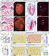 Figure 2
Cardiac-specific Slc5a6-KO (Slc5a6cKO) mouse recapitulates a cardiomyopathy phenotype. (A and B) Hearts from E15.5 control embryos (A) were comparable to Slc5a6cKO mutant embryos (B). (C–F) Slc5a6cKO mutant hearts collected at 26 weeks were visibly enlarged (D and F) compared with controls (C and E). (G and H) WGA staining was performed on coronal heart sections of 14-week control (G) and Slc5a6cKO mutant hearts (H). (I) Cardiomyocytes (CM) were significantly larger in both ventricles in the Slc5a6cKO mutant (n = 4) compared with the control (n = 6) hearts. (J) No difference in heart weight (HW) to body weight (BW) ratio was observed between control (n = 8) and Slc5a6cKO mutant (n = 6) mice at 20 weeks. (K–M) Fibrosis was increased in Slc5a6cKO mutant hearts (n = 3; L and M) compared with control hearts (n = 3; K and M) at 20 weeks. (N–P) Following vitamin supplementation, no increase in fibrosis in Slc5a6cKO mutants (n = 3) compared to control (n = 3) mice was observed. Data are represented as mean ± SEM. ns, nonsignificant. **P < 0.01, ***P < 0.001 by 1-way ANOVA with Bonferroni’s correction for multiple comparisons (I) or unpaired t-test for two sample comparisons (M, J, and P). RV, right ventricle; LV, left ventricle; RA, right atria; LA, left atria; Con, control; cKO, Slc5a6cKO; ConV, vitamin-supplemented control; cKOV, vitamin-supplemented Slc5a6cKO. Scale bars: 500 μm (A and B), 2 mm (C and D), 1 mm (E and F), 50 μm (G and H), and 100 μm (K, L, N, and O).
Thus, Slc5a6cKO mice exhibit the key features of cardiomyopathy, with a cardiac enlargement at 26 weeks, preceded by cardiomyocyte hypertrophy, fibrosis, and activation of fetal gene expression.
Cardiac function is severely reduced in Slc5a6cKO mice. Cardiac magnetic resonance (CMR) imaging and 3-lead electrocardiography (ECG) readings were used to assess the functional consequences of structural abnormalities. CMR analysis at 14 weeks revealed a significant reduction in stroke volume (SV) and the percentage change in left ventricular wall thickness in Slc5a6cKO mice (Supplemental Figure 3, A–F). At 20 weeks, quantification of left ventricular volumes (Figure 3A) showed significant decreases in EF, SV, and cardiac output (CO), and an increase in end systolic volume (ESV) in Slc5a6cKO mutants (Figure 3, B–E, and Supplemental Table 1). No change in end diastolic volume (EDV) was observed (Figure 3F). A significant change in left ventricular wall thickness between diastole and systole, confirming normal systolic thickening of the left ventricular wall, was observed in the control hearts, but was absent in the Slc5a6cKO mutants (Figure 3G). Together, these findings are consistent with an early systolic dysfunction phenotype in Slc5a6cKO hearts at 20 weeks that precedes overt chamber dilation.
 Figure 3
Impaired cardiac contractility and ventricular remodeling in Slc5a6cKO mice are prevented by vitamin supplementation. (A–G) Cardiac magnetic resonance (CMR) imaging at 20 weeks. (A) Mouse cardiac CMR images of short-axis slice through the mid-heart showing left ventricular diastole and systole, with the epicardium outlined in red and endocardium in yellow. Significant decreases in ejection fraction (B), stroke volume (C), cardiac output (D), end systolic volume (ESV) (E), and percentage change in left ventricular (LV) wall thickness at diastole and systole (G) were detected in Slc5a6cKO mutants (n = 4) compared with controls (n = 6). The end diastolic volume (EDV) showed no difference (F). After vitamin supplementation, no apparent differences in cardiac function were seen (n = 2 for each genotype). Con, control; cKO, Slc5a6cKO; ConV, vitamin-supplemented control; cKOV, vitamin-supplemented Slc5a6cKO. Data are represented as mean ± SEM. ns, nonsignificant. *P < 0.05, **P < 0.01, ***P < 0.001 by 1-way ANOVA with Bonferroni’s correction for multiple comparisons (B–G) or nonparametric Kruskal-Wallis test with Dunn’s correction for multiple comparisons.
ECG was performed on control and Slc5a6cKO mice at 8 and 20 weeks to monitor cardiac electrical activity as an indicator of cardiac function in vivo, to track cardiac conduction changes longitudinally. As in human ECG, in mice, atrial depolarization appears as the P wave followed by the PR interval, representing the time taken for the electrical impulse to travel from the atria to the ventricles. Ventricular depolarization forms the QRS complex. In mice, the J wave appears as a distinct deflection immediately following the QRS complex, and transitions into the T wave, marking the progression from early to late ventricular repolarization (23). These parameters are all clear in the ECG traces from 20-week control mice (Figure 4A). In comparison, the PR interval in 20-week Slc5a6cKO mice was visually extended, and the J wave was missing (arrow in Figure 4B). Slc5a6cKO mutants showed a significantly reduced heart rate at both ages (Figure 4, E and I). Correspondingly, PR intervals were significantly prolonged, increasing from 0.051 ± 0.001 seconds at 8 weeks to 0.063 ± 0.002 seconds (P = 0.0001) at 20 weeks in Slc5a6cKO mice, compared with controls (Figure 4, F and J, and Supplemental Figure 3G), indicating worsening atrioventricular (AV) conduction.
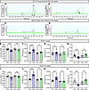 Figure 4
Slc5a6cKO mice develop progressive electrical conduction abnormalities that are prevented by vitamin supplementation. (A–D) Representative average ECG traces from 20-week mice. Control (A) and vitamin-supplemented control (C) mice have a well-defined P wave, QRS complex, and J wave, whereas Slc5a6cKO mice (B) presented with a longer PR interval (dotted line) and loss of the J wave (arrow). ECG traces from vitamin-supplemented Slc5a6cKO mutants (D) were comparable to control mice. (E–L) Quantification of ECG traces from 8-week mice (E–H) and 20-week mice (I–L) on a normal diet, showed a significant decrease in heart rate, increase in PR interval, widening of the QRS complex (20-weeks only), and loss of the J wave amplitude in Slc5a6cKO mutants. No differences were observed in the vitamin-supplemented Slc5a6cKO mice compared to vitamin-supplemented control mice (8 weeks: Con n = 13, cKO n = 12, ConV n = 12, cKOV n = 10; 20 weeks: Con n = 10, cKO n = 14, ConV n = 12, cKOV n = 10). Con, control; cKO, Slc5a6cKO; ConV, vitamin-supplemented control; cKOV, vitamin-supplemented Slc5a6cKO. Data are represented as mean ± SEM. ns, nonsignificant. **P < 0.01, ***P < 0.001, ****P < 0.0001 by 1-way ANOVA with Bonferroni’s correction for multiple comparisons (E and J) or nonparametric Kruskal-Wallis test with Dunn’s correction for multiple comparisons (F–I, K, and L).
QRS morphology also deteriorated with age. Although QRS duration was normal at 8 weeks, it became significantly widened by 20 weeks (Figure 4, G and K). In contrast, R wave amplitude was significantly reduced at both ages (Supplemental Figure 3, H and I). J waves were present in only 23.5% of Slc5a6cKO mutants at 8 weeks and were completely absent by 20 weeks (Figure 4, H and L, Supplemental Figure 3, O and P, and Table 1). In addition, S waves were absent in 28.6% of the Slc5a6cKO mutants (Supplemental Figure 3Q and Table 1).
AV conduction and arrhythmia burden also deteriorated with age. By 20 weeks, high-degree AV block with missed QRS complexes occurred in 21.4% of Slc5a6cKO mutants, and ventricular ectopy was observed in 28.6% of Slc5a6cKO mutants, and neither were observed in any other genotypes (Supplemental Figure 3, R and S). In 3 mice, ventricular ectopic beats constituted 20% or more of all QRS complexes, indicating substantial ventricular electrical instability. Thus, all Slc5a6cKO mutants had ECG abnormalities at 20 weeks, with about a quarter demonstrating severe and life-threatening arrhythmia.
The cardiac-specific deletion of Slc5a6, therefore, caused anatomical, histological, and functional cardiac changes consistent with cardiomyopathy, resulting in progressive cardiac dysfunction and sudden death by 26 weeks.
Impaired biotin and pantothenic acid transport in Slc5a6cKO mice. Biotin, a cofactor for carboxylases, and pantothenic acid, required for the synthesis of coenzyme A (CoA), are both transported by SMVT (24, 25). To show that vitamin transport was reduced in Slc5a6cKO hearts, we assessed these vitamin-dependent metabolic pathways.
Analysis of heart tissue by liquid chromatography coupled with quadrupole time-of-flight mass spectrometry (LC-MS-qTOF) revealed significantly increased levels of 3-HIA and 2-methylcitric acid (2-MCA) in Slc5a6cKO hearts, indicative of MCC and propionyl-CoA carboxylase (PCC) deficiencies, respectively (Figure 5, A and B).
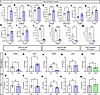 Figure 5
Reduced vitamin transport in hearts from Slc5a6cKO hearts. (A–M) Metabolic analysis revealed significant increases in 3-HIA and 2-MCA in Slc5a6cKO mutants (n = 6) compared with controls (n = 5) (A and B). (C–I) Acylcarnitine analysis of plasma showed significant increases in C0, C5OH, C5OH/C0 ratio, C3DC, C6, C8, and C14 in Slc5a6cKO mutants (n = 6) compared with controls (n = 5). (J–M) Reduced pantothenic acid uptake was observed in Slc5a6cKO mutants (J) as well as a significant decrease in PA, PPA, PP, and CoAG. (N–W) In heart protein samples (n = 3–4 for each genotype), a decrease in PCC/MCC biotinylation was observed in Slc5a6cKO mutants at 5 and 20 weeks (N and P), with no corresponding change in liver samples (O and Q). In vitamin-supplemented Slc5a6cKO mutants, PCC/MCC biotinylation levels in the hearts were comparable to controls (R). No change in PC was observed in the hearts or livers (S–W). Data are represented as mean ± SEM. ns, nonsignificant. *P < 0.05, **P < 0.01, ***P < 0.001, ****P < 0.0001 by unpaired t test for 2-sample comparisons. 3-HIA, 3-hydroxyisovaleric acid; 2-MCA, 2-methylcitric acid; C0, free carnitine; C5OH, 3-hydroxyisovaleryl-carnitine; C3DC, malonylcarnitine; C6, hexanoylcarnitine; C8, octanoylcarnitine; C14, tetradecanoylcarnitine; PA, pantothenic acid; PPA, phosphopantothenic acid; PP, phosphopantetheine; CoAG, CoA-glutathione; MCC, 3-methylcrotonyl-CoA carboxylase; PCC, propionyl-CoA carboxylase; PC, pyruvate carboxylase; Con, control; cKO, Slc5a6cKO; ConV, vitamin-supplemented control; cKOV, vitamin-supplemented Slc5a6cKO.
Flow injection analysis coupled with tandem MS (FIA-MS/MS) of the plasma acylcarnitine profile further supported disrupted carboxylase function. Free carnitine (C0), 3-hydroxyisovaleryl-carnitine (C5OH), and the C5OH/C0 ratio were all significantly increased, which are hallmarks of MCC and biotinidase deficiency (Figure 5, C–E). Additional increases in C3DC, C6, C8, and C14 (Figure 5, F–I) reflect accumulation of mitochondrial acyl-CoA intermediates caused by impaired β-oxidation and leucine catabolism, forcing the excess acyl groups to be diverted into the carnitine pool and exported as acylcarnitines.
Additionally, cardiac levels of pantothenic acid and its derivatives phosphopantothenic acid and phosphopantetheine were significantly decreased, leading to impaired CoA synthesis, as evidenced by reduced CoA-glutathione (Figure 5, J–M, and Supplemental Table 2).
Western blot analysis confirmed a significant reduction in MCC and PCC biotinylation in Slc5a6cKO hearts at 5 weeks (before any structural or functional cardiac differences were observed) and 20 weeks (experimental endpoint) (Figure 5, N and P, and Supplemental Figure 4, A and C), which was unchanged in the liver (Figure 5, O and Q, and Supplemental Figure 4, B and D). Pyruvate carboxylase (PC) levels were unaffected (Figure 5, S–W, and Supplemental Figure 4, A–D). Consistent with findings in patients, the Slc5a6cKO mice showed elevated C5OH and 3-HIA, hence reproducing the hallmark metabolic changes seen in SLC5A6 deficiency (10, 15). Together, these results indicate a heart-specific reduction in biotin and pantothenic acid transport in Slc5a6cKO mice.
Vitamin supplementation ameliorates the DCM phenotype. As Patient II-5 in Family 2 responded well to early vitamin treatment, we tested whether vitamin supplementation improved cardiac function in Slc5a6cKO mice by supplementing drinking water and diet with biotin and pantothenic acid: biotin at approximately 120 mg/kg and pantothenic acid at approximately 2,089 mg/kg, compared with 0.4 mg/kg and 33 mg/kg available in the normal standard diet. This represents an approximately 300- and approximately 63-fold increase, respectively. These doses are equivalent to those used in humans using body surface area scaling and remain substantially below the LC50 thresholds for both vitamins (Supplemental Table 3) (26). Supplementation was initiated in parental mice before conception, continued throughout gestation and provided long-term to the offspring. Maintenance of normal levels of MCC and PCC biotinylation in 20-week hearts of supplemented Slc5a6cKO mice (Figure 5R and Supplemental Figure 4E) indicated successful vitamin uptake.
Vitamin-supplemented Slc5a6cKO mice survived to 40 weeks, at which time the mice were electively culled. Vitamin supplementation did not affect body weight at 20 or 40 weeks (Supplemental Figure 5, A–F), no excessive fibrosis in Slc5a6cKO hearts was seen (Figure 2, N–P, and Supplemental Figure 5, G–K), and the expression of Nppa and Myh6 remained normal at 20 weeks (Supplemental Figure 2, R and S). There were no discernible differences in cardiac function (EF, SV, CO, ESV, EDV) or percentage change in left ventricular wall thickness, between vitamin-supplemented control and vitamin-supplemented Slc5a6cKO mice (Figure 3, B–G), and all ECG parameters were no longer significantly different between control and Slc5a6cKO mice at 8 and 20 weeks (Figure 4, D–L, and Supplemental Figure 3, H and I). No AV block or ventricular ectopy was observed in vitamin-supplemented Slc5a6cKO mice (Table 1). At 40 weeks, all the cardiac electrical measurements were similar between control mice on a normal diet and vitamin-supplemented control and Slc5a6cKO mice (Supplemental Figure 3, J–N). Vitamin supplementation, therefore, prevented the cardiomyopathy phenotype observed in Slc5a6cKO mice and had no long-term impact on cardiac function.
Three-dimensional mitochondrial morphology is increased in Slc5a6cKO hearts. As changes in mitochondrial morphology are associated with metabolic dysfunction and disease progression (27, 28), we analyzed the ultrastructure of cardiac mitochondria using TEM. As conventional 2-dimensional descriptors do not fully capture changes in mitochondrial morphology, we quantified morphology using the mitochondrial complexity index (MCI) (29), a 3-dimensional (3D) metric that integrates measures of mitochondrial volume, surface irregularity, and branching patterns. The MCI increases with greater branching, elongation, and irregularity of mitochondrial shape.
At 20 weeks, control hearts presented with predominantly spherically shaped mitochondria, with minimal branching or complex shapes, and formed parallel clusters along longitudinal myofibers (Figure 6, A and C, and Supplemental Figure 6, A–D). In contrast, mitochondria in Slc5a6cKO hearts were irregularly shaped and arranged randomly between myofibrils, with evidence of mitochondrial degradation (Figure 6, B and D, and Supplemental Figure 6, E–L). The MCI was significantly increased in Slc5a6cKO hearts, with a corresponding significant decrease in volume (Figure 6, I and J, and Supplemental Table 4). The Slc5a6cKO hearts, therefore, had smaller mitochondria with more complex shapes, including excessive elongation, branching, and nanotunnels (Supplemental Figure 6, M–P). This is further represented by the significant difference in the lines of best fit for controls versus Slc5a6cKO hearts on the bivariate plot (P < 0.0001; Figure 6K) and a shift to the right in the cumulative frequency distributions (Figure 6L).
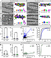 Figure 6
Increased mitochondrial complexity following loss of Slc5a6 is prevented by vitamin supplementation. (A–H) Mitochondria imaging at 20 weeks by TEM (A, B, E, and F) and 3D reconstructions (C, D, G, and H). Mitochondria in controls, vitamin-supplemented controls, and vitamin-supplemented Slc5a6cKO hearts were all comparable (A, C, and E–H), whereas mitochondria in Slc5a6cKO hearts (B and D) were unevenly arranged and irregularly shaped with evidence of mitochondrial degradation (arrows in B). Representative individual mitochondria of the minimum (min), first quartile (1Q), median (med), third quartile (3Q), and maximum (max) MCI are shown (total number of mitochondria analyzed: Con = 468, cKO = 530, ConV = 241 and cKOV = 240). (I–L) The MCI was significantly increased in 20-week Slc5a6cKO mice on a normal diet (I) and the mitochondrial volume was significantly decreased (J). Bivariate analysis of MCI versus volume shows a significant difference in the regression line (K) and the cumulative frequency distribution curves do not overlap (L). (M–P) In vitamin-supplemented Slc5a6cKO mice, the MCI was significantly reduced (M) and there was no difference in mitochondrial volume (N) and complexity (O and P). Data are represented as mean ± SEM. ns, nonsignificant. **P < 0.01, ****P < 0.0001 by nonparametric Mann-Whitney test for two sample comparisons (I, J, M, and N) and the slopes of the regression lines were compared using an 1-way Analysis of Covariance (ANCOVA) (K and O). Con, control; cKO, Slc5a6cKO; ConV, vitamin-supplemented control; cKOV, vitamin-supplemented Slc5a6cKO. Scale bars: 500 nm (A, B, E, and F) and 1 μm (C, D, G, and H).
To establish whether the changes in mitochondrial morphology were associated with early indicators of disease, the mitochondria were also investigated at 8 weeks. The overall arrangement of the parallel clusters of the mitochondria along the myofibrils was comparable between control and Slc5a6cKO mice (Supplemental Figure 7, A and B). There was, however, a significant increase in the MCI of mitochondria in Slc5a6cKO hearts compared with controls, indicating the mitochondria had a more complex shape, although there was no change in volume (Supplemental Figure 7, C, D, and I–L, and Supplemental Table 4). No significant change in mitochondrial DNA copy number was observed (Supplemental Figure 7, Q and R).
Cardiac mitochondrial morphology was then assessed in vitamin-supplemented mice. At 20 weeks, the overall organization and alignment of mitochondria was similar between vitamin-supplemented control and Slc5a6cKO hearts. There was a significant reduction in MCI of mitochondria in Slc5a6cKO hearts compared with controls, indicating that mitochondria were uniform in size and less complex, and there was no change in volume (Figure 6, E–H, M, and N, and Supplemental Table 4), suggesting that vitamins prevented the abnormal mitochondrial morphology. The bivariate plot of mitochondrial MCI versus volume and cumulative MCI frequency showed no differences between vitamin-supplemented control and Slc5a6cKO hearts at 20 weeks (Figure 6, O and P). At 8 weeks, there was a small yet significant decrease in MCI but not volume (Supplemental Figure 7, E–H, M, and N, and Supplemental Table 4). Interestingly, in vitamin-supplemented mice at 8 weeks, the correlation between the spread of the mitochondria was similar between control and Slc5a6cKO hearts, but still significantly different (P = 0.0084; Supplemental Figure 7, O and P).
Thus, the subtle change in mitochondrial complexity in Slc5a6cKO hearts at 8 weeks coincides with the early indication of cardiac dysfunction in Slc5a6cKO mice. As DCM progresses towards functional decline at 20 weeks, the mitochondria become more complex in shape and is associated with a corresponding increase in mitochondria degradation. Vitamin supplementation mitigates this effect in Slc5a6cKO mice.
Early disruption of the cardiac proteome precedes cardiomyopathy in Slc5a6cKO mice. To elucidate the cellular mechanism driving cardiomyopathy progression in the Slc5a6cKO mice, we performed an unbiased proteomics analysis to identify altered signaling pathways at 8 weeks before the overt cardiomyopathy phenotype was evident. Protein was extracted from hearts of control and Slc5a6cKO mice maintained on a normal diet and a vitamin-supplemented diet (n = 5 per group).
Analysis of differential protein expression, using principal component analysis and a hierarchical heatmap plot, showed that the Slc5a6cKO samples formed a distinct group compared with the control and vitamin-supplemented samples (Figure 7, A and B).
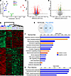 Figure 7
Proteomic analysis revealed altered metabolism and increased fibrosis correlate with early indicators of cardiomyopathy in Slc5a6cKO mice. (A–E) Proteomic analysis at 8 weeks. PCA plot (A) and heatmap (B) illustrate that the proteome of the Slc5a6cKO mutants was distinct from the 3 other groups (control, vitamin-supplemented control, and vitamin-supplemented Slc5a6cKO hearts; n = 5 per group). (C and D) Volcano plots showing proteins with a significant (q < 0.05) fold change. (C) Under a normal diet, 1459 proteins with a fold change of ≤ –0.5 and ≥ 0.5 (red dots) and 478 proteins with a fold change of > –0.5 and < 0.5 (blue dots) were seen. (D) With vitamin supplementation, only 153 proteins with a fold change of ≤ –0.5 and ≥ 0.5 (orange dots) and 179 proteins with a fold change of > –0.5 and < 0.5 (green dots) were seen. (E) Venn diagram illustrating 124 of the proteins with a fold change of ≤ –0.5 and ≥ 0.5 remained significantly differentially expressed following vitamin supplementation. (F and G) IPA analysis of up- and downregulated proteins. For the mice on a normal diet, the majority of the enriched downregulated pathways in Slc5a6cKO mutant mice are linked to different components of the energy metabolism pathways in mitochondria (shown by asterisks in F) (F). In comparison, vitamin-supplemented Slc5a6cKO mutant mice only had downregulation of peroxisome-related pathways (G).
In Slc5a6cKO hearts on a normal diet, a total of 1,459 proteins showed significant differential expression (599 downregulated, 860 upregulated, P < 0.05) compared with controls (Figure 7C). For the vitamin-supplemented mice, only 153 proteins showed significant differential expression (81 downregulated, 72 upregulated, P < 0.05) in Slc5a6cKO hearts compared with controls (Figure 7D). Of the proteins differentially expressed under a normal diet, only 124 of those proteins remained significantly differentially expressed following vitamin supplementation (Figure 7E). There was no significant difference in protein expression between control mice on the normal diet or the vitamin-supplemented diet.
Pathway analysis was performed on the significantly up- and downregulated proteins combined. From Slc5a6cKO mice on a normal diet, in the top 15 significantly enriched pathways (Figure 7F), the upregulated pathways included the “sirtuin signaling pathway” and “collagen biosynthesis and modifying enzymes,” which are both linked to increasing fibrosis (30, 31), and “granzyme A signaling,” which correlates with the upregulation of mitochondrial dysfunction pathways (32). Downregulated pathways were predominantly related to energy metabolism: “respiratory electron transport,” “mitochondrial translation,” “oxidation phosphorylation,” “complex I biogenesis,” “branched-chain amino acid [BCAA] catabolism” (specifically valine and isoleucine degradation), and “fatty acid β-oxidation.” The hematoma resolution signaling pathway is linked to genes involved in peroxisomal β-oxidation (33). In contrast, vitamin supplementation dramatically reduced the number of enriched pathways in Slc5a6cKO hearts (Figure 7G), with downregulation only identified in peroxisome-related pathways. There was no upregulation of other known vitamin-related transporters, including monocarboxylate transporter 1 (MCT1), which transports biotin in specific cell types such as immune cells and skin, in Slc5a6cKO hearts on a normal or vitamin-supplemented diet.
Energy metabolism involves several interconnected pathways. Analysis of different pathways showed that the majority of proteins linked to the TCA cycle, fatty acid β-oxidation, BCAA catabolism, and the respiratory electron transport chain (ETC) were all downregulated in the Slc5a6cKO hearts on a normal diet, with a corresponding increase in proteins involved in glycolysis (Figure 8A), which is a common compensatory response in heart failure (34).
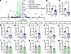 Figure 8
Broad suppression of mitochondrial energy metabolism pathways and electron transport chain components in Slc5a6cKO hearts. (A) Summary of 5 different pathways involved in energy metabolism identified in the proteomics analysis of 8-week hearts. The majority of proteins in the TCA cycle, fatty acid β-oxidation, branched-chain amino acid (BCAA) catabolism, and electron transport chain (ETC) are downregulated in Slc5a6cKO mutants, with an increase in proteins associated with glycolysis. (B and C) Quantification of Western blot analyses showed reduced abundance of representative β-oxidation enzymes HADHA and MCAD in 5-week Slc5a6cKO hearts compared with controls. (D–M) qPCR analysis of genes representing a subunit from each complex (CI–CV) of the ETC (CI: Ndufb8, CII: Sdhb, CIII: Uqcrc2, CIV: Mtco2 and CV: Atp5a1) in 8-week hearts. There was a significant downregulation in CI, CII, CIV, and CV in Slc5a6cKO hearts (D–H) and no corresponding decrease in vitamin-treated hearts (I–M) (n = 5 hearts for each genotype). Data are represented as mean ± SEM. ns, nonsignificant. *P < 0.05, **P < 0.01, ***P < 0.001, ****P < 0.0001 by unpaired t test for 2 sample comparisons. Con, control; cKO, Slc5a6cKO; ConV, vitamin-supplemented control; cKOV, vitamin-supplemented Slc5a6cKO.
Western blot analysis of heart tissue confirmed the downregulation of 2 keys enzymes required in β-oxidation, medium-chain acyl-CoA dehydrogenase (MCAD) and hydroxyacyl-CoA dehydrogenase trifunctional multienzyme complex subunit α (HADHA) (Figure 8, B and C, and Supplemental Figure 8). Each mitochondrial complex in the ETC, complex I to V, is made from multiple subunits. Notably, from the subunits detected in the proteomics experiment, the components exhibited widespread reduced expression in Slc5a6cKO mutants compared with controls, with the following percentage of subunits showing decreased expression: complex I 90.2%, complex II 100%, complex III 77.8%, complex IV 77%, and complex V 76.5% (q < 0.05, all fold changes; Supplemental Table 5). Remarkably, vitamin supplementation substantially prevented these deficits, reducing the proportion of downregulated subunits to only 2 in complex I (NDUFA11 and NDUFS6) and 1 in complex IV (MT-CO3) in vitamin-supplemented Slc5a6cKO hearts compared with controls. Quantitative real-time PCR (qPCR) analysis of representative genes from the subunits of each complex (Supplemental Table 5) confirmed significant downregulation of subunits in complexes I, II, IV, and V in Slc5a6cKO hearts (Figure 8, D–H), with no significant differences detected in vitamin-treated hearts (Figure 8, I–M).
Integrated analysis of proteomic and metabolomic datasets from 8-week-old control and Slc5a6cKO hearts revealed coordinated changes across multiple energy-metabolism pathways, including glycolysis, pyruvate oxidation, β-oxidation, TCA cycle, and BCAA catabolism (Supplemental Figure 9 and Figure 9). All 5 components of the pyruvate dehydrogenase (PDH) complex were downregulated, potentially reducing the capacity for pyruvate oxidation via this lipoic acid– and CoA-dependent pathway. In parallel, reduced abundance of key fatty acid transport and β-oxidation enzymes, together with accumulation of medium- and long-chain acylcarnitines, is consistent with incomplete mitochondrial fatty acid oxidation and diminished contribution to acetyl-CoA production. The TCA cycle also exhibited coordinated downregulation of multiple enzymatic components, suggesting reduced cycle capacity to oxidize acetyl-CoA. Finally, BCAA catabolism was broadly suppressed, with reduced expression of enzymes involved in leucine, isoleucine, and valine oxidation and evidence of propionyl-CoA overflow, including accumulation of 2-methylcitrate.
 Figure 9
Integrated model of altered cardiac energy metabolism pathways in Slc5a6cKO hearts. Schematic overview integrating proteomic and metabolomic findings, illustrating affected energy metabolism pathways and factors, in Slc5a6cKO hearts (outlined in red). Vitamins and their role in the pathways are color coded: biotin, yellow; lipoic acid, green; and pantothenic acid, blue.


