
Research ArticleNeuroscienceOphthalmology
Open Access |  10.1172/jci.insight.202223
Optic nerve regeneration requires the intracellular domain of LIFRα/CD118
Qian Jiang,1,2 Cong Wang,1,2 Yuerong Ren,1,2 Peiyun Duan,1,2 Ke Tian,1,2 Xiangwei Duan,1,2 Binghan Cai,1,2 Changzhong Xu,1,2 Ke Liu,1,2 Jian Li,3 Larry Benowitz,4 Ningli Wang,5 Bing Jiang,1,2 and Lili Xie1,2
1Department of Ophthalmology, The Second Xiangya Hospital, Central South University, Changsha, Hunan, China.
2Hunan Clinical Research Center of Ophthalmic Disease, Changsha, Hunan, China.
3Division of Rheumatology, Inflammation and Immunity, Brigham and Women’s Hospital and Department of Medicine, Harvard Medical School, Boston, Massachusetts, USA.
4Departments of Neurosurgery and Ophthalmology, Boston Children’s Hospital and Harvard Medical School, Boston, Massachusetts, USA.
5Department of Ophthalmology, Beijing Tongren Hospital, Capital Medical University, Beijing, China.
Address correspondence to: Lili Xie or Bing Jiang, Department of Ophthalmology, The Second Xiangya Hospital, Central South University, Changsha, Hunan 410011, China. Phone: 86.18390801107; Email: lili.xie@csu.edu.cn (LX). Phone: 86.13875868866; Email: drjiangb@csu.edu.cn (BJ).
Find articles by Jiang, Q. in: PubMed | Google Scholar
1Department of Ophthalmology, The Second Xiangya Hospital, Central South University, Changsha, Hunan, China.
2Hunan Clinical Research Center of Ophthalmic Disease, Changsha, Hunan, China.
3Division of Rheumatology, Inflammation and Immunity, Brigham and Women’s Hospital and Department of Medicine, Harvard Medical School, Boston, Massachusetts, USA.
4Departments of Neurosurgery and Ophthalmology, Boston Children’s Hospital and Harvard Medical School, Boston, Massachusetts, USA.
5Department of Ophthalmology, Beijing Tongren Hospital, Capital Medical University, Beijing, China.
Address correspondence to: Lili Xie or Bing Jiang, Department of Ophthalmology, The Second Xiangya Hospital, Central South University, Changsha, Hunan 410011, China. Phone: 86.18390801107; Email: lili.xie@csu.edu.cn (LX). Phone: 86.13875868866; Email: drjiangb@csu.edu.cn (BJ).
Find articles by Wang, C. in: PubMed | Google Scholar
1Department of Ophthalmology, The Second Xiangya Hospital, Central South University, Changsha, Hunan, China.
2Hunan Clinical Research Center of Ophthalmic Disease, Changsha, Hunan, China.
3Division of Rheumatology, Inflammation and Immunity, Brigham and Women’s Hospital and Department of Medicine, Harvard Medical School, Boston, Massachusetts, USA.
4Departments of Neurosurgery and Ophthalmology, Boston Children’s Hospital and Harvard Medical School, Boston, Massachusetts, USA.
5Department of Ophthalmology, Beijing Tongren Hospital, Capital Medical University, Beijing, China.
Address correspondence to: Lili Xie or Bing Jiang, Department of Ophthalmology, The Second Xiangya Hospital, Central South University, Changsha, Hunan 410011, China. Phone: 86.18390801107; Email: lili.xie@csu.edu.cn (LX). Phone: 86.13875868866; Email: drjiangb@csu.edu.cn (BJ).
Find articles by Ren, Y. in: PubMed | Google Scholar
1Department of Ophthalmology, The Second Xiangya Hospital, Central South University, Changsha, Hunan, China.
2Hunan Clinical Research Center of Ophthalmic Disease, Changsha, Hunan, China.
3Division of Rheumatology, Inflammation and Immunity, Brigham and Women’s Hospital and Department of Medicine, Harvard Medical School, Boston, Massachusetts, USA.
4Departments of Neurosurgery and Ophthalmology, Boston Children’s Hospital and Harvard Medical School, Boston, Massachusetts, USA.
5Department of Ophthalmology, Beijing Tongren Hospital, Capital Medical University, Beijing, China.
Address correspondence to: Lili Xie or Bing Jiang, Department of Ophthalmology, The Second Xiangya Hospital, Central South University, Changsha, Hunan 410011, China. Phone: 86.18390801107; Email: lili.xie@csu.edu.cn (LX). Phone: 86.13875868866; Email: drjiangb@csu.edu.cn (BJ).
Find articles by Duan, P. in: PubMed | Google Scholar
1Department of Ophthalmology, The Second Xiangya Hospital, Central South University, Changsha, Hunan, China.
2Hunan Clinical Research Center of Ophthalmic Disease, Changsha, Hunan, China.
3Division of Rheumatology, Inflammation and Immunity, Brigham and Women’s Hospital and Department of Medicine, Harvard Medical School, Boston, Massachusetts, USA.
4Departments of Neurosurgery and Ophthalmology, Boston Children’s Hospital and Harvard Medical School, Boston, Massachusetts, USA.
5Department of Ophthalmology, Beijing Tongren Hospital, Capital Medical University, Beijing, China.
Address correspondence to: Lili Xie or Bing Jiang, Department of Ophthalmology, The Second Xiangya Hospital, Central South University, Changsha, Hunan 410011, China. Phone: 86.18390801107; Email: lili.xie@csu.edu.cn (LX). Phone: 86.13875868866; Email: drjiangb@csu.edu.cn (BJ).
Find articles by Tian, K. in: PubMed | Google Scholar
1Department of Ophthalmology, The Second Xiangya Hospital, Central South University, Changsha, Hunan, China.
2Hunan Clinical Research Center of Ophthalmic Disease, Changsha, Hunan, China.
3Division of Rheumatology, Inflammation and Immunity, Brigham and Women’s Hospital and Department of Medicine, Harvard Medical School, Boston, Massachusetts, USA.
4Departments of Neurosurgery and Ophthalmology, Boston Children’s Hospital and Harvard Medical School, Boston, Massachusetts, USA.
5Department of Ophthalmology, Beijing Tongren Hospital, Capital Medical University, Beijing, China.
Address correspondence to: Lili Xie or Bing Jiang, Department of Ophthalmology, The Second Xiangya Hospital, Central South University, Changsha, Hunan 410011, China. Phone: 86.18390801107; Email: lili.xie@csu.edu.cn (LX). Phone: 86.13875868866; Email: drjiangb@csu.edu.cn (BJ).
Find articles by Duan, X. in: PubMed | Google Scholar
1Department of Ophthalmology, The Second Xiangya Hospital, Central South University, Changsha, Hunan, China.
2Hunan Clinical Research Center of Ophthalmic Disease, Changsha, Hunan, China.
3Division of Rheumatology, Inflammation and Immunity, Brigham and Women’s Hospital and Department of Medicine, Harvard Medical School, Boston, Massachusetts, USA.
4Departments of Neurosurgery and Ophthalmology, Boston Children’s Hospital and Harvard Medical School, Boston, Massachusetts, USA.
5Department of Ophthalmology, Beijing Tongren Hospital, Capital Medical University, Beijing, China.
Address correspondence to: Lili Xie or Bing Jiang, Department of Ophthalmology, The Second Xiangya Hospital, Central South University, Changsha, Hunan 410011, China. Phone: 86.18390801107; Email: lili.xie@csu.edu.cn (LX). Phone: 86.13875868866; Email: drjiangb@csu.edu.cn (BJ).
Find articles by Cai, B. in: PubMed | Google Scholar
1Department of Ophthalmology, The Second Xiangya Hospital, Central South University, Changsha, Hunan, China.
2Hunan Clinical Research Center of Ophthalmic Disease, Changsha, Hunan, China.
3Division of Rheumatology, Inflammation and Immunity, Brigham and Women’s Hospital and Department of Medicine, Harvard Medical School, Boston, Massachusetts, USA.
4Departments of Neurosurgery and Ophthalmology, Boston Children’s Hospital and Harvard Medical School, Boston, Massachusetts, USA.
5Department of Ophthalmology, Beijing Tongren Hospital, Capital Medical University, Beijing, China.
Address correspondence to: Lili Xie or Bing Jiang, Department of Ophthalmology, The Second Xiangya Hospital, Central South University, Changsha, Hunan 410011, China. Phone: 86.18390801107; Email: lili.xie@csu.edu.cn (LX). Phone: 86.13875868866; Email: drjiangb@csu.edu.cn (BJ).
Find articles by Xu, C. in: PubMed | Google Scholar
1Department of Ophthalmology, The Second Xiangya Hospital, Central South University, Changsha, Hunan, China.
2Hunan Clinical Research Center of Ophthalmic Disease, Changsha, Hunan, China.
3Division of Rheumatology, Inflammation and Immunity, Brigham and Women’s Hospital and Department of Medicine, Harvard Medical School, Boston, Massachusetts, USA.
4Departments of Neurosurgery and Ophthalmology, Boston Children’s Hospital and Harvard Medical School, Boston, Massachusetts, USA.
5Department of Ophthalmology, Beijing Tongren Hospital, Capital Medical University, Beijing, China.
Address correspondence to: Lili Xie or Bing Jiang, Department of Ophthalmology, The Second Xiangya Hospital, Central South University, Changsha, Hunan 410011, China. Phone: 86.18390801107; Email: lili.xie@csu.edu.cn (LX). Phone: 86.13875868866; Email: drjiangb@csu.edu.cn (BJ).
Find articles by Liu, K. in: PubMed | Google Scholar
1Department of Ophthalmology, The Second Xiangya Hospital, Central South University, Changsha, Hunan, China.
2Hunan Clinical Research Center of Ophthalmic Disease, Changsha, Hunan, China.
3Division of Rheumatology, Inflammation and Immunity, Brigham and Women’s Hospital and Department of Medicine, Harvard Medical School, Boston, Massachusetts, USA.
4Departments of Neurosurgery and Ophthalmology, Boston Children’s Hospital and Harvard Medical School, Boston, Massachusetts, USA.
5Department of Ophthalmology, Beijing Tongren Hospital, Capital Medical University, Beijing, China.
Address correspondence to: Lili Xie or Bing Jiang, Department of Ophthalmology, The Second Xiangya Hospital, Central South University, Changsha, Hunan 410011, China. Phone: 86.18390801107; Email: lili.xie@csu.edu.cn (LX). Phone: 86.13875868866; Email: drjiangb@csu.edu.cn (BJ).
Find articles by Li, J. in: PubMed | Google Scholar
1Department of Ophthalmology, The Second Xiangya Hospital, Central South University, Changsha, Hunan, China.
2Hunan Clinical Research Center of Ophthalmic Disease, Changsha, Hunan, China.
3Division of Rheumatology, Inflammation and Immunity, Brigham and Women’s Hospital and Department of Medicine, Harvard Medical School, Boston, Massachusetts, USA.
4Departments of Neurosurgery and Ophthalmology, Boston Children’s Hospital and Harvard Medical School, Boston, Massachusetts, USA.
5Department of Ophthalmology, Beijing Tongren Hospital, Capital Medical University, Beijing, China.
Address correspondence to: Lili Xie or Bing Jiang, Department of Ophthalmology, The Second Xiangya Hospital, Central South University, Changsha, Hunan 410011, China. Phone: 86.18390801107; Email: lili.xie@csu.edu.cn (LX). Phone: 86.13875868866; Email: drjiangb@csu.edu.cn (BJ).
Find articles by
Benowitz, L.
in:
PubMed
|
Google Scholar
|

1Department of Ophthalmology, The Second Xiangya Hospital, Central South University, Changsha, Hunan, China.
2Hunan Clinical Research Center of Ophthalmic Disease, Changsha, Hunan, China.
3Division of Rheumatology, Inflammation and Immunity, Brigham and Women’s Hospital and Department of Medicine, Harvard Medical School, Boston, Massachusetts, USA.
4Departments of Neurosurgery and Ophthalmology, Boston Children’s Hospital and Harvard Medical School, Boston, Massachusetts, USA.
5Department of Ophthalmology, Beijing Tongren Hospital, Capital Medical University, Beijing, China.
Address correspondence to: Lili Xie or Bing Jiang, Department of Ophthalmology, The Second Xiangya Hospital, Central South University, Changsha, Hunan 410011, China. Phone: 86.18390801107; Email: lili.xie@csu.edu.cn (LX). Phone: 86.13875868866; Email: drjiangb@csu.edu.cn (BJ).
Find articles by Wang, N. in: PubMed | Google Scholar
1Department of Ophthalmology, The Second Xiangya Hospital, Central South University, Changsha, Hunan, China.
2Hunan Clinical Research Center of Ophthalmic Disease, Changsha, Hunan, China.
3Division of Rheumatology, Inflammation and Immunity, Brigham and Women’s Hospital and Department of Medicine, Harvard Medical School, Boston, Massachusetts, USA.
4Departments of Neurosurgery and Ophthalmology, Boston Children’s Hospital and Harvard Medical School, Boston, Massachusetts, USA.
5Department of Ophthalmology, Beijing Tongren Hospital, Capital Medical University, Beijing, China.
Address correspondence to: Lili Xie or Bing Jiang, Department of Ophthalmology, The Second Xiangya Hospital, Central South University, Changsha, Hunan 410011, China. Phone: 86.18390801107; Email: lili.xie@csu.edu.cn (LX). Phone: 86.13875868866; Email: drjiangb@csu.edu.cn (BJ).
Find articles by Jiang, B. in: PubMed | Google Scholar
1Department of Ophthalmology, The Second Xiangya Hospital, Central South University, Changsha, Hunan, China.
2Hunan Clinical Research Center of Ophthalmic Disease, Changsha, Hunan, China.
3Division of Rheumatology, Inflammation and Immunity, Brigham and Women’s Hospital and Department of Medicine, Harvard Medical School, Boston, Massachusetts, USA.
4Departments of Neurosurgery and Ophthalmology, Boston Children’s Hospital and Harvard Medical School, Boston, Massachusetts, USA.
5Department of Ophthalmology, Beijing Tongren Hospital, Capital Medical University, Beijing, China.
Address correspondence to: Lili Xie or Bing Jiang, Department of Ophthalmology, The Second Xiangya Hospital, Central South University, Changsha, Hunan 410011, China. Phone: 86.18390801107; Email: lili.xie@csu.edu.cn (LX). Phone: 86.13875868866; Email: drjiangb@csu.edu.cn (BJ).
Find articles by Xie, L. in: PubMed | Google Scholar
Published September 1, 2026 - More info
JCI Insight. 2026;11(19):e202223. https://doi.org/10.1172/jci.insight.202223.
© 2026 Jiang et al. This work is licensed under the Creative Commons Attribution 4.0 International License. To view a copy of this license, visit http://creativecommons.org/licenses/by/4.0/.
Received: November 7, 2025; Accepted: August 21, 2026


-
Results
LIFR expression correlates with RGC growth potential
LIFR expression after optic nerve injury and during regeneration. To investigate whether the expression of any receptors correlates with RGCs’ growth state, we first examined receptor expression profiles in RGCs after injury and under regenerative conditions. RGCs were FACS-sorted from mature mice at 1, 3, or 5 days after optic nerve crush (ONC) with or without intraocular injection of zymosan, a yeast cell wall preparation that induces optic nerve regeneration by elevating intraocular inflammation (Supplemental Figure 1D; supplemental material available online with this article; https://doi.org/10.1172/jci.insight.202223DS1) and levels of immune cell–derived growth factors (e.g., oncomodulin, SDF1) (8–10, 17, 18). These studies used Vglut2-tdTomato mice in which RGCs were genetically labeled, as verified by immunostaining against the RGC-selective marker βIII-tubulin (Supplemental Figure 1A). Using qPCR to assess expression profiles of 53 receptors previously found by RNA-seq to be expressed in normal adult RGCs (6), only the LIF receptor (Lifr) showed a significant decrease after ONC and partial recovery with the pro-regenerative treatment (Table 1). In a follow-up study, we found that Lifr mRNA in mature RGCs was about half that seen at P5 (P < 0.01; Supplemental Figure 1B) and, in conformity with the results reported above, was further reduced by 57% from normal adult levels at day 3 after ONC (P < 0.01) and by 97% at day 5 after ONC (P < 0.0001) (Supplemental Figure 1C). ONC-induced reduction of Lifr mRNA was partially alleviated by intraocular zymosan (compared with ONC-only groups, intravitreal zymosan enhanced Lifr mRNA levels in adult RGCs 4.0-fold at day 3, P < 0.0001; and 3.6-fold at day 5, P < 0.001) (Supplemental Figure 1D). At the protein level, compared with RGCs from mice at P1, LIFR intensity declined by approximately 30% 7 days after birth (P < 0.0001; Figure 1, A and B) but did not decrease further at later time points (all P > 0.05; Figure 1, A and B, and Supplemental Figure 1, E and F; RGCs immunostained for RNA-binding protein with multiple splicing, RBPMS). In parallel with the decline in Lifr mRNA levels, optic nerve injury further reduced LIFR intensity in mature RGCs by 25%–32% (25.3% reduction at day 1 after ONC, P < 0.05; 31.6% reduction at day 5 after ONC, P < 0.01) (Figure 1, A and B). The consistent disparity between the mRNA and protein changes suggests that the LIFR protein may turn over slowly.
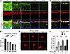 Figure 1
LIFR expression in RGCs. (A) Retinal cross sections from P1, P7, and mature (6 weeks) mice; and 1, 3, and 5 days after optical nerve crush (dpc) immunostained for LIFR (green), RBPMS (red), and DAPI (blue). GCL, ganglion cell layer; IPL, inner plexiform layer. Scale bar: 30 μm. This experiment was conducted once. (B) Quantification of LIFR intensity in RGCs from mice at time points shown in A. Statistical significance was determined by 1-way ANOVA followed by Tukey’s multiple-comparison test (*P < 0.05, **P < 0.01, ****P < 0.0001; n = 5–6 eyes per group). (C) Retinal cross sections from mice 2 weeks after AAV2-Ctrl or AAV2-LIFR injection immunostained with antibodies to LIFR (green), RBPMS (red), and DAPI (blue). Scale bar: 30 μm. This experiment was conducted once. (D) Representative images showing neurite outgrowth in cultured RGCs (red) from Vglut2-tdTomato mice with indicated treatments at day 3. Scale bar: 50 μm. This experiment was conducted once. (E) Quantitation of neurite outgrowth in RGCs. Statistical significance was determined by unpaired 2-tailed t test (**P < 0.01, ****P < 0.0001; n = 4–5 wells per group). Data represent mean ± SEM.
To characterize developmental changes in LIFR expression in greater detail, we assessed LIFR protein levels in rats at E17, E21, P1, P2, P8, and P11, time points previously used to study developmental changes in RGCs’ growth potential (19). In conformity with the finding that embryonic RGCs have a stronger intrinsic capacity for axon growth than postnatal RGCs (19), we saw a 16.5% decrease in LIFR expression between E17 and E21 that remained at a similar level through P2 (at P1 and P2, P < 0.05), then declined further by P8 (55.9% decline relative to E17 at P8, 52.2% decline at P11, both P < 0.0001; Supplemental Figure 1, G and H).
LIFR overexpression is sufficient to stimulate neurite outgrowth in adult RGCs. To evaluate the efficacy of LIFR overexpression, we delivered CAG promoter-driven adeno-associated virus serotype 2.2 (AAV2-LIFR) via intravitreal injection. This viral vector preferentially targets RGCs, with minor off-target transduction of amacrine cells and glia (11, 20, 21). Retinal flat-mount analysis showed that intravitreal AAV2 transduced approximately 82.7% of RBPMS+ RGCs (Supplemental Figure 2A). Within RBPMS+ RGCs, AAV2-LIFR induced a 3.2-fold elevation in LIFR protein abundance compared with the empty vector controls (P < 0.01; Figure 1C and Supplemental Figure 2B). Western blot analysis of whole retinas obtained 2 weeks after viral injection failed to detect a significant rise in total LIFR protein (P > 0.05; Supplemental Figure 2, C and D), presumably because RGCs represent less than 1% of all cells in the retina, but at the mRNA level, whole-retina qPCR showed a modest yet significant 13.5% upregulation of endogenous Lifr transcripts after AAV2-LIFR delivery, consistent with our quantitative immunofluorescence measurements (P < 0.05; Supplemental Figure 2E).
Next, to test the consequences of LIFR overexpression, we again overexpressed LIFR (versus a control construct) in adult Vglut2-tdTomato mice (in which RGCs are genetically labeled with tdTomato), and then prepared dissociated retinal cultures 2 weeks later. After 3 days in culture, mature RGCs overexpressing LIFR showed a 2.4-fold increase in neurite outgrowth compared with controls (P < 0.01; Figure 1, D and E). Regarding the cognate ligand, in the absence of receptor overexpression, LIF at either 100 or 300 ng/mL had no detectable effect on neurite outgrowth nor RGC survival (P > 0.05; Figure 1, D and E, and Supplemental Figure 2F). However, with LIFR overexpression, LIF approximately doubled levels of neurite outgrowth: 120% increase with LIF at 100 ng/mL (P < 0.0001) and 140% increase with LIF at 300 ng/mL (P < 0.01; Figure 1, D and E). In one case, we detected a 24.3% decrease in RGC survival in the presence of the ligand (AAV2-LIFR + LIF, 100 ng/mL, P < 0.05; Supplemental Figure 2F).
Intraocular zymosan stimulation robustly upregulates Lif mRNA levels in infiltrating neutrophils. Because neutrophils recruited by intraocular zymosan secrete trophic factors that promote optic nerve regeneration (9, 10, 17), we tested whether these cells also express Lif transcripts. By immunochemistry for the neutrophil marker Gr1, we confirmed the presence of numerous neutrophils in the vitreous chamber from 12 hours to 3 days after intraocular zymosan, which declined by 5 days (Supplemental Figure 3A). We isolated neutrophils from the vitreous using magnetic beads coated with an anti-Ly6G antibody at different time points after intraocular zymosan and assessed neutrophil purity by flow cytometry after incubation with an anti-Gr1 primary antibody conjugated with FITC. Neutrophils, characterized by Giemsa-stained multilobed nuclei, accounted for 93.8% of the isolated cells (Supplemental Figure 3B). Because the number of isolated neutrophils was limited, we quantified Lif mRNA by absolute qPCR. Compared with the sham-treated group, Lif mRNA levels increased dramatically within the first 5 days after zymosan injection (50.4-fold increase at day 1, P > 0.05; 214.8-fold increase at day 3, P < 0.0001; 52.6-fold increase at day 5, P > 0.05; Supplemental Figure 3C). Thus, infiltrative neutrophils express Lif mRNA in addition to the trophic factors reported previously (7, 9, 14, 18).
LIFR overexpression induces optic nerve regeneration with minimal reliance on its canonical ligands
AAV2-LIFR promotes axon regeneration. To investigate the effect of LIFR overexpression in vivo, we injected AAV2-LIFR intraocularly into adult mice 2 weeks prior to ONC. Even in the absence of exogenous LIF, LIFR overexpression augmented axon regeneration 4.5-fold over mice receiving a control vector when evaluated 2 weeks after nerve injury (P < 0.0001; Figure 2, A and B) without altering RGC survival (P > 0.05; Figure 2, A and C). As previously reported (6, 8, 9, 22), RGC survival in whole-mounted retina was visualized by immunostaining against βIII-tubulin (antibody TUJ1). We verified that TUJ1 immunoreactivity was restricted to RBPMS+ RGCs (Supplemental Figure 4A).
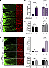 Figure 2
LIFR overexpression promotes axon regeneration in the mature optic nerve. (A) Regenerating axons and RGC survival in whole-mounted retinas visualized by fluorescent CTB (green) and immunostaining against βIII-tubulin (red), respectively, 2 weeks after ONC with the indicated treatments. An asterisk indicates the injury site. Scale bar: 100 μm (left), 50 μm (right). This experiment was conducted twice. (B) Quantification of regenerating axons. Statistical significance was determined by unpaired 2-tailed t test (n.s. P > 0.05, ****P < 0.0001; n = 7–15 nerves per group). (C) Quantification of RGC survival. Statistical significance was determined by unpaired 2-tailed t test (n.s. P > 0.05; n = 7–16 eyes per group). (D) Representative images for regenerating axons and RGC survival in whole-mounted retinas with indicated treatment. The asterisk indicates the injury site. Scale bar: 100 μm (left), 50 μm (right). This experiment was conducted once. (E) Quantification of regenerating axons. Statistical significance was determined by unpaired 2-tailed t test (n.s. P > 0.05, ****P < 0.0001; n = 5–6 nerves per group). (F) Quantification of RGC survival. Statistical significance was determined by unpaired 2-tailed t test (n.s. P > 0.05; n = 5–6 eyes per group). Data represent mean ± SEM.
AAV2-LIFR–induced regeneration is sustained after neutralization of the LIFR ligand. To determine whether the effect of LIFR overexpression requires the presence of LIF, the cognate ligand, we introduced a LIF-neutralizing antibody to diminish endogenous levels of the protein. LIF concentration in aqueous humor and serum is approximately 4 pg/mL (0.15 pM: human) (23), and we used a more than 10,000-fold excess of LIF neutralizing antibody (400 ng/mL, ~2.7 nM) to reduce levels of the protein. As before, AAV2-LIFR was introduced intraocularly 2 weeks before ONC, and the LIF neutralizing antibody (400 ng/mL) was injected intravitreally twice, once at 5 days before and again immediately after ONC. LIF neutralizing antibody did not alter AAV2-LIFR–induced axon regeneration (P > 0.05; Figure 2, A and B) and had no impact on RGC survival (P > 0.05; Figure 2, A and C). We also tested the effect of applying neutralizing antibodies against 2 alternative ligands for LIFR, cardiotrophin-1 (anti-CTF1; 400 ng/mL) and oncostatin M (anti-OSM; 3000 ng/mL) in the vitreous. Neither antibody altered AAV2-LIFR–induced axon regeneration (P > 0.05; Supplemental Figure 4, B–D).
LIFR overexpression augments the effects of LIF. In complementary studies, we investigated whether LIFR overexpression improves RGCs’ response to the cognate ligand. Whereas a single intraocular injection of LIF (1 μg/eye) immediately after ONC had no detectable effect (P > 0.05 compared with BSA, 1 μg/eye; Figure 2, D and E), it had a clear effect when LIFR was overexpressed (intraocular AAV2-LIFR: 3.2-fold increase compared with LIF plus AAV2-Ctrl, Figure 2, D and E; 2.4-fold increase compared with AAV2-LIFR: both P < 0.0001, Figure 2, A, B, D, and E). RGC survival was unaffected in all cases (P > 0.05; Figure 2, A, C, D, and F).
AAV2-LIFR enhances RGCs’ response to unrelated growth factors
We next examined whether receptor overexpression also increases RGCs’ responsiveness to unrelated trophic factors, including IGF1 (1 μg/eye), stromal cell–derived factor 1 (SDF1, 0.9 μg/eye), and oncomodulin (Ocm, 90 ng/eye), all of which have been shown to promote optic nerve regeneration (7, 9, 11, 14). As reported earlier (7, 9, 14), single intraocular injections of either IGF1 or SDF1 stimulated moderate levels of axon regeneration (IGF1: P < 0.01, Figure 3, A and B; SDF1: P < 0.0001, Figure 3, A and D), and strongly increased RGC survival (72.4% increase for IGF1, P < 0.001, Figure 3C; 81.3% increase for SDF1, P < 0.0001, Figure 3E), whereas Ocm (plus CPT-cAMP) had little effect in the absence of complementary factors (e.g., Pten deletion, SDF1 expression) (7) (P > 0.05, Figure 3F). However, when combined with LIFR overexpression, all 3 factors roughly doubled levels of axon regeneration compared with receptor overexpression alone (P < 0.05; Figure 3, A, B, D, and F). Thus, LIFR elevation enhances RGCs’ responsiveness to both the cognate ligand and to unrelated trophic factors. None of the combinations improved RGC survival above the levels achieved with individual factors alone (P > 0.05; Figure 3, A, C, E, and G).
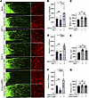 Figure 3
AAV2-LIFR enhances RGCs’ responsiveness to unrelated trophic factors. (A) Regenerating axons and RGC survival visualized by fluorescent CTB transport (green) and immunostaining against βIII-tubulin (red), respectively, 2 weeks after ONC with indicated treatments. Asterisk indicates injury site. Scale bar: 100 μm (left), 50 μm (right). This experiment was conducted once. (B, D, and F) Quantification of axon regeneration after IGF1 (B), SDF1 (D), and Ocm plus cAMP (F) treatment. Dashed lines in B, D, and F indicate regeneration in the BSA-treated control group. Statistical significance was determined by 1-way ANOVA followed by Tukey’s multiple-comparison test (n.s. P > 0.05, *P < 0.05, ***P < 0.001, ****P < 0.0001; n = 6–14 nerves per group). (C, E, and G) Quantification of RGC survival after IGF1 (C), SDF1 (E), and Ocm plus cAMP (G) treatment. Dashed lines in C, E, and G indicate survival in the BSA-treated control group. Statistical significance was determined by 1-way ANOVA followed by Tukey’s multiple-comparison test; n.s. P > 0.05, *P < 0.05, **P < 0.01; n = 5–12 eyes per group (C), 6–7 eyes per group (E), and 6–17 eyes per group (G). Data represent mean ± SEM.
Long-term characterization of LIFR-induced regenerative effects after nerve injury
To determine whether the regenerative benefits conferred by LIFR overexpression endure beyond the acute post-injury phase, we extended our experimental endpoint to 8 weeks after ONC. Six experimental cohorts were included for comparison: AAV2-Ctrl; AAV2-LIFR; phosphatase and tensin homolog (Pten) deletion; AAV2-LIFR combined with Pten deletion; Pten deletion combined with the Zn2+ chelator TPEN; and the triple intervention group comprising Pten deletion, AAV2-LIFR, and intraocular TPEN.
At 8 weeks after nerve injury, a single treatment with AAV2-LIFR induced modest axon regeneration (Figure 4A), which was likely attributed to its failure to sustain robust RGC survival. Consistent with our short-term observations, AAV2-LIFR did not significantly improve RGC survival compared with AAV2-Ctrl at this time point (P > 0.05; Supplemental Figure 5, A and B). We further assessed visual function in the AAV2-Ctrl and AAV2-LIFR groups. Pattern electroretinography (pERG) recordings showed that AAV2-LIFR increased the N35-P50 amplitude to 1.7-fold of that in the AAV2-Ctrl group (P < 0.05; Figure 4, C and D). In contrast, the pERG P50-N95 amplitude remained unaltered after AAV2-LIFR administration (P > 0.05; Figure 4, C and E). Moreover, standalone AAV2-LIFR failed to produce significant improvements in pattern visual evoked potential (pVEP) or optomotor response (OMR) at 8 weeks after ONC (P > 0.05; Supplemental Figure 5, C–F).
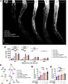 Figure 4
Long-term assessment of AAV2-LIFR–mediated optic nerve regeneration and visual function after optic nerve injury. (A) Representative images of axon regeneration visualized by CTB labeling (gray) at 8 weeks after ONC with the indicated treatments. The dashed line indicates the injury site. Scale bar: 400 μm. This experiment was conducted once. (B) Quantification of regenerating axons at 250, 500, 1,000, and 2,000 μm distal to the crush site with the indicated treatments. Statistical significance was determined by 2-way ANOVA followed by Tukey’s multiple-comparison test (*P < 0.05, **P < 0.01, ***P < 0.001, ****P < 0.0001; n = 5–11 nerves per group). (C) Representative pERG waveforms recorded at 8 weeks after ONC from each treatment cohort. N35, P50, and N95 peaks are labeled. Scale bars: vertical, 2.5 μV; horizontal, 80 ms. This experiment was conducted once. (D and E) Quantification of pERG N35-P50 (D) and P50-N95 (E) amplitudes. Statistical significance was determined by 1-way ANOVA followed by Tukey’s multiple-comparison test (n.s. P > 0.05, *P < 0.05, **P < 0.01, ***P < 0.001; n = 7–13 eyes per group). Data represent mean ± SEM.
Previous studies have validated that combined Pten deletion and intraocular TPEN treatment can durably promote RGC survival and axon regeneration (24). In line with these findings, we observed persistent and robust regenerative outcomes in the Pten deletion plus TPEN group. On this potent pro-regenerative background, supplementary AAV2-LIFR produced a modest, nonsignificant increase in axon regeneration, reaching 1.2-, 1.1-, 1.2-, and 1.8-fold the levels observed with Pten deletion plus TPEN at 250, 500, 1,000, and 2,000 μm distal to the injury site, respectively (P > 0.05; Figure 4, A and B). Additionally, AAV2-LIFR supplementation did not further increase RGC survival or alter the detected electrophysiological and behavioral functional outcomes in the context of Pten deletion combined with TPEN background (P > 0.05; Figure 4, C–E, and Supplemental Figure 5, A–F). These results suggest that AAV2-LIFR may confer an additional long-distance growth advantage in the Pten deletion plus TPEN background, though this promotional effect was subtle and did not reach statistical significance under our experimental conditions.
The most prominent supplementary beneficial effect of AAV2-LIFR was observed in the Pten deletion background. Pten deletion is a robust pro-regenerative manipulation that activates PI3K/AKT signaling (13, 25) and elicits a sustained regenerative response at 8 weeks after ONC (Figure 4A). Notably, the combination of AAV2-LIFR and Pten deletion exerted a synergistic pro-regenerative effect, markedly augmenting the regenerative capacity of Pten deletion alone. Axon regeneration in the combined group was increased to 1.8-fold at 250 μm, 1.9-fold at 500 μm, 1.8-fold at 1,000 μm, and 1.3-fold at 2,000 μm of the levels in the sole Pten deletion group (P < 0.0001 at 250 and 500 μm; P > 0.05 at 1,000 and 2,000 μm; Figure 4, A and B). This benefit was also validated by the retinal electrophysiological assay. In the Pten deletion background, AAV2-LIFR increased the pERG N35-P50 amplitude to 1.5-fold of that in the single Pten deletion group (P < 0.01; Figure 4, C and D). Despite no observable improvements in pVEP amplitude and OMR function after AAV2-LIFR supplementation in the Pten deletion model (P > 0.05; Supplemental Figure 5, C–F), our results confirm that LIFR overexpression can further potentiate Pten deletion–induced axon regeneration and partially restore RGC-associated retinal electrophysiological function at 8 weeks after ONC.
Longer-range anatomical analyses further characterized the degree of neural repair and regeneration. In both the single Pten deletion and combined treatment groups, axons labeled with cholera toxin subunit B (CTB) successfully extended to the optic chiasm, with only a small number of axons crossing the chiasmatic midline (P > 0.05; Supplemental Figure 5, G–I). However, none of the pro-regenerative intervention groups exhibited obvious CTB-positive axonal innervation in downstream visual relay regions, including the lateral geniculate nucleus and superior colliculus, in the examined pro-regenerative groups (Supplemental Figure 5J). Collectively, these long-term experimental data demonstrate that the combination of AAV2-LIFR and Pten deletion yields the most prominent supplementary therapeutic benefits. LIFR-targeted treatment can serve as an effective adjuvant strategy to enhance structural axonal regeneration and improve retinal electrophysiological function in conducive pro-regenerative settings. Nonetheless, full functional visual recovery and robust central target reinnervation require further optimized therapeutic interventions.
LIFR knockdown suppresses optic nerve regeneration
To further investigate the contribution of LIFR to optic nerve regeneration, we knocked down expression of the receptor in RGCs of mature mice using an AAV2 expressing a targeted shRNA. After 2 weeks, IHC confirmed a reduction in LIFR intensity in RBPMS+ RGCs to background, that is, the level seen when the primary antibody was omitted during immunostaining (P < 0.05; Figure 5, A and B). Global LIFR knockdown was also investigated across the entire retina by immunoblotting and qPCR at 2 weeks after viral injection. Western blot analysis of whole retinal lysates revealed no statistically meaningful decline in total LIFR protein abundance compared with control retinas (P > 0.05; Supplemental Figure 6, A and B), whereas whole-retina qPCR showed a modest but significant 11% reduction in Lifr transcript levels after AAV2-shLIFR delivery (P < 0.05; Supplemental Figure 6C). This transcriptional repression aligned with quantitative fluorescence measurements performed in RBPMS+ RGCs. AAV2-shLIFR reduced zymosan-induced axon regeneration to the baseline level seen after ONC alone (P < 0.0001; Figure 5, C and D), and somewhat unexpectedly, decreased zymosan-induced RGC survival to the baseline level (P < 0.001; Figure 5, C and E). To explore the importance of LIFR more generally, we also tested the effect of AAV2-shLIFR on regeneration induced by knockdown of PTEN. AAV2-shLIFR fully abolished both axon regeneration and the neuroprotective effect of PTEN knockdown (P < 0.001; Figure 5, F–H).
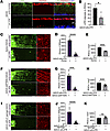 Figure 5
LIFR knockdown suppresses optic nerve regeneration. (A) Retinal cross sections from mice 2 weeks after AAV2-shScramble or AAV2-shLIFR injection immunostained for LIFR (green) and RBPMS (red) and stained with DAPI (blue). GCL, ganglion cell layer; IPL, inner plexiform layer. Scale bar: 30 μm. This experiment was conducted once. (B) Quantification of LIFR intensity in RGCs. Dashed line indicates LIFR intensity when the first antibody is omitted during immunostaining. Statistical significance was determined by unpaired 2-tailed t test (*P < 0.05; n = 3–4 eyes per group). (C, F, and I) Representative images of axon regeneration (green) and RGC survival (red) after LIFR knockdown combined with zymosan (C), AAV2-shPTEN (F), or AAV2-miR-21-sponge (I). An asterisk indicates the injury site. Scale bars: 100 μm (left) and 50 μm (right). The experiments in C and F were conducted once, and experiments in I were conducted twice. (D, G, and J) Quantification of axon regeneration after LIFR knockdown combined with zymosan (D), AAV2-shPTEN (G), or AAV2-miR-21-sponge (J). Statistical significance was determined by an unpaired 2-tailed t test; ***P < 0.001, ****P < 0.0001; n = 8–12 nerves (D), 7–9 nerves (G), and 11–17 nerves (J) per group. (E, H, and K) Quantification of RGC survival after LIFR knockdown combined with zymosan (E), AAV2-shPten (H), or AAV2-miR-21-sponge (K). Statistical significance was determined by an unpaired 2-tailed t test; n.s. P > 0.05, ***P < 0.001; n = 4 eyes (E), 7–8 eyes (H), and 5–7 eyes (K) per group. Data represent mean ± SEM.
miRNAs are small noncoding RNAs that play important regulatory roles by targeting mRNAs for cleavage or translational repression (26). miR-21 has been reported to interact with Lifr mRNA directly (27, 28), which we verified by showing that miR-21 binds to the 3′ UTR of Lifr mRNA (dual-luciferase reporter assay; Supplemental Figure 6, D and E). We found that miR-21 was highly expressed in RBPMS+ RGCs using FISH (Supplemental Figure 6F). We next constructed an AAV2 expressing miR-21 antisense sequences (AAV2-miR-21-sponge) to sequester miR-21 away from its endogenous targets. Like LIFR overexpression, AAV2-miR-21-sponge enhanced axon regeneration 4.5-fold over baseline (P < 0.0001 compared with AAV2-Ctrl-sponge; Supplemental Figure 6, G and H), with no significant impact on RGC survival (P > 0.05; Supplemental Figure 6, G and I). However, genetic knockdown of LIFR via intravitreal AAV2-shLIFR administration 3 weeks prior to ONC completely abrogated the pro-regenerative effect induced by intravitreal AAV2-miR-21-sponge delivered 2 weeks before ONC (P < 0.0001; Figure 5, I and J). LIFR knockdown did not significantly alter RGC survival in the AAV2-miR-21-sponge–treated group (P > 0.05; Figure 5, I and K) These findings further validate the role of LIFR as an essential regulator of RGC axonal regeneration.
Transcriptomic analyses
To investigate the molecular consequences of LIFR overexpression, RGCs from Vglut2-tdTomato mice were FACS-isolated 2 weeks after intraocular injection of AAV2-LIFR or AAV2-Ctrl, and then analyzed by bulk RNA-seq. AAV2-LIFR had pronounced transcriptional effects on adult RGCs, as indicated by principal component analysis (PCA; Supplemental Figure 7A) and the vast number of differentially expressed genes (DEGs) (4,020 DEGs, q < 0.05, |log2FC|>1, 2,112 upregulated and 1,908 downregulated, AAV2-LIFR vs. AAV2-Ctrl; Figure 6A and Supplemental Figure 7C). AAV2-LIFR upregulated genes encoding many growth factors (e.g., Egf, Ngf, Igf2, Ccl5) and receptors (Igf1r, Igf2r), along with the expected increase in Lifr, while downregulating the expression of genes associated with inhibition of regeneration (Rtn4r [Nogo receptor, NgR], Rtn4rl2 [NgR2], Lingo1, Cspg5), growth cone collapse (Sema6c, Sema4f), and cell death (Nlrp1a, Faf1) (Figure 6B). qPCR validation performed on FACS-isolated RGCs confirmed significant expression alteration of representative RNA-seq hits, including Lifr (P < 0.05), Ngf (P < 0.01), and Faf1 (P < 0.001) (Supplemental Figure 7, E–G). Gene Ontology (GO) enrichment analysis revealed that LIFR overexpression upregulated groups of genes relevant to positive regulation of the MAPK cascade, positive responses to cytokine stimulus, cytokine receptor activity, positive regulation of axon regeneration, and regulation of receptor signaling pathways via JAK/STAT and downregulated groups of genes associated with autophagic cell death, programmed necrotic cell death, intrinsic apoptotic signaling, and negative regulation of axonogenesis, axon extension, and regeneration (Figure 6C).
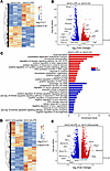 Figure 6
Transcriptional effects. (A and D) Heatmap showing the magnitude of upregulated (orange to red) and downregulated (blue) genes (q < 0.05, |log2FC| > 1) in RGCs 2 weeks after intraocular AAV2-LIFR (A, AAV2-LIFR vs. AAV2-Ctrl) or AAV2-shLIFR (D, AAV2-shLIFR vs. AAV2-shScramble, 3 replicates in each group). (B and E) Volcano plot showing individual upregulated (red) and downregulated (blue) genes in RGCs after transfection with AAV2-LIFR (B, AAV2-LIFR vs. AAV2-Ctrl) or AAV2-shLIFR (E, AAV2-shLIFR vs. AAV2-shScramble). (C) GO enrichment analysis of differentially expressed genes with indicated comparison in RGCs. Upregulated (red) and downregulated (blue) GO terms (AAV2-LIFR vs. AAV2-Ctrl).
In complementary studies, we explored the transcriptional effect of knocking down LIFR (via AAV2-shLIFR) in RGCs, again using bulk RNA-seq. As with LIFR overexpression, LIFR knockdown had robust transcriptional effects as revealed by PCA, heatmap analysis (Supplemental Figure 7B and Figure 6D), and the large number of DEGs (4,708 DEGs, q < 0.05, |log2FC|>1, 2,065 upregulated and 2,643 downregulated, AAV2-shLIFR vs. AAV2-shScramble; Figure 6D and Supplemental Figure 7D). In a reversal of the effects of LIFR overexpression, LIFR knockdown enhanced the expression of myelin-associated inhibitors of axon growth and their receptors (Rtn4 [Nogo], Rtn4r [NgR], and Mbp), while downregulating anti-apoptotic genes (Bcl2a1a, Bcl2a1b, Bmpr1b) and genes encoding growth factors, receptors, and their effectors (Igf1, Isl2, Il6ra, Nefl, Nefh, Akt2, and Jak3) (Figure 6E). At the same time, GO enrichment analysis showed that LIFR knockdown led to the upregulation of groups associated with programmed cell death, damaged DNA binding, and negative regulation of axonogenesis and axon regeneration, along with downregulation of terms relevant to the positive regulation of MAPK cascade, cellular response to IGF and NGF (and LIF), cytokine-mediated signaling pathways, positive regulation of axon regeneration, and RGC axon guidance (Supplemental Figure 7H).
Supporting evidence for LIFR regulation of signal transduction pathways
We further investigated the role of LIFR in neuroprotective and pro-regenerative signaling pathways by IHC. As before, we manipulated levels of LIFR expression by intraocular injections of either AAV2-LIFR or AAV2-shLIFR, and then evaluated levels of phosphorylated (active) ERK (pERK), phosphorylated protein kinase B (pAKT), phosphorylated ribosomal protein S6 (pS6), and phosphorylated STAT 3 (pSTAT3) in RBPMS+ RGCs 2 weeks later using appropriate antibodies. In conformity with the transcriptomic results, LIFR overexpression increased pERK levels in RGCs (P < 0.01 compared with the AAV2-Ctrl group; Figure 7, A and C), whereas LIFR knockdown reduced pERK levels in RGCs (by 33.7%, P < 0.01 compared with AAV2-shScramble; Figure 7, A and C). AAV2-shLIFR also reduced levels of pAKT and pS6, 2 markers of mTOR pathway activation, by 45.7% and 47.0%, respectively (all P < 0.001 compared with the AAV2-shScramble group; Figure 7, A, D, and E). LIFR overexpression also seemed to reduce pAKT and pS6 levels, though this did not attain statistical significance (P > 0.05; Figure 7, A, D, and E). No significant differences were observed for pSTAT3 levels in RGCs as a result of LIFR overexpression or knockdown (P > 0.05; Figure 7, B and F).
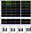 Figure 7
LIFR activates cell-signaling pathways related to CNS axon regeneration. (A) Retinal cross sections from mice 2 weeks after intraocular AAV2-Ctrl, AAV2-LIFR, AAV2-shScramble, AAV2-shLIFR, or 3 days after BSA or LIF injection, immunostained for pERK (green), pAKT (green), pS6 (green), RBPMS (red), and DAPI (blue). GCL, ganglion cell layer; IPL, inner plexiform layer. Scale bar: 30 μm. This experiment was conducted once. (B) Retinal cross sections immunostained for pSTAT3 (green), RBPMS (red), and DAPI (blue) after the same treatments as in A. Scale bar: 30 μm. This experiment was conducted once. (C–F) Quantification of pERK (C), pAKT (D), pS6 (E), and pSTAT3 (F) intensity in RBPMS+ RGCs with indicated treatments. Statistical significance was determined by unpaired 2-tailed t test (n.s. P > 0.05, **P < 0.01, ***P < 0.001; n = 4 per group). Data represent mean ± SEM.
To compare the effects of LIFR overexpression with those of LIF itself, we also evaluated expression of the above signal molecules by IHC 3 days after intraocular injections of recombinant LIF protein (1 μg/eye). Intraocular LIF did not alter pERK, pAKT, or pS6 expression (compared with BSA control, P > 0.05; Figure 7, A and C–E), whereas, as expected, it increased pSTAT3 levels (P < 0.001; Figure 7, B and F). This finding suggests the possibility that LIFR overexpression and LIF might activate distinct downstream signaling pathways, although the mechanisms involved are unclear.
Given that LIFR manipulation alters pERK levels and MAPK-related transcriptional signatures, we tested whether activation of MAPK signaling could rescue the regenerative deficits induced by LIFR knockdown. BRAF is an upstream activator of the MEK/ERK cascade, and activated BRAF signaling is an established driver of CNS axon regeneration (29). In the context of LIFR depletion via AAV2-shLIFR, AAV2-mediated BRAF overexpression largely restored axonal regeneration, increasing the number of regenerating axons 62.4-fold (P < 0.001; Supplemental Figure 8, A and B) distal to the injury site relative to AAV2-shLIFR alone. In addition, AAV2-BRAF increased RGC survival 1.3-fold compared with the AAV2-shLIFR group (P < 0.01; Supplemental Figure 8, A and C). Collectively, these data indicate that MAPK pathway activation represents a major component of LIFR-dependent optic nerve regeneration, and BRAF overexpression can only partially reverse the impaired regenerative phenotype caused by LIFR knockdown.
The intracellular domain of LIFR is sufficient to promote optic nerve regeneration
Because our experiments showed that the benefits of LIFR overexpression have limited dependence on its cognate ligands, we investigated whether a truncated form of the receptor lacking extracellular ligand-binding sites is sufficient to stimulate regeneration. For this, we designed an AAV vector expressing a truncated LIFR (AAV2-LIFRΔ) that includes only the signal peptide, transmembrane, and intracellular domains. Two weeks after AAV transduction, anti-FLAG immunoblotting of whole retinal lysates readily detected full-length LIFR protein in retinas receiving AAV2-LIFR, whereas no corresponding band was observed in the AAV2-LIFRΔ group (Supplemental Figure 9A). Quantification further verified markedly diminished FLAG signals in AAV2-LIFRΔ–treated retinas relative to retinas transduced with full-length AAV2-LIFR (P < 0.01; Supplemental Figure 9B), confirming that the truncated AAV2-LIFRΔ construct fails to produce measurable full-length LIFR protein. Two weeks after intraocular injection of AAV2-LIFRΔ, we injured the optic nerve and evaluated regeneration after another 2 weeks. AAV2-LIFRΔ elevated axon regeneration 3.2-fold compared with AAV2-Ctrl (P < 0.001; Figure 8, A and B), which was only slightly lower than overexpression of the full-length receptor (26.0% lower compared with AAV2-LIFR: P > 0.05; Figure 8, A and B). Like AAV2-LIFR, AAV2-LIFRΔ did not affect RGC survival (P > 0.05; Figure 8, A and C). These results indicate that extracellular domain–truncated LIFR retains substantial pro-regenerative capacity, highlighting the critical functional contribution of LIFR’s transmembrane and intracellular domains and the independence of LIFR-mediated regeneration of its canonical ligand.
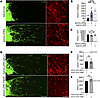 Figure 8
LIFR intracellular domain promotes regeneration. (A) Representative images of regenerating axons (CTB, green) and RGC survival (TUJ1, red) in mice treated with AAV2-LIFRΔ or AAV2-Ctrl. Asterisks indicate the injury site. Scale bars: 100 μm (left) and 75 μm (right). This experiment was conducted once. (B and C) Quantification of axon regeneration (B) and RGC survival (C) after AAV2-LIFRΔ treatment. Statistical significance was determined by 1-way ANOVA followed by Tukey’s multiple-comparison test; n.s. P > 0.05, ***P < 0.001, ****P < 0.0001; n = 6–13 nerves (B) and 4–6 retinas (C) per group. (D) Representative images of regenerating axons (CTB, green) and RGC survival (TUJ1, red) in mice treated with AAV2-LIFR-ΔBox1 or AAV2-LIFR-Y969F. Asterisk indicates the injury site. Scale bars: 100 μm (left) and 50 μm (right). This experiment was conducted once. (E and F) Quantification of axon regeneration (E) and RGC survival (F) after AAV2-LIFR-ΔBox1 or AAV2-LIFR-Y969F treatment. Dashed lines indicate the levels induced by AAV2-LIFR. Statistical significance was determined by unpaired 2-tailed t test; n.s. P > 0.05; n = 6–7 nerves (E) and 6–7 retinas (F) per group. Data represent mean ± SEM.
Prior structure-function studies of LIFR have shown that cytoplasmic regions of LIFR and gp130 act cooperatively to mediate downstream signal transduction (30), with the Box1 motif within the LIFRα cytoplasmic tail serving as a functionally indispensable region (31). In addition, modular tyrosine-based motifs in cytokine receptor complexes, including those containing LIFR, are known to recruit STAT proteins and other downstream effectors to fine-tune signaling cascades (32). Therefore, we constructed 2 full-length LIFR mutant vectors: one with a specific deletion of the Box1 motif (AAV2-LIFR-ΔBox1) and another carrying a tyrosine-to-phenylalanine point mutation at the Y969 residue (AAV2-LIFR-Y969F). We found that both mutants exhibited substantially impaired pro-regenerative activity relative to WT LIFR. The number of regenerating axons elicited by each mutant was reduced to 44% of that observed in the AAV2-LIFR group. No significant difference was detected between the AAV2-LIFR-ΔBox1 and AAV2-LIFR-Y969F mutant groups (P > 0.05; Figure 8, D and E). Meanwhile, RGC survival in both mutant groups remained comparable to that observed in the AAV2-LIFR group, with no significant difference between each mutant group and the AAV2-LIFR baseline (P > 0.05; Figure 8, D and F). These results indicate that Box1 and Y969 are required to mediate the maximal pro-regenerative output of LIFR; nevertheless, disruption of either motif alone fails to fully ablate LIFR-dependent axonal regenerative capacity.
We also explored the role of LIFR on different RGC subtypes by immunostaining for SMI32 (against neurofilament heavy chain for αRGCs), cocaine- and amphetamine-regulated transcript (CART) (for on-off direction-selective RGCs, ooDSGCs), and melanopsin (intrinsically photosensitive RGCs, ipRGCs). IHC verified that all 3 markers colocalized with the pan-RGC marker, RBPMS (Supplemental Figure 9C). Again, AAV2-LIFRΔ was intraocularly injected 2 weeks before nerve injury, and the survival of the 3 RGC subtypes was assessed after another 1 or 2 weeks. Despite the absence of an overall effect on RGC survival, AAV2-LIFRΔ strongly increased ipRGC survival (by 47.6% at 1 week and 48.3% at 2 weeks after injury, respectively, both P < 0.05; Supplemental Figure 9, D and E), decreased the survival of ooDSGCs (by 40.4% at 1 week and 41.9% at 2 weeks after injury; P < 0.01, P < 0.05, respectively; Supplemental Figure 9, D and F), and did not affect the survival of αRGCs (P > 0.05; Supplemental Figure 9, D and G). Thus, LIFRΔ appears to alter RGC survival in a subtype-specific way. Figure 9 schematically illustrates our major findings.
 Figure 9
Schematic summary of the major findings. The expression of LIFR determines the regenerative potential and trophic responsiveness of RGCs. Compared with neonatal RGCs, LIFR is reduced in adult RGCs and is further reduced in adult RGCs after optic nerve injury. A pro-regenerative treatment (e.g., sterile intraocular inflammation) restores its expression to the normal mature level (top). LIFR overexpression in RGCs (intraocular AAV2-LIFR) promotes optic nerve regeneration independent of the ligand, LIF, and amplifies the effects of unrelated growth factors (e.g., IGF1, SDF1, Ocm: bottom left). In contrast, LIFR knockdown abolishes regeneration induced by potent stimuli such as PTEN knockdown, intraocular zymosan, or miR-21 inhibition with AAV2-miR-21-sponge (bottom center). A truncation of the extracellular domain of LIFR (LIFRΔ) also promotes regeneration (bottom right).
Finally, we characterized the expression of LIFR in sections of a postmortem human retina from a 17-year-old male. IHC revealed abundant LIFR expression in RBPMS+ RGCs in the human retina (Figure 10).
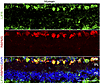 Figure 10
LIFR expression in human RGCs. Cross section from a human postmortem retina immunostained for LIFR (green), RBPMS (red), and DAPI (blue). GCL, ganglion cell layer; IPL, inner plexiform layer; INL, inner nuclear layer. Scale bar: 60 μm.


