
Research ArticleGeneticsOncology
Open Access |  10.1172/jci.insight.203005
Comparative analysis of distinct genomic landscapes in young-onset gBRCA1/2 breast cancer
Mwangala P. Akamandisa,1,2 Mingyi Xia,1 Wilson Cheah,3 Bradley Wubbenhorst,1 Kurt P. D’Andrea,1 Mengyao Fan,1 Jake S. Shilan,1 Dana Pueschl,1 Anupma Nayak,4 Hayley McKenzie,3 William Tapper,3 Ellen R. Copson,3 Ramsey I. Cutress,3 Susan M. Domchek,2,5,6 Diana M. Eccles,3 and Katherine L. Nathanson1,2,6
1Division of Translational Medicine and Human Genetics and
2Abramson Cancer Center, Perelman School of Medicine, University of Pennsylvania, Philadelphia, Pennsylvania, USA.
3University of Southampton, Southampton, United Kingdom.
4Department of Pathology and Laboratory Medicine,
5Division of Hematology and Oncology, and
6Basser Center for BRCA, Perelman School of Medicine, University of Pennsylvania, Philadelphia, Pennsylvania, USA.
Address correspondence to: Katherine L. Nathanson, 356 BRB II/III, 421 Curie Boulevard, University of Pennsylvania, Philadelphia, Pennsylvania 19104, USA. Phone: 215.662.4740; Email: knathans@upenn.edu.
Find articles by Akamandisa, M. in: PubMed | Google Scholar
1Division of Translational Medicine and Human Genetics and
2Abramson Cancer Center, Perelman School of Medicine, University of Pennsylvania, Philadelphia, Pennsylvania, USA.
3University of Southampton, Southampton, United Kingdom.
4Department of Pathology and Laboratory Medicine,
5Division of Hematology and Oncology, and
6Basser Center for BRCA, Perelman School of Medicine, University of Pennsylvania, Philadelphia, Pennsylvania, USA.
Address correspondence to: Katherine L. Nathanson, 356 BRB II/III, 421 Curie Boulevard, University of Pennsylvania, Philadelphia, Pennsylvania 19104, USA. Phone: 215.662.4740; Email: knathans@upenn.edu.
Find articles by Xia, M. in: PubMed | Google Scholar
1Division of Translational Medicine and Human Genetics and
2Abramson Cancer Center, Perelman School of Medicine, University of Pennsylvania, Philadelphia, Pennsylvania, USA.
3University of Southampton, Southampton, United Kingdom.
4Department of Pathology and Laboratory Medicine,
5Division of Hematology and Oncology, and
6Basser Center for BRCA, Perelman School of Medicine, University of Pennsylvania, Philadelphia, Pennsylvania, USA.
Address correspondence to: Katherine L. Nathanson, 356 BRB II/III, 421 Curie Boulevard, University of Pennsylvania, Philadelphia, Pennsylvania 19104, USA. Phone: 215.662.4740; Email: knathans@upenn.edu.
Find articles by Cheah, W. in: PubMed | Google Scholar
1Division of Translational Medicine and Human Genetics and
2Abramson Cancer Center, Perelman School of Medicine, University of Pennsylvania, Philadelphia, Pennsylvania, USA.
3University of Southampton, Southampton, United Kingdom.
4Department of Pathology and Laboratory Medicine,
5Division of Hematology and Oncology, and
6Basser Center for BRCA, Perelman School of Medicine, University of Pennsylvania, Philadelphia, Pennsylvania, USA.
Address correspondence to: Katherine L. Nathanson, 356 BRB II/III, 421 Curie Boulevard, University of Pennsylvania, Philadelphia, Pennsylvania 19104, USA. Phone: 215.662.4740; Email: knathans@upenn.edu.
Find articles by
Wubbenhorst, B.
in:
PubMed
|
Google Scholar
|

1Division of Translational Medicine and Human Genetics and
2Abramson Cancer Center, Perelman School of Medicine, University of Pennsylvania, Philadelphia, Pennsylvania, USA.
3University of Southampton, Southampton, United Kingdom.
4Department of Pathology and Laboratory Medicine,
5Division of Hematology and Oncology, and
6Basser Center for BRCA, Perelman School of Medicine, University of Pennsylvania, Philadelphia, Pennsylvania, USA.
Address correspondence to: Katherine L. Nathanson, 356 BRB II/III, 421 Curie Boulevard, University of Pennsylvania, Philadelphia, Pennsylvania 19104, USA. Phone: 215.662.4740; Email: knathans@upenn.edu.
Find articles by D’Andrea, K. in: PubMed | Google Scholar
1Division of Translational Medicine and Human Genetics and
2Abramson Cancer Center, Perelman School of Medicine, University of Pennsylvania, Philadelphia, Pennsylvania, USA.
3University of Southampton, Southampton, United Kingdom.
4Department of Pathology and Laboratory Medicine,
5Division of Hematology and Oncology, and
6Basser Center for BRCA, Perelman School of Medicine, University of Pennsylvania, Philadelphia, Pennsylvania, USA.
Address correspondence to: Katherine L. Nathanson, 356 BRB II/III, 421 Curie Boulevard, University of Pennsylvania, Philadelphia, Pennsylvania 19104, USA. Phone: 215.662.4740; Email: knathans@upenn.edu.
Find articles by Fan, M. in: PubMed | Google Scholar
1Division of Translational Medicine and Human Genetics and
2Abramson Cancer Center, Perelman School of Medicine, University of Pennsylvania, Philadelphia, Pennsylvania, USA.
3University of Southampton, Southampton, United Kingdom.
4Department of Pathology and Laboratory Medicine,
5Division of Hematology and Oncology, and
6Basser Center for BRCA, Perelman School of Medicine, University of Pennsylvania, Philadelphia, Pennsylvania, USA.
Address correspondence to: Katherine L. Nathanson, 356 BRB II/III, 421 Curie Boulevard, University of Pennsylvania, Philadelphia, Pennsylvania 19104, USA. Phone: 215.662.4740; Email: knathans@upenn.edu.
Find articles by Shilan, J. in: PubMed | Google Scholar
1Division of Translational Medicine and Human Genetics and
2Abramson Cancer Center, Perelman School of Medicine, University of Pennsylvania, Philadelphia, Pennsylvania, USA.
3University of Southampton, Southampton, United Kingdom.
4Department of Pathology and Laboratory Medicine,
5Division of Hematology and Oncology, and
6Basser Center for BRCA, Perelman School of Medicine, University of Pennsylvania, Philadelphia, Pennsylvania, USA.
Address correspondence to: Katherine L. Nathanson, 356 BRB II/III, 421 Curie Boulevard, University of Pennsylvania, Philadelphia, Pennsylvania 19104, USA. Phone: 215.662.4740; Email: knathans@upenn.edu.
Find articles by Pueschl, D. in: PubMed | Google Scholar
1Division of Translational Medicine and Human Genetics and
2Abramson Cancer Center, Perelman School of Medicine, University of Pennsylvania, Philadelphia, Pennsylvania, USA.
3University of Southampton, Southampton, United Kingdom.
4Department of Pathology and Laboratory Medicine,
5Division of Hematology and Oncology, and
6Basser Center for BRCA, Perelman School of Medicine, University of Pennsylvania, Philadelphia, Pennsylvania, USA.
Address correspondence to: Katherine L. Nathanson, 356 BRB II/III, 421 Curie Boulevard, University of Pennsylvania, Philadelphia, Pennsylvania 19104, USA. Phone: 215.662.4740; Email: knathans@upenn.edu.
Find articles by Nayak, A. in: PubMed | Google Scholar
1Division of Translational Medicine and Human Genetics and
2Abramson Cancer Center, Perelman School of Medicine, University of Pennsylvania, Philadelphia, Pennsylvania, USA.
3University of Southampton, Southampton, United Kingdom.
4Department of Pathology and Laboratory Medicine,
5Division of Hematology and Oncology, and
6Basser Center for BRCA, Perelman School of Medicine, University of Pennsylvania, Philadelphia, Pennsylvania, USA.
Address correspondence to: Katherine L. Nathanson, 356 BRB II/III, 421 Curie Boulevard, University of Pennsylvania, Philadelphia, Pennsylvania 19104, USA. Phone: 215.662.4740; Email: knathans@upenn.edu.
Find articles by McKenzie, H. in: PubMed | Google Scholar
1Division of Translational Medicine and Human Genetics and
2Abramson Cancer Center, Perelman School of Medicine, University of Pennsylvania, Philadelphia, Pennsylvania, USA.
3University of Southampton, Southampton, United Kingdom.
4Department of Pathology and Laboratory Medicine,
5Division of Hematology and Oncology, and
6Basser Center for BRCA, Perelman School of Medicine, University of Pennsylvania, Philadelphia, Pennsylvania, USA.
Address correspondence to: Katherine L. Nathanson, 356 BRB II/III, 421 Curie Boulevard, University of Pennsylvania, Philadelphia, Pennsylvania 19104, USA. Phone: 215.662.4740; Email: knathans@upenn.edu.
Find articles by Tapper, W. in: PubMed | Google Scholar
1Division of Translational Medicine and Human Genetics and
2Abramson Cancer Center, Perelman School of Medicine, University of Pennsylvania, Philadelphia, Pennsylvania, USA.
3University of Southampton, Southampton, United Kingdom.
4Department of Pathology and Laboratory Medicine,
5Division of Hematology and Oncology, and
6Basser Center for BRCA, Perelman School of Medicine, University of Pennsylvania, Philadelphia, Pennsylvania, USA.
Address correspondence to: Katherine L. Nathanson, 356 BRB II/III, 421 Curie Boulevard, University of Pennsylvania, Philadelphia, Pennsylvania 19104, USA. Phone: 215.662.4740; Email: knathans@upenn.edu.
Find articles by Copson, E. in: PubMed | Google Scholar
1Division of Translational Medicine and Human Genetics and
2Abramson Cancer Center, Perelman School of Medicine, University of Pennsylvania, Philadelphia, Pennsylvania, USA.
3University of Southampton, Southampton, United Kingdom.
4Department of Pathology and Laboratory Medicine,
5Division of Hematology and Oncology, and
6Basser Center for BRCA, Perelman School of Medicine, University of Pennsylvania, Philadelphia, Pennsylvania, USA.
Address correspondence to: Katherine L. Nathanson, 356 BRB II/III, 421 Curie Boulevard, University of Pennsylvania, Philadelphia, Pennsylvania 19104, USA. Phone: 215.662.4740; Email: knathans@upenn.edu.
Find articles by Cutress, R. in: PubMed | Google Scholar
1Division of Translational Medicine and Human Genetics and
2Abramson Cancer Center, Perelman School of Medicine, University of Pennsylvania, Philadelphia, Pennsylvania, USA.
3University of Southampton, Southampton, United Kingdom.
4Department of Pathology and Laboratory Medicine,
5Division of Hematology and Oncology, and
6Basser Center for BRCA, Perelman School of Medicine, University of Pennsylvania, Philadelphia, Pennsylvania, USA.
Address correspondence to: Katherine L. Nathanson, 356 BRB II/III, 421 Curie Boulevard, University of Pennsylvania, Philadelphia, Pennsylvania 19104, USA. Phone: 215.662.4740; Email: knathans@upenn.edu.
Find articles by Domchek, S. in: PubMed | Google Scholar
1Division of Translational Medicine and Human Genetics and
2Abramson Cancer Center, Perelman School of Medicine, University of Pennsylvania, Philadelphia, Pennsylvania, USA.
3University of Southampton, Southampton, United Kingdom.
4Department of Pathology and Laboratory Medicine,
5Division of Hematology and Oncology, and
6Basser Center for BRCA, Perelman School of Medicine, University of Pennsylvania, Philadelphia, Pennsylvania, USA.
Address correspondence to: Katherine L. Nathanson, 356 BRB II/III, 421 Curie Boulevard, University of Pennsylvania, Philadelphia, Pennsylvania 19104, USA. Phone: 215.662.4740; Email: knathans@upenn.edu.
Find articles by Eccles, D. in: PubMed | Google Scholar
1Division of Translational Medicine and Human Genetics and
2Abramson Cancer Center, Perelman School of Medicine, University of Pennsylvania, Philadelphia, Pennsylvania, USA.
3University of Southampton, Southampton, United Kingdom.
4Department of Pathology and Laboratory Medicine,
5Division of Hematology and Oncology, and
6Basser Center for BRCA, Perelman School of Medicine, University of Pennsylvania, Philadelphia, Pennsylvania, USA.
Address correspondence to: Katherine L. Nathanson, 356 BRB II/III, 421 Curie Boulevard, University of Pennsylvania, Philadelphia, Pennsylvania 19104, USA. Phone: 215.662.4740; Email: knathans@upenn.edu.
Find articles by
Nathanson, K.
in:
PubMed
|
Google Scholar
|
Published April 28, 2026 - More info
JCI Insight. 2026;11(12):e203005. https://doi.org/10.1172/jci.insight.203005.
© 2026 Akamandisa et al. This work is licensed under the Creative Commons Attribution 4.0 International License. To view a copy of this license, visit http://creativecommons.org/licenses/by/4.0/.
Received: December 8, 2025; Accepted: April 21, 2026

-
Results
Cohort description. We evaluated WES results of 136 treatment-naive matched tumor-germline samples from gBRCA1/2-positive women diagnosed with young-onset breast cancer participating in the POSH study (Figure 1, Supplemental Figure 1, and Supplemental Tables 1 and 2; supplemental material available online with this article; https://doi.org/10.1172/jci.insight.203005DS1). Eighty-six (63.2%) and 50 (36.8%) women had gBRCA1 and gBRCA2 germline PVs, respectively. The median age at diagnosis was 36 years, and 92.6% of women were age ≤40 (Table 1). Overall, 65 women (47.8%) had ER-positive tumors. Demographic, tumor pathology, and treatment data are shown in Table 1.
Molecular features in breast cancers. We evaluated the genomic landscapes of the breast cancers, including asLOH, HRD, tumor mutational burden (TMB), and single-base substitution (SBS). Tumors had high rates of asLOH when stratified by gene (93% for gBRCA1 and 96% for gBRCA2) and by ER status (94.4% for ER negative and 93.8% for ER positive) (Supplemental Table 3). HRD scores were significantly higher in tumors with asLOH compared with tumors without asLOH (nonLOH) in both gBRCA1 (57.4 ± 1.3 vs. 22.6 ± 6.1, P < 0.0001) and gBRCA2 (43.7 ± 1.5 vs. 23.5 ± 6.5, P = 0.005) PV carriers and when grouped by ER status (ER-negative tumors: 57.6 ± 1.3 vs. 22.8 ± 9.2, P < 0.0001; ER-positive tumors: 46.4 ± 1.6 vs. 22.8 ± 4.1, P < 0.001) (Figure 2A and Supplemental Table 3). gBRCA1 tumors with asLOH had significantly higher HRD scores than gBRCA2 tumors with asLOH (57.4 ± 1.3 vs. 43.7 ± 1.5, P < 0.0001) (Figure 2A).
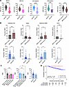 Figure 2
Molecular features of breast cancer in germline BRCA1 and BRCA2 pathogenic variant carriers with overall survival. (A) Homologous recombination deficiency (HRD) scores in tumors with allele-specific loss of heterozygosity (asLOH) compared with those without, stratified by germline variant gene and ER status using 2-sided t tests. Dotted black line is HRD score = 42. (B) Median of the proportion of single-base substitution signature 3 (SBS3), SBS1, SBS26, and SBS18 in tumors with asLOH by gene and in tumors with asLOH and those without, compared using Mann-Whitney non-parametric tests. Error bars are interquartile range (IQR). HR, homologous recombination; MMR, DNA mismatch repair. (C) Median tumor mutational burden (TMB) by asLOH status, germline gene variant, and ER status, compared using Kruskal-Wallis and Mann-Whitney non-parametric tests. Error bars are IQR. (D) Overall survival by asLOH status, evaluated using the Kaplan-Meier estimator and log-rank test. *P < 0.05, **P < 0.01, ***P < 0.001, ****P <.0001.
SBS3, the signature associated with gBRCA1/2 PVs (37), was the most abundant signature in all tumors with an average contribution of 32.8% (Supplemental Figure 2). SBS1 (aging-associated), SBS26 (mismatch repair deficiency–associated), and SBS18 (reactive oxygen species–associated) were the next most abundant signatures (37). gBRCA1 tumors had significantly higher median percentage proportional contributions of SBS1 (12.9 vs. 7.3, P = 0.013) and SBS18 (1.4 vs. 0, P = 0.007), and significantly lower proportions of SBS3 (27.3 vs. 42.6, P = 0.002) and SBS26 (5.9 vs. 9.4, P = 0.049), than gBRCA2 tumors. The median percentage proportional contributions of SBS3 and SBS18 were significantly higher in tumors with asLOH than in nonLOH tumors (SBS3: 38.5 vs. 15.9, P = 0.023; SBS18: 0 vs 0, P = 0.023) (Figure 2B and Supplemental Table 4).
Tumors with gBRCA1 asLOH had higher median TMB than those with gBRCA2 asLOH (3.4, IQR 1.6–11.2, vs. 1.5, IQR 0.9–3.4, P = 0.001). ER-positive tumors with asLOH had significantly lower median TMB than ER-negative tumors with asLOH (1.7, IQR 0.9–3.9, vs. 3.4, IQR 1.6–11.0, P = 0.008) (Figure 2C). However, there was no difference in median TMB between tumors with and without asLOH. Tumors with TMB ≥ 10 have been found to be responsive to treatment with the anti–PD-1 monoclonal antibody pembrolizumab (46, 47). In our cohort, slightly more tumors with asLOH (24 of 128 [18.8%]) had TMB ≥ 10 than those without asLOH (1 of 8 [12%]), but it was not notable.
We evaluated breast cancer OS by tumor molecular features. Survival in women with nonLOH tumors was 100% throughout the 8.2-year median follow-up period but not statistically significantly different from that of women with tumors with asLOH (Figure 2D). Favorable survival for women with nonLOH tumors remained when the analysis was stratified by germline variant gene or by ER status (Supplemental Figure 3). The clinical characteristics of the nonLOH tumors are shown in Supplemental Table 5. Although not significantly so, women with HRD-high (≥42) tumors and those with SBS26 tended to do worse (Supplemental Figure 4, A and B). Women with tumors with SBS3 had significantly lower OS than those without (HR 4.46, P = 0.033). Similarly, when stratified by gene and ER status, women with SBS3 tumors had a decreased OS, albeit not to a significant degree (Supplemental Figure 4, C–G).
Copy number variation in tumors. WES enabled copy number analysis, which revealed chromosome arm chr1q and chr8q gains, both prevalent in breast cancers (48), in all tumors (Figure 3A). Eighty-eight and 108 tumors had single-copy loss of RB1 and TP53, respectively. Copy loss of RB1 and BRCA2, both located on chromosome 13, occurred on the same segment significantly more frequently than expected by chance (56/88, P = 0.010), whereas copy loss of TP53 and BRCA1, both on chromosome 17, occurred on the same segment significantly less frequently than expected by chance (22/108, P < 0.001) (Figure 3A). Thus, it appears that RB1 and BRCA2 loss are non-independent, whereas copy number loss of TP53 and BRCA1 are independent of each other. The loss of RB1 (RB1 asLOH) occurred significantly more frequently in gBRCA2 asLOH tumors than in gBRCA1 asLOH tumors; however, there was no difference in the frequency of RB1 PVs or RB1 biallelic loss. Conversely, TP53 asLOH was not different between gBRCA1 asLOH and gBRCA2 asLOH tumors, although gBRCA1 asLOH tumors had significantly more TP53 PVs and TP53 biallelic loss than gBRCA2 asLOH tumors (Figure 3A).
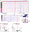 Figure 3
Copy number variation in tumor samples. (A) Landscape of copy number variants in tumors. Inset shows zoomed-in images of chromosomes 13 and 17 and the quantified values of times that RB1 shares a copy number loss segment with BRCA2 and TP53 shares one with BRCA1, evaluated using a χ2 test. Vertical bar graph shows the frequency of RB1 and TP53 pathogenic variants (PVs), asLOH, and biallelic loss in BRCA1 and BRCA2 tumors with asLOH, evaluated using Firth logistic regression. *P < 0.05, ***P < 0.001. (B) Enrichment of chromosome arm gains and losses in tumors with BRCA1 asLOH compared with those with BRCA2 asLOH, adjusted for ER status. Enrichment was performed using Firth logistic regression. Dashed line is –log10(P value) = 1.3.
We observed differences in chromosome arm copy number between tumors with gBRCA1 asLOH and gBRCA2 asLOH (Supplemental Figure 5). After adjustment for ER status (49), chr6q, chr19p, chr2q, chr7q, and chr10p gains and chr5q copy number were enriched significantly in tumors with gBRCA1 asLOH compared with those with gBRCA2 asLOH (Figure 3B).
Enrichment of gene-level alterations. To further define the complete genomic landscape of the tumors, we assessed gene-level somatic alterations (pathogenic single-nucleotide variants/indels and CNVs) in 598 cancer-associated genes (Supplemental Table 2). We found 4,146 pathogenic single-nucleotide variants (SNVs)/indels and 4,205 pathogenic CNVs in 572/598 genes (Supplemental Table 6). Pathogenic SNVs/indels in the 50 most commonly altered genes are shown in Supplemental Figure 6.
When we compared pathogenic SNVs/indels and CNVs in tumors from gBRCA1 PV carriers with those from gBRCA2 PV carriers, with adjustment for ER status, we observed a larger number of somatic alterations significantly enriched in gBRCA1 tumors (Figure 4A and Supplemental Table 7). Pathway analysis revealed enrichment of pathogenic SNVs/indels/CNVs in genes in multiple hallmark pathways (50), including the reactive oxygen species (ROS) pathway, DNA repair, and epithelial-mesenchymal transition, in gBRCA1 tumors compared with gBRCA2 tumors (Figure 4A).
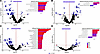 Figure 4
Differential enrichment of somatic variants, inclusive of pathogenic single-nucleotide variants, indels, and copy number alterations, and hallmark pathway enrichment in breast cancers. Enrichment of somatic variants and enrichment of Hallmark Pathway alterations by germline variant gene adjusted for ER status (A); by ER status adjusted for germline variant gene (B); by HRD (high ≥42) adjusted for asLOH (C); and by asLOH stratified by germline gene variant (D). Enrichment analysis was performed using Firth logistic regression. Dashed line is –log10(P value) = 1.3.
With adjustment for gBRCA1/2 PV carrier status, ER-positive tumors had greater enrichment of pathogenic somatic SNVs/indels/CNVs compared with ER-negative tumors (Figure 4B and Supplemental Table 8) and were enriched for alterations in genes in the coagulation and mTORC1 signaling hallmark pathways. ER-negative tumors were enriched for alterations in the interferon-γ response pathway (Figure 4B). The total number of genes with SNVs/indels/CNVs and with SNVs/indels was higher in ER-negative tumors compared with ER-positive tumors when the analysis was performed without adjustment for gBRCA1/2 PV status, as seen in previous studies (Supplemental Figure 7) (51).
More genes were enriched in SNVs/indels and CNVs in HRD-high tumors compared with HRD-low tumors, adjusted for LOH status (Figure 4C and Supplemental Table 9). Genes in the E2F targets and WNT/β-catenin signaling pathways were the most highly enriched in HRD-high tumors (Figure 4C). Tumors without asLOH were enriched for alterations in the oxidative phosphorylation pathway compared with those with asLOH (Figure 4D and Supplemental Table 10).
Presence of CDK4/6 resistance alterations. Genetic alterations implicated in acquired and intrinsic resistance include alterations in RB1, TP53, AURKA, MYC, CCNE1, SPEN, FAT1, ARID1A, PTEN, FGFR1, and EGFR (40, 45, 52–58). We evaluated the presence of somatic alterations (SNVs/indels; and CNVs, including single-copy loss) implicated in resistance to CDK4/6i in ER-positive, HER2-negative tumors within the POSH cohort, and we found that all tumors contained at least one alteration implicated in resistance (Figure 5) (54). Genes implicated in CDK4/6i resistance exhibited various patterns of alteration in gBRCA1 and gBRCA2 PV carriers in ER-positive, HER2-negative tumors from the POSH cohort (Figure 5). Alterations in RB1, primarily consisting of single-copy loss (asLOH), were more frequent in gBRCA2 tumors (33/38, 86.8%) than in gBRCA1 tumors (8/12, 66.7%). Additionally, 4 tumors in gBRCA2 PV carriers had frameshift PVs in RB1. PVs in TP53 were more frequent in gBRCA1 tumors (6/12, 50%) than in gBRCA2 tumors (5/38, 13%), although the overall prevalence of TP53 alterations was similarly high in both groups (83.3% in gBRCA1 tumors and 86.8% in gBRCA2 tumors) when single-copy-number loss was considered. Other CDK4/6i resistance–associated alterations observed in gBRCA1 and gBRCA2 tumors were in SPEN (gBRCA1: 50%; gBRCA2: 52.6%), AURKA (gBRCA1: 25%; gBRCA2: 26.3%), ARID1A (gBRCA1: 33.3%; gBRCA2: 50.0%), FAT1 (gBRCA1: 83.3%; gBRCA2: 55.3%), PTEN (gBRCA1: 50.0%; gBRCA2: 34.2%), MYC (gBRCA1: 58.3%; gBRCA2: 47.4%), EGFR (gBRCA1: 8.3%; gBRCA2: 2.6%), and CCNE1 (gBRCA1: 8.3%; gBRCA2: 0%).
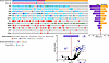 Figure 5
Enrichment of somatic variants associated with resistance to CDK4/6 inhibitors in ER-positive, HER2-negative tumors from germline BRCA1/2 carriers in POSH compared with noncarriers from TCGA. Oncoprint of somatic variants in tumors, frequency of somatic variants, and enrichment of variants in POSH versus TCGA samples. Enrichment analysis was performed using Firth logistic regression. Dashed line is –log10(P value) = 1.3; q is the false discovery rate–adjusted P value.
To determine whether gBRCA1/2 carriers were more likely than noncarriers to have somatic alterations in CDK4/6i resistance–associated genes, we compared rates of alteration between carriers from POSH and noncarriers from The Cancer Genome Atlas (TCGA) (Supplemental Figure 8), all with ER-positive, HER2-negative tumors. To mitigate any potential bias in sequencing coverage across the 2 cohorts, we limited our analysis to genes with at least 1 alteration detected in the TCGA cohort (Supplemental Table 8). We found statistically significant enrichment of alterations in several genes in tumors from the POSH cohort compared with those from TCGA, including RB1 (OR 6.3, 95% CI 2.8–15.4, adjusted P value [Padj] = 0.001), TP53 (OR 4.6, 95% CI 1.9–12.1, Padj = 0.017), FAT1 (OR 3.9, 95% CI 1.84–8.7, Padj = 0.013), and MYC (OR 4.0, 95% CI 1.8–9.1, Padj = 0.017), all of which are associated with resistance to CDK4/6i (Figure 5).


