
Research ArticleClinical ResearchImmunology
Open Access |  10.1172/jci.insight.203021
Performance of expanded diagnostic criteria for APECED in independent cohorts and implications for earlier diagnosis
Elise M.N. Ferré,1 Joseph Pechacek,1 Monica M. Schmitt,1 Taura Webb,1 Heather Moorman,1 Thomas DiMaggio,1 Princess Barber,1 Vasielios Oikonomou,1 Stacey R. Rose,1 Peter D. Burbelo,2 Lindsey B. Rosen,3 Amy P. Hsu,3 Jennifer Stoddard,4 Shakuntala Rampertaap,4 Sergio D. Rosenzweig,4 Anjali Rai,5 Maria Teresa Magone,6 Chantal Cousineau-Krieger,6 Niki M. Moutsopoulos,7 Pamela J. Gardner,8 Heidi H. Kong,9 Leslie Castelo-Soccio,9 Ariane Soldatos,10 Katherine R. Calvo,11 Meryl Waldman,12 Behdad Afzali,12 Stefania Pittaluga,13 David Kleiner,13 Steven M. Holland,3 Kevin Fennelly,14 Jill Rothschild,15 Bryce A. Seifert,16 Magdalena Walkiewicz-Yvon,16 Theo Heller,5 Karen Winer,17 and Michail S. Lionakis1
1Fungal Pathogenesis Section, Laboratory of Clinical Immunology and Microbiology (LCIM), National Institute of Allergy and Infectious Diseases (NIAID),
2Adeno-Associated Virus Biology Section, National Institute of Dental and Craniofacial Research (NIDCR),
3Immunopathogenesis Section, LCIM, NIAID,
4Immunology Service, Department of Laboratory Medicine (DLM), Clinical Center,
5Translational Hepatology Section, Liver Diseases Branch, National Institute of Diabetes and Digestive and Kidney Diseases (NIDDK),
6Consult Services Section, National Eye Institute,
7Human Barrier Immunity Section, Laboratory of Host Immunity & Microbiome, NIAID,
8Dental Consult Services, NIDCR,
9Cutaneous Microbiome and Inflammation Section, National Institute of Arthritis and Musculoskeletal and Skin Diseases,
10Pediatric Neurology Consultation Service, Office of the Clinical Director, National Institute of Neurological Disorders and Stroke,
11Hematology Laboratory Service, DLM, Clinical Center,
12Clinical Nephrology Section, Kidney Diseases Branch, NIDDK,
13Laboratory of Pathology, National Cancer Institute,
14Laboratory of Chronic Airway Infection, Critical Care Medicine and Pulmonary Branch, National Heart, Lung, and Blood Institute,
15Pediatric Hospitalist Section, Department of Pediatrics,
16Division of Intramural Research, NIAID, and
17Pediatric Growth and Nutrition Branch, National Institute of Child Health and Human Development, NIH, Bethesda, Maryland, USA.
Address correspondence to: Michail S. Lionakis, Fungal Pathogenesis Section, Laboratory of Clinical Immunology and Microbiology (LCIM), National Institute of Allergy & Infectious Diseases (NIAID), NIH, 9000 Rockville Pike, Building 10, Room 11N216B, Bethesda, Maryland, 20892, USA. Phone: 301.443.5089; Email: lionakism@mail.nih.gov.
Authorship note: EMNF, JP, and MMS contributed equally to this work.
Find articles by Ferré, E. in: PubMed | Google Scholar
1Fungal Pathogenesis Section, Laboratory of Clinical Immunology and Microbiology (LCIM), National Institute of Allergy and Infectious Diseases (NIAID),
2Adeno-Associated Virus Biology Section, National Institute of Dental and Craniofacial Research (NIDCR),
3Immunopathogenesis Section, LCIM, NIAID,
4Immunology Service, Department of Laboratory Medicine (DLM), Clinical Center,
5Translational Hepatology Section, Liver Diseases Branch, National Institute of Diabetes and Digestive and Kidney Diseases (NIDDK),
6Consult Services Section, National Eye Institute,
7Human Barrier Immunity Section, Laboratory of Host Immunity & Microbiome, NIAID,
8Dental Consult Services, NIDCR,
9Cutaneous Microbiome and Inflammation Section, National Institute of Arthritis and Musculoskeletal and Skin Diseases,
10Pediatric Neurology Consultation Service, Office of the Clinical Director, National Institute of Neurological Disorders and Stroke,
11Hematology Laboratory Service, DLM, Clinical Center,
12Clinical Nephrology Section, Kidney Diseases Branch, NIDDK,
13Laboratory of Pathology, National Cancer Institute,
14Laboratory of Chronic Airway Infection, Critical Care Medicine and Pulmonary Branch, National Heart, Lung, and Blood Institute,
15Pediatric Hospitalist Section, Department of Pediatrics,
16Division of Intramural Research, NIAID, and
17Pediatric Growth and Nutrition Branch, National Institute of Child Health and Human Development, NIH, Bethesda, Maryland, USA.
Address correspondence to: Michail S. Lionakis, Fungal Pathogenesis Section, Laboratory of Clinical Immunology and Microbiology (LCIM), National Institute of Allergy & Infectious Diseases (NIAID), NIH, 9000 Rockville Pike, Building 10, Room 11N216B, Bethesda, Maryland, 20892, USA. Phone: 301.443.5089; Email: lionakism@mail.nih.gov.
Authorship note: EMNF, JP, and MMS contributed equally to this work.
Find articles by Pechacek, J. in: PubMed | Google Scholar
1Fungal Pathogenesis Section, Laboratory of Clinical Immunology and Microbiology (LCIM), National Institute of Allergy and Infectious Diseases (NIAID),
2Adeno-Associated Virus Biology Section, National Institute of Dental and Craniofacial Research (NIDCR),
3Immunopathogenesis Section, LCIM, NIAID,
4Immunology Service, Department of Laboratory Medicine (DLM), Clinical Center,
5Translational Hepatology Section, Liver Diseases Branch, National Institute of Diabetes and Digestive and Kidney Diseases (NIDDK),
6Consult Services Section, National Eye Institute,
7Human Barrier Immunity Section, Laboratory of Host Immunity & Microbiome, NIAID,
8Dental Consult Services, NIDCR,
9Cutaneous Microbiome and Inflammation Section, National Institute of Arthritis and Musculoskeletal and Skin Diseases,
10Pediatric Neurology Consultation Service, Office of the Clinical Director, National Institute of Neurological Disorders and Stroke,
11Hematology Laboratory Service, DLM, Clinical Center,
12Clinical Nephrology Section, Kidney Diseases Branch, NIDDK,
13Laboratory of Pathology, National Cancer Institute,
14Laboratory of Chronic Airway Infection, Critical Care Medicine and Pulmonary Branch, National Heart, Lung, and Blood Institute,
15Pediatric Hospitalist Section, Department of Pediatrics,
16Division of Intramural Research, NIAID, and
17Pediatric Growth and Nutrition Branch, National Institute of Child Health and Human Development, NIH, Bethesda, Maryland, USA.
Address correspondence to: Michail S. Lionakis, Fungal Pathogenesis Section, Laboratory of Clinical Immunology and Microbiology (LCIM), National Institute of Allergy & Infectious Diseases (NIAID), NIH, 9000 Rockville Pike, Building 10, Room 11N216B, Bethesda, Maryland, 20892, USA. Phone: 301.443.5089; Email: lionakism@mail.nih.gov.
Authorship note: EMNF, JP, and MMS contributed equally to this work.
Find articles by Schmitt, M. in: PubMed | Google Scholar
1Fungal Pathogenesis Section, Laboratory of Clinical Immunology and Microbiology (LCIM), National Institute of Allergy and Infectious Diseases (NIAID),
2Adeno-Associated Virus Biology Section, National Institute of Dental and Craniofacial Research (NIDCR),
3Immunopathogenesis Section, LCIM, NIAID,
4Immunology Service, Department of Laboratory Medicine (DLM), Clinical Center,
5Translational Hepatology Section, Liver Diseases Branch, National Institute of Diabetes and Digestive and Kidney Diseases (NIDDK),
6Consult Services Section, National Eye Institute,
7Human Barrier Immunity Section, Laboratory of Host Immunity & Microbiome, NIAID,
8Dental Consult Services, NIDCR,
9Cutaneous Microbiome and Inflammation Section, National Institute of Arthritis and Musculoskeletal and Skin Diseases,
10Pediatric Neurology Consultation Service, Office of the Clinical Director, National Institute of Neurological Disorders and Stroke,
11Hematology Laboratory Service, DLM, Clinical Center,
12Clinical Nephrology Section, Kidney Diseases Branch, NIDDK,
13Laboratory of Pathology, National Cancer Institute,
14Laboratory of Chronic Airway Infection, Critical Care Medicine and Pulmonary Branch, National Heart, Lung, and Blood Institute,
15Pediatric Hospitalist Section, Department of Pediatrics,
16Division of Intramural Research, NIAID, and
17Pediatric Growth and Nutrition Branch, National Institute of Child Health and Human Development, NIH, Bethesda, Maryland, USA.
Address correspondence to: Michail S. Lionakis, Fungal Pathogenesis Section, Laboratory of Clinical Immunology and Microbiology (LCIM), National Institute of Allergy & Infectious Diseases (NIAID), NIH, 9000 Rockville Pike, Building 10, Room 11N216B, Bethesda, Maryland, 20892, USA. Phone: 301.443.5089; Email: lionakism@mail.nih.gov.
Authorship note: EMNF, JP, and MMS contributed equally to this work.
Find articles by Webb, T. in: PubMed | Google Scholar
1Fungal Pathogenesis Section, Laboratory of Clinical Immunology and Microbiology (LCIM), National Institute of Allergy and Infectious Diseases (NIAID),
2Adeno-Associated Virus Biology Section, National Institute of Dental and Craniofacial Research (NIDCR),
3Immunopathogenesis Section, LCIM, NIAID,
4Immunology Service, Department of Laboratory Medicine (DLM), Clinical Center,
5Translational Hepatology Section, Liver Diseases Branch, National Institute of Diabetes and Digestive and Kidney Diseases (NIDDK),
6Consult Services Section, National Eye Institute,
7Human Barrier Immunity Section, Laboratory of Host Immunity & Microbiome, NIAID,
8Dental Consult Services, NIDCR,
9Cutaneous Microbiome and Inflammation Section, National Institute of Arthritis and Musculoskeletal and Skin Diseases,
10Pediatric Neurology Consultation Service, Office of the Clinical Director, National Institute of Neurological Disorders and Stroke,
11Hematology Laboratory Service, DLM, Clinical Center,
12Clinical Nephrology Section, Kidney Diseases Branch, NIDDK,
13Laboratory of Pathology, National Cancer Institute,
14Laboratory of Chronic Airway Infection, Critical Care Medicine and Pulmonary Branch, National Heart, Lung, and Blood Institute,
15Pediatric Hospitalist Section, Department of Pediatrics,
16Division of Intramural Research, NIAID, and
17Pediatric Growth and Nutrition Branch, National Institute of Child Health and Human Development, NIH, Bethesda, Maryland, USA.
Address correspondence to: Michail S. Lionakis, Fungal Pathogenesis Section, Laboratory of Clinical Immunology and Microbiology (LCIM), National Institute of Allergy & Infectious Diseases (NIAID), NIH, 9000 Rockville Pike, Building 10, Room 11N216B, Bethesda, Maryland, 20892, USA. Phone: 301.443.5089; Email: lionakism@mail.nih.gov.
Authorship note: EMNF, JP, and MMS contributed equally to this work.
Find articles by Moorman, H. in: PubMed | Google Scholar
1Fungal Pathogenesis Section, Laboratory of Clinical Immunology and Microbiology (LCIM), National Institute of Allergy and Infectious Diseases (NIAID),
2Adeno-Associated Virus Biology Section, National Institute of Dental and Craniofacial Research (NIDCR),
3Immunopathogenesis Section, LCIM, NIAID,
4Immunology Service, Department of Laboratory Medicine (DLM), Clinical Center,
5Translational Hepatology Section, Liver Diseases Branch, National Institute of Diabetes and Digestive and Kidney Diseases (NIDDK),
6Consult Services Section, National Eye Institute,
7Human Barrier Immunity Section, Laboratory of Host Immunity & Microbiome, NIAID,
8Dental Consult Services, NIDCR,
9Cutaneous Microbiome and Inflammation Section, National Institute of Arthritis and Musculoskeletal and Skin Diseases,
10Pediatric Neurology Consultation Service, Office of the Clinical Director, National Institute of Neurological Disorders and Stroke,
11Hematology Laboratory Service, DLM, Clinical Center,
12Clinical Nephrology Section, Kidney Diseases Branch, NIDDK,
13Laboratory of Pathology, National Cancer Institute,
14Laboratory of Chronic Airway Infection, Critical Care Medicine and Pulmonary Branch, National Heart, Lung, and Blood Institute,
15Pediatric Hospitalist Section, Department of Pediatrics,
16Division of Intramural Research, NIAID, and
17Pediatric Growth and Nutrition Branch, National Institute of Child Health and Human Development, NIH, Bethesda, Maryland, USA.
Address correspondence to: Michail S. Lionakis, Fungal Pathogenesis Section, Laboratory of Clinical Immunology and Microbiology (LCIM), National Institute of Allergy & Infectious Diseases (NIAID), NIH, 9000 Rockville Pike, Building 10, Room 11N216B, Bethesda, Maryland, 20892, USA. Phone: 301.443.5089; Email: lionakism@mail.nih.gov.
Authorship note: EMNF, JP, and MMS contributed equally to this work.
Find articles by DiMaggio, T. in: PubMed | Google Scholar
1Fungal Pathogenesis Section, Laboratory of Clinical Immunology and Microbiology (LCIM), National Institute of Allergy and Infectious Diseases (NIAID),
2Adeno-Associated Virus Biology Section, National Institute of Dental and Craniofacial Research (NIDCR),
3Immunopathogenesis Section, LCIM, NIAID,
4Immunology Service, Department of Laboratory Medicine (DLM), Clinical Center,
5Translational Hepatology Section, Liver Diseases Branch, National Institute of Diabetes and Digestive and Kidney Diseases (NIDDK),
6Consult Services Section, National Eye Institute,
7Human Barrier Immunity Section, Laboratory of Host Immunity & Microbiome, NIAID,
8Dental Consult Services, NIDCR,
9Cutaneous Microbiome and Inflammation Section, National Institute of Arthritis and Musculoskeletal and Skin Diseases,
10Pediatric Neurology Consultation Service, Office of the Clinical Director, National Institute of Neurological Disorders and Stroke,
11Hematology Laboratory Service, DLM, Clinical Center,
12Clinical Nephrology Section, Kidney Diseases Branch, NIDDK,
13Laboratory of Pathology, National Cancer Institute,
14Laboratory of Chronic Airway Infection, Critical Care Medicine and Pulmonary Branch, National Heart, Lung, and Blood Institute,
15Pediatric Hospitalist Section, Department of Pediatrics,
16Division of Intramural Research, NIAID, and
17Pediatric Growth and Nutrition Branch, National Institute of Child Health and Human Development, NIH, Bethesda, Maryland, USA.
Address correspondence to: Michail S. Lionakis, Fungal Pathogenesis Section, Laboratory of Clinical Immunology and Microbiology (LCIM), National Institute of Allergy & Infectious Diseases (NIAID), NIH, 9000 Rockville Pike, Building 10, Room 11N216B, Bethesda, Maryland, 20892, USA. Phone: 301.443.5089; Email: lionakism@mail.nih.gov.
Authorship note: EMNF, JP, and MMS contributed equally to this work.
Find articles by Barber, P. in: PubMed | Google Scholar
1Fungal Pathogenesis Section, Laboratory of Clinical Immunology and Microbiology (LCIM), National Institute of Allergy and Infectious Diseases (NIAID),
2Adeno-Associated Virus Biology Section, National Institute of Dental and Craniofacial Research (NIDCR),
3Immunopathogenesis Section, LCIM, NIAID,
4Immunology Service, Department of Laboratory Medicine (DLM), Clinical Center,
5Translational Hepatology Section, Liver Diseases Branch, National Institute of Diabetes and Digestive and Kidney Diseases (NIDDK),
6Consult Services Section, National Eye Institute,
7Human Barrier Immunity Section, Laboratory of Host Immunity & Microbiome, NIAID,
8Dental Consult Services, NIDCR,
9Cutaneous Microbiome and Inflammation Section, National Institute of Arthritis and Musculoskeletal and Skin Diseases,
10Pediatric Neurology Consultation Service, Office of the Clinical Director, National Institute of Neurological Disorders and Stroke,
11Hematology Laboratory Service, DLM, Clinical Center,
12Clinical Nephrology Section, Kidney Diseases Branch, NIDDK,
13Laboratory of Pathology, National Cancer Institute,
14Laboratory of Chronic Airway Infection, Critical Care Medicine and Pulmonary Branch, National Heart, Lung, and Blood Institute,
15Pediatric Hospitalist Section, Department of Pediatrics,
16Division of Intramural Research, NIAID, and
17Pediatric Growth and Nutrition Branch, National Institute of Child Health and Human Development, NIH, Bethesda, Maryland, USA.
Address correspondence to: Michail S. Lionakis, Fungal Pathogenesis Section, Laboratory of Clinical Immunology and Microbiology (LCIM), National Institute of Allergy & Infectious Diseases (NIAID), NIH, 9000 Rockville Pike, Building 10, Room 11N216B, Bethesda, Maryland, 20892, USA. Phone: 301.443.5089; Email: lionakism@mail.nih.gov.
Authorship note: EMNF, JP, and MMS contributed equally to this work.
Find articles by Oikonomou, V. in: PubMed | Google Scholar
1Fungal Pathogenesis Section, Laboratory of Clinical Immunology and Microbiology (LCIM), National Institute of Allergy and Infectious Diseases (NIAID),
2Adeno-Associated Virus Biology Section, National Institute of Dental and Craniofacial Research (NIDCR),
3Immunopathogenesis Section, LCIM, NIAID,
4Immunology Service, Department of Laboratory Medicine (DLM), Clinical Center,
5Translational Hepatology Section, Liver Diseases Branch, National Institute of Diabetes and Digestive and Kidney Diseases (NIDDK),
6Consult Services Section, National Eye Institute,
7Human Barrier Immunity Section, Laboratory of Host Immunity & Microbiome, NIAID,
8Dental Consult Services, NIDCR,
9Cutaneous Microbiome and Inflammation Section, National Institute of Arthritis and Musculoskeletal and Skin Diseases,
10Pediatric Neurology Consultation Service, Office of the Clinical Director, National Institute of Neurological Disorders and Stroke,
11Hematology Laboratory Service, DLM, Clinical Center,
12Clinical Nephrology Section, Kidney Diseases Branch, NIDDK,
13Laboratory of Pathology, National Cancer Institute,
14Laboratory of Chronic Airway Infection, Critical Care Medicine and Pulmonary Branch, National Heart, Lung, and Blood Institute,
15Pediatric Hospitalist Section, Department of Pediatrics,
16Division of Intramural Research, NIAID, and
17Pediatric Growth and Nutrition Branch, National Institute of Child Health and Human Development, NIH, Bethesda, Maryland, USA.
Address correspondence to: Michail S. Lionakis, Fungal Pathogenesis Section, Laboratory of Clinical Immunology and Microbiology (LCIM), National Institute of Allergy & Infectious Diseases (NIAID), NIH, 9000 Rockville Pike, Building 10, Room 11N216B, Bethesda, Maryland, 20892, USA. Phone: 301.443.5089; Email: lionakism@mail.nih.gov.
Authorship note: EMNF, JP, and MMS contributed equally to this work.
Find articles by Rose, S. in: PubMed | Google Scholar
1Fungal Pathogenesis Section, Laboratory of Clinical Immunology and Microbiology (LCIM), National Institute of Allergy and Infectious Diseases (NIAID),
2Adeno-Associated Virus Biology Section, National Institute of Dental and Craniofacial Research (NIDCR),
3Immunopathogenesis Section, LCIM, NIAID,
4Immunology Service, Department of Laboratory Medicine (DLM), Clinical Center,
5Translational Hepatology Section, Liver Diseases Branch, National Institute of Diabetes and Digestive and Kidney Diseases (NIDDK),
6Consult Services Section, National Eye Institute,
7Human Barrier Immunity Section, Laboratory of Host Immunity & Microbiome, NIAID,
8Dental Consult Services, NIDCR,
9Cutaneous Microbiome and Inflammation Section, National Institute of Arthritis and Musculoskeletal and Skin Diseases,
10Pediatric Neurology Consultation Service, Office of the Clinical Director, National Institute of Neurological Disorders and Stroke,
11Hematology Laboratory Service, DLM, Clinical Center,
12Clinical Nephrology Section, Kidney Diseases Branch, NIDDK,
13Laboratory of Pathology, National Cancer Institute,
14Laboratory of Chronic Airway Infection, Critical Care Medicine and Pulmonary Branch, National Heart, Lung, and Blood Institute,
15Pediatric Hospitalist Section, Department of Pediatrics,
16Division of Intramural Research, NIAID, and
17Pediatric Growth and Nutrition Branch, National Institute of Child Health and Human Development, NIH, Bethesda, Maryland, USA.
Address correspondence to: Michail S. Lionakis, Fungal Pathogenesis Section, Laboratory of Clinical Immunology and Microbiology (LCIM), National Institute of Allergy & Infectious Diseases (NIAID), NIH, 9000 Rockville Pike, Building 10, Room 11N216B, Bethesda, Maryland, 20892, USA. Phone: 301.443.5089; Email: lionakism@mail.nih.gov.
Authorship note: EMNF, JP, and MMS contributed equally to this work.
Find articles by
Burbelo, P.
in:
PubMed
|
Google Scholar
|

1Fungal Pathogenesis Section, Laboratory of Clinical Immunology and Microbiology (LCIM), National Institute of Allergy and Infectious Diseases (NIAID),
2Adeno-Associated Virus Biology Section, National Institute of Dental and Craniofacial Research (NIDCR),
3Immunopathogenesis Section, LCIM, NIAID,
4Immunology Service, Department of Laboratory Medicine (DLM), Clinical Center,
5Translational Hepatology Section, Liver Diseases Branch, National Institute of Diabetes and Digestive and Kidney Diseases (NIDDK),
6Consult Services Section, National Eye Institute,
7Human Barrier Immunity Section, Laboratory of Host Immunity & Microbiome, NIAID,
8Dental Consult Services, NIDCR,
9Cutaneous Microbiome and Inflammation Section, National Institute of Arthritis and Musculoskeletal and Skin Diseases,
10Pediatric Neurology Consultation Service, Office of the Clinical Director, National Institute of Neurological Disorders and Stroke,
11Hematology Laboratory Service, DLM, Clinical Center,
12Clinical Nephrology Section, Kidney Diseases Branch, NIDDK,
13Laboratory of Pathology, National Cancer Institute,
14Laboratory of Chronic Airway Infection, Critical Care Medicine and Pulmonary Branch, National Heart, Lung, and Blood Institute,
15Pediatric Hospitalist Section, Department of Pediatrics,
16Division of Intramural Research, NIAID, and
17Pediatric Growth and Nutrition Branch, National Institute of Child Health and Human Development, NIH, Bethesda, Maryland, USA.
Address correspondence to: Michail S. Lionakis, Fungal Pathogenesis Section, Laboratory of Clinical Immunology and Microbiology (LCIM), National Institute of Allergy & Infectious Diseases (NIAID), NIH, 9000 Rockville Pike, Building 10, Room 11N216B, Bethesda, Maryland, 20892, USA. Phone: 301.443.5089; Email: lionakism@mail.nih.gov.
Authorship note: EMNF, JP, and MMS contributed equally to this work.
Find articles by Rosen, L. in: PubMed | Google Scholar
1Fungal Pathogenesis Section, Laboratory of Clinical Immunology and Microbiology (LCIM), National Institute of Allergy and Infectious Diseases (NIAID),
2Adeno-Associated Virus Biology Section, National Institute of Dental and Craniofacial Research (NIDCR),
3Immunopathogenesis Section, LCIM, NIAID,
4Immunology Service, Department of Laboratory Medicine (DLM), Clinical Center,
5Translational Hepatology Section, Liver Diseases Branch, National Institute of Diabetes and Digestive and Kidney Diseases (NIDDK),
6Consult Services Section, National Eye Institute,
7Human Barrier Immunity Section, Laboratory of Host Immunity & Microbiome, NIAID,
8Dental Consult Services, NIDCR,
9Cutaneous Microbiome and Inflammation Section, National Institute of Arthritis and Musculoskeletal and Skin Diseases,
10Pediatric Neurology Consultation Service, Office of the Clinical Director, National Institute of Neurological Disorders and Stroke,
11Hematology Laboratory Service, DLM, Clinical Center,
12Clinical Nephrology Section, Kidney Diseases Branch, NIDDK,
13Laboratory of Pathology, National Cancer Institute,
14Laboratory of Chronic Airway Infection, Critical Care Medicine and Pulmonary Branch, National Heart, Lung, and Blood Institute,
15Pediatric Hospitalist Section, Department of Pediatrics,
16Division of Intramural Research, NIAID, and
17Pediatric Growth and Nutrition Branch, National Institute of Child Health and Human Development, NIH, Bethesda, Maryland, USA.
Address correspondence to: Michail S. Lionakis, Fungal Pathogenesis Section, Laboratory of Clinical Immunology and Microbiology (LCIM), National Institute of Allergy & Infectious Diseases (NIAID), NIH, 9000 Rockville Pike, Building 10, Room 11N216B, Bethesda, Maryland, 20892, USA. Phone: 301.443.5089; Email: lionakism@mail.nih.gov.
Authorship note: EMNF, JP, and MMS contributed equally to this work.
Find articles by
Hsu, A.
in:
PubMed
|
Google Scholar
|
1Fungal Pathogenesis Section, Laboratory of Clinical Immunology and Microbiology (LCIM), National Institute of Allergy and Infectious Diseases (NIAID),
2Adeno-Associated Virus Biology Section, National Institute of Dental and Craniofacial Research (NIDCR),
3Immunopathogenesis Section, LCIM, NIAID,
4Immunology Service, Department of Laboratory Medicine (DLM), Clinical Center,
5Translational Hepatology Section, Liver Diseases Branch, National Institute of Diabetes and Digestive and Kidney Diseases (NIDDK),
6Consult Services Section, National Eye Institute,
7Human Barrier Immunity Section, Laboratory of Host Immunity & Microbiome, NIAID,
8Dental Consult Services, NIDCR,
9Cutaneous Microbiome and Inflammation Section, National Institute of Arthritis and Musculoskeletal and Skin Diseases,
10Pediatric Neurology Consultation Service, Office of the Clinical Director, National Institute of Neurological Disorders and Stroke,
11Hematology Laboratory Service, DLM, Clinical Center,
12Clinical Nephrology Section, Kidney Diseases Branch, NIDDK,
13Laboratory of Pathology, National Cancer Institute,
14Laboratory of Chronic Airway Infection, Critical Care Medicine and Pulmonary Branch, National Heart, Lung, and Blood Institute,
15Pediatric Hospitalist Section, Department of Pediatrics,
16Division of Intramural Research, NIAID, and
17Pediatric Growth and Nutrition Branch, National Institute of Child Health and Human Development, NIH, Bethesda, Maryland, USA.
Address correspondence to: Michail S. Lionakis, Fungal Pathogenesis Section, Laboratory of Clinical Immunology and Microbiology (LCIM), National Institute of Allergy & Infectious Diseases (NIAID), NIH, 9000 Rockville Pike, Building 10, Room 11N216B, Bethesda, Maryland, 20892, USA. Phone: 301.443.5089; Email: lionakism@mail.nih.gov.
Authorship note: EMNF, JP, and MMS contributed equally to this work.
Find articles by Stoddard, J. in: PubMed | Google Scholar
1Fungal Pathogenesis Section, Laboratory of Clinical Immunology and Microbiology (LCIM), National Institute of Allergy and Infectious Diseases (NIAID),
2Adeno-Associated Virus Biology Section, National Institute of Dental and Craniofacial Research (NIDCR),
3Immunopathogenesis Section, LCIM, NIAID,
4Immunology Service, Department of Laboratory Medicine (DLM), Clinical Center,
5Translational Hepatology Section, Liver Diseases Branch, National Institute of Diabetes and Digestive and Kidney Diseases (NIDDK),
6Consult Services Section, National Eye Institute,
7Human Barrier Immunity Section, Laboratory of Host Immunity & Microbiome, NIAID,
8Dental Consult Services, NIDCR,
9Cutaneous Microbiome and Inflammation Section, National Institute of Arthritis and Musculoskeletal and Skin Diseases,
10Pediatric Neurology Consultation Service, Office of the Clinical Director, National Institute of Neurological Disorders and Stroke,
11Hematology Laboratory Service, DLM, Clinical Center,
12Clinical Nephrology Section, Kidney Diseases Branch, NIDDK,
13Laboratory of Pathology, National Cancer Institute,
14Laboratory of Chronic Airway Infection, Critical Care Medicine and Pulmonary Branch, National Heart, Lung, and Blood Institute,
15Pediatric Hospitalist Section, Department of Pediatrics,
16Division of Intramural Research, NIAID, and
17Pediatric Growth and Nutrition Branch, National Institute of Child Health and Human Development, NIH, Bethesda, Maryland, USA.
Address correspondence to: Michail S. Lionakis, Fungal Pathogenesis Section, Laboratory of Clinical Immunology and Microbiology (LCIM), National Institute of Allergy & Infectious Diseases (NIAID), NIH, 9000 Rockville Pike, Building 10, Room 11N216B, Bethesda, Maryland, 20892, USA. Phone: 301.443.5089; Email: lionakism@mail.nih.gov.
Authorship note: EMNF, JP, and MMS contributed equally to this work.
Find articles by Rampertaap, S. in: PubMed | Google Scholar
1Fungal Pathogenesis Section, Laboratory of Clinical Immunology and Microbiology (LCIM), National Institute of Allergy and Infectious Diseases (NIAID),
2Adeno-Associated Virus Biology Section, National Institute of Dental and Craniofacial Research (NIDCR),
3Immunopathogenesis Section, LCIM, NIAID,
4Immunology Service, Department of Laboratory Medicine (DLM), Clinical Center,
5Translational Hepatology Section, Liver Diseases Branch, National Institute of Diabetes and Digestive and Kidney Diseases (NIDDK),
6Consult Services Section, National Eye Institute,
7Human Barrier Immunity Section, Laboratory of Host Immunity & Microbiome, NIAID,
8Dental Consult Services, NIDCR,
9Cutaneous Microbiome and Inflammation Section, National Institute of Arthritis and Musculoskeletal and Skin Diseases,
10Pediatric Neurology Consultation Service, Office of the Clinical Director, National Institute of Neurological Disorders and Stroke,
11Hematology Laboratory Service, DLM, Clinical Center,
12Clinical Nephrology Section, Kidney Diseases Branch, NIDDK,
13Laboratory of Pathology, National Cancer Institute,
14Laboratory of Chronic Airway Infection, Critical Care Medicine and Pulmonary Branch, National Heart, Lung, and Blood Institute,
15Pediatric Hospitalist Section, Department of Pediatrics,
16Division of Intramural Research, NIAID, and
17Pediatric Growth and Nutrition Branch, National Institute of Child Health and Human Development, NIH, Bethesda, Maryland, USA.
Address correspondence to: Michail S. Lionakis, Fungal Pathogenesis Section, Laboratory of Clinical Immunology and Microbiology (LCIM), National Institute of Allergy & Infectious Diseases (NIAID), NIH, 9000 Rockville Pike, Building 10, Room 11N216B, Bethesda, Maryland, 20892, USA. Phone: 301.443.5089; Email: lionakism@mail.nih.gov.
Authorship note: EMNF, JP, and MMS contributed equally to this work.
Find articles by
Rosenzweig, S.
in:
PubMed
|
Google Scholar
|
1Fungal Pathogenesis Section, Laboratory of Clinical Immunology and Microbiology (LCIM), National Institute of Allergy and Infectious Diseases (NIAID),
2Adeno-Associated Virus Biology Section, National Institute of Dental and Craniofacial Research (NIDCR),
3Immunopathogenesis Section, LCIM, NIAID,
4Immunology Service, Department of Laboratory Medicine (DLM), Clinical Center,
5Translational Hepatology Section, Liver Diseases Branch, National Institute of Diabetes and Digestive and Kidney Diseases (NIDDK),
6Consult Services Section, National Eye Institute,
7Human Barrier Immunity Section, Laboratory of Host Immunity & Microbiome, NIAID,
8Dental Consult Services, NIDCR,
9Cutaneous Microbiome and Inflammation Section, National Institute of Arthritis and Musculoskeletal and Skin Diseases,
10Pediatric Neurology Consultation Service, Office of the Clinical Director, National Institute of Neurological Disorders and Stroke,
11Hematology Laboratory Service, DLM, Clinical Center,
12Clinical Nephrology Section, Kidney Diseases Branch, NIDDK,
13Laboratory of Pathology, National Cancer Institute,
14Laboratory of Chronic Airway Infection, Critical Care Medicine and Pulmonary Branch, National Heart, Lung, and Blood Institute,
15Pediatric Hospitalist Section, Department of Pediatrics,
16Division of Intramural Research, NIAID, and
17Pediatric Growth and Nutrition Branch, National Institute of Child Health and Human Development, NIH, Bethesda, Maryland, USA.
Address correspondence to: Michail S. Lionakis, Fungal Pathogenesis Section, Laboratory of Clinical Immunology and Microbiology (LCIM), National Institute of Allergy & Infectious Diseases (NIAID), NIH, 9000 Rockville Pike, Building 10, Room 11N216B, Bethesda, Maryland, 20892, USA. Phone: 301.443.5089; Email: lionakism@mail.nih.gov.
Authorship note: EMNF, JP, and MMS contributed equally to this work.
Find articles by Rai, A. in: PubMed | Google Scholar
1Fungal Pathogenesis Section, Laboratory of Clinical Immunology and Microbiology (LCIM), National Institute of Allergy and Infectious Diseases (NIAID),
2Adeno-Associated Virus Biology Section, National Institute of Dental and Craniofacial Research (NIDCR),
3Immunopathogenesis Section, LCIM, NIAID,
4Immunology Service, Department of Laboratory Medicine (DLM), Clinical Center,
5Translational Hepatology Section, Liver Diseases Branch, National Institute of Diabetes and Digestive and Kidney Diseases (NIDDK),
6Consult Services Section, National Eye Institute,
7Human Barrier Immunity Section, Laboratory of Host Immunity & Microbiome, NIAID,
8Dental Consult Services, NIDCR,
9Cutaneous Microbiome and Inflammation Section, National Institute of Arthritis and Musculoskeletal and Skin Diseases,
10Pediatric Neurology Consultation Service, Office of the Clinical Director, National Institute of Neurological Disorders and Stroke,
11Hematology Laboratory Service, DLM, Clinical Center,
12Clinical Nephrology Section, Kidney Diseases Branch, NIDDK,
13Laboratory of Pathology, National Cancer Institute,
14Laboratory of Chronic Airway Infection, Critical Care Medicine and Pulmonary Branch, National Heart, Lung, and Blood Institute,
15Pediatric Hospitalist Section, Department of Pediatrics,
16Division of Intramural Research, NIAID, and
17Pediatric Growth and Nutrition Branch, National Institute of Child Health and Human Development, NIH, Bethesda, Maryland, USA.
Address correspondence to: Michail S. Lionakis, Fungal Pathogenesis Section, Laboratory of Clinical Immunology and Microbiology (LCIM), National Institute of Allergy & Infectious Diseases (NIAID), NIH, 9000 Rockville Pike, Building 10, Room 11N216B, Bethesda, Maryland, 20892, USA. Phone: 301.443.5089; Email: lionakism@mail.nih.gov.
Authorship note: EMNF, JP, and MMS contributed equally to this work.
Find articles by Magone, M. in: PubMed | Google Scholar
1Fungal Pathogenesis Section, Laboratory of Clinical Immunology and Microbiology (LCIM), National Institute of Allergy and Infectious Diseases (NIAID),
2Adeno-Associated Virus Biology Section, National Institute of Dental and Craniofacial Research (NIDCR),
3Immunopathogenesis Section, LCIM, NIAID,
4Immunology Service, Department of Laboratory Medicine (DLM), Clinical Center,
5Translational Hepatology Section, Liver Diseases Branch, National Institute of Diabetes and Digestive and Kidney Diseases (NIDDK),
6Consult Services Section, National Eye Institute,
7Human Barrier Immunity Section, Laboratory of Host Immunity & Microbiome, NIAID,
8Dental Consult Services, NIDCR,
9Cutaneous Microbiome and Inflammation Section, National Institute of Arthritis and Musculoskeletal and Skin Diseases,
10Pediatric Neurology Consultation Service, Office of the Clinical Director, National Institute of Neurological Disorders and Stroke,
11Hematology Laboratory Service, DLM, Clinical Center,
12Clinical Nephrology Section, Kidney Diseases Branch, NIDDK,
13Laboratory of Pathology, National Cancer Institute,
14Laboratory of Chronic Airway Infection, Critical Care Medicine and Pulmonary Branch, National Heart, Lung, and Blood Institute,
15Pediatric Hospitalist Section, Department of Pediatrics,
16Division of Intramural Research, NIAID, and
17Pediatric Growth and Nutrition Branch, National Institute of Child Health and Human Development, NIH, Bethesda, Maryland, USA.
Address correspondence to: Michail S. Lionakis, Fungal Pathogenesis Section, Laboratory of Clinical Immunology and Microbiology (LCIM), National Institute of Allergy & Infectious Diseases (NIAID), NIH, 9000 Rockville Pike, Building 10, Room 11N216B, Bethesda, Maryland, 20892, USA. Phone: 301.443.5089; Email: lionakism@mail.nih.gov.
Authorship note: EMNF, JP, and MMS contributed equally to this work.
Find articles by Cousineau-Krieger, C. in: PubMed | Google Scholar
1Fungal Pathogenesis Section, Laboratory of Clinical Immunology and Microbiology (LCIM), National Institute of Allergy and Infectious Diseases (NIAID),
2Adeno-Associated Virus Biology Section, National Institute of Dental and Craniofacial Research (NIDCR),
3Immunopathogenesis Section, LCIM, NIAID,
4Immunology Service, Department of Laboratory Medicine (DLM), Clinical Center,
5Translational Hepatology Section, Liver Diseases Branch, National Institute of Diabetes and Digestive and Kidney Diseases (NIDDK),
6Consult Services Section, National Eye Institute,
7Human Barrier Immunity Section, Laboratory of Host Immunity & Microbiome, NIAID,
8Dental Consult Services, NIDCR,
9Cutaneous Microbiome and Inflammation Section, National Institute of Arthritis and Musculoskeletal and Skin Diseases,
10Pediatric Neurology Consultation Service, Office of the Clinical Director, National Institute of Neurological Disorders and Stroke,
11Hematology Laboratory Service, DLM, Clinical Center,
12Clinical Nephrology Section, Kidney Diseases Branch, NIDDK,
13Laboratory of Pathology, National Cancer Institute,
14Laboratory of Chronic Airway Infection, Critical Care Medicine and Pulmonary Branch, National Heart, Lung, and Blood Institute,
15Pediatric Hospitalist Section, Department of Pediatrics,
16Division of Intramural Research, NIAID, and
17Pediatric Growth and Nutrition Branch, National Institute of Child Health and Human Development, NIH, Bethesda, Maryland, USA.
Address correspondence to: Michail S. Lionakis, Fungal Pathogenesis Section, Laboratory of Clinical Immunology and Microbiology (LCIM), National Institute of Allergy & Infectious Diseases (NIAID), NIH, 9000 Rockville Pike, Building 10, Room 11N216B, Bethesda, Maryland, 20892, USA. Phone: 301.443.5089; Email: lionakism@mail.nih.gov.
Authorship note: EMNF, JP, and MMS contributed equally to this work.
Find articles by Moutsopoulos, N. in: PubMed | Google Scholar
1Fungal Pathogenesis Section, Laboratory of Clinical Immunology and Microbiology (LCIM), National Institute of Allergy and Infectious Diseases (NIAID),
2Adeno-Associated Virus Biology Section, National Institute of Dental and Craniofacial Research (NIDCR),
3Immunopathogenesis Section, LCIM, NIAID,
4Immunology Service, Department of Laboratory Medicine (DLM), Clinical Center,
5Translational Hepatology Section, Liver Diseases Branch, National Institute of Diabetes and Digestive and Kidney Diseases (NIDDK),
6Consult Services Section, National Eye Institute,
7Human Barrier Immunity Section, Laboratory of Host Immunity & Microbiome, NIAID,
8Dental Consult Services, NIDCR,
9Cutaneous Microbiome and Inflammation Section, National Institute of Arthritis and Musculoskeletal and Skin Diseases,
10Pediatric Neurology Consultation Service, Office of the Clinical Director, National Institute of Neurological Disorders and Stroke,
11Hematology Laboratory Service, DLM, Clinical Center,
12Clinical Nephrology Section, Kidney Diseases Branch, NIDDK,
13Laboratory of Pathology, National Cancer Institute,
14Laboratory of Chronic Airway Infection, Critical Care Medicine and Pulmonary Branch, National Heart, Lung, and Blood Institute,
15Pediatric Hospitalist Section, Department of Pediatrics,
16Division of Intramural Research, NIAID, and
17Pediatric Growth and Nutrition Branch, National Institute of Child Health and Human Development, NIH, Bethesda, Maryland, USA.
Address correspondence to: Michail S. Lionakis, Fungal Pathogenesis Section, Laboratory of Clinical Immunology and Microbiology (LCIM), National Institute of Allergy & Infectious Diseases (NIAID), NIH, 9000 Rockville Pike, Building 10, Room 11N216B, Bethesda, Maryland, 20892, USA. Phone: 301.443.5089; Email: lionakism@mail.nih.gov.
Authorship note: EMNF, JP, and MMS contributed equally to this work.
Find articles by Gardner, P. in: PubMed | Google Scholar
1Fungal Pathogenesis Section, Laboratory of Clinical Immunology and Microbiology (LCIM), National Institute of Allergy and Infectious Diseases (NIAID),
2Adeno-Associated Virus Biology Section, National Institute of Dental and Craniofacial Research (NIDCR),
3Immunopathogenesis Section, LCIM, NIAID,
4Immunology Service, Department of Laboratory Medicine (DLM), Clinical Center,
5Translational Hepatology Section, Liver Diseases Branch, National Institute of Diabetes and Digestive and Kidney Diseases (NIDDK),
6Consult Services Section, National Eye Institute,
7Human Barrier Immunity Section, Laboratory of Host Immunity & Microbiome, NIAID,
8Dental Consult Services, NIDCR,
9Cutaneous Microbiome and Inflammation Section, National Institute of Arthritis and Musculoskeletal and Skin Diseases,
10Pediatric Neurology Consultation Service, Office of the Clinical Director, National Institute of Neurological Disorders and Stroke,
11Hematology Laboratory Service, DLM, Clinical Center,
12Clinical Nephrology Section, Kidney Diseases Branch, NIDDK,
13Laboratory of Pathology, National Cancer Institute,
14Laboratory of Chronic Airway Infection, Critical Care Medicine and Pulmonary Branch, National Heart, Lung, and Blood Institute,
15Pediatric Hospitalist Section, Department of Pediatrics,
16Division of Intramural Research, NIAID, and
17Pediatric Growth and Nutrition Branch, National Institute of Child Health and Human Development, NIH, Bethesda, Maryland, USA.
Address correspondence to: Michail S. Lionakis, Fungal Pathogenesis Section, Laboratory of Clinical Immunology and Microbiology (LCIM), National Institute of Allergy & Infectious Diseases (NIAID), NIH, 9000 Rockville Pike, Building 10, Room 11N216B, Bethesda, Maryland, 20892, USA. Phone: 301.443.5089; Email: lionakism@mail.nih.gov.
Authorship note: EMNF, JP, and MMS contributed equally to this work.
Find articles by
Kong, H.
in:
PubMed
|
Google Scholar
|
1Fungal Pathogenesis Section, Laboratory of Clinical Immunology and Microbiology (LCIM), National Institute of Allergy and Infectious Diseases (NIAID),
2Adeno-Associated Virus Biology Section, National Institute of Dental and Craniofacial Research (NIDCR),
3Immunopathogenesis Section, LCIM, NIAID,
4Immunology Service, Department of Laboratory Medicine (DLM), Clinical Center,
5Translational Hepatology Section, Liver Diseases Branch, National Institute of Diabetes and Digestive and Kidney Diseases (NIDDK),
6Consult Services Section, National Eye Institute,
7Human Barrier Immunity Section, Laboratory of Host Immunity & Microbiome, NIAID,
8Dental Consult Services, NIDCR,
9Cutaneous Microbiome and Inflammation Section, National Institute of Arthritis and Musculoskeletal and Skin Diseases,
10Pediatric Neurology Consultation Service, Office of the Clinical Director, National Institute of Neurological Disorders and Stroke,
11Hematology Laboratory Service, DLM, Clinical Center,
12Clinical Nephrology Section, Kidney Diseases Branch, NIDDK,
13Laboratory of Pathology, National Cancer Institute,
14Laboratory of Chronic Airway Infection, Critical Care Medicine and Pulmonary Branch, National Heart, Lung, and Blood Institute,
15Pediatric Hospitalist Section, Department of Pediatrics,
16Division of Intramural Research, NIAID, and
17Pediatric Growth and Nutrition Branch, National Institute of Child Health and Human Development, NIH, Bethesda, Maryland, USA.
Address correspondence to: Michail S. Lionakis, Fungal Pathogenesis Section, Laboratory of Clinical Immunology and Microbiology (LCIM), National Institute of Allergy & Infectious Diseases (NIAID), NIH, 9000 Rockville Pike, Building 10, Room 11N216B, Bethesda, Maryland, 20892, USA. Phone: 301.443.5089; Email: lionakism@mail.nih.gov.
Authorship note: EMNF, JP, and MMS contributed equally to this work.
Find articles by Castelo-Soccio, L. in: PubMed | Google Scholar
1Fungal Pathogenesis Section, Laboratory of Clinical Immunology and Microbiology (LCIM), National Institute of Allergy and Infectious Diseases (NIAID),
2Adeno-Associated Virus Biology Section, National Institute of Dental and Craniofacial Research (NIDCR),
3Immunopathogenesis Section, LCIM, NIAID,
4Immunology Service, Department of Laboratory Medicine (DLM), Clinical Center,
5Translational Hepatology Section, Liver Diseases Branch, National Institute of Diabetes and Digestive and Kidney Diseases (NIDDK),
6Consult Services Section, National Eye Institute,
7Human Barrier Immunity Section, Laboratory of Host Immunity & Microbiome, NIAID,
8Dental Consult Services, NIDCR,
9Cutaneous Microbiome and Inflammation Section, National Institute of Arthritis and Musculoskeletal and Skin Diseases,
10Pediatric Neurology Consultation Service, Office of the Clinical Director, National Institute of Neurological Disorders and Stroke,
11Hematology Laboratory Service, DLM, Clinical Center,
12Clinical Nephrology Section, Kidney Diseases Branch, NIDDK,
13Laboratory of Pathology, National Cancer Institute,
14Laboratory of Chronic Airway Infection, Critical Care Medicine and Pulmonary Branch, National Heart, Lung, and Blood Institute,
15Pediatric Hospitalist Section, Department of Pediatrics,
16Division of Intramural Research, NIAID, and
17Pediatric Growth and Nutrition Branch, National Institute of Child Health and Human Development, NIH, Bethesda, Maryland, USA.
Address correspondence to: Michail S. Lionakis, Fungal Pathogenesis Section, Laboratory of Clinical Immunology and Microbiology (LCIM), National Institute of Allergy & Infectious Diseases (NIAID), NIH, 9000 Rockville Pike, Building 10, Room 11N216B, Bethesda, Maryland, 20892, USA. Phone: 301.443.5089; Email: lionakism@mail.nih.gov.
Authorship note: EMNF, JP, and MMS contributed equally to this work.
Find articles by Soldatos, A. in: PubMed | Google Scholar
1Fungal Pathogenesis Section, Laboratory of Clinical Immunology and Microbiology (LCIM), National Institute of Allergy and Infectious Diseases (NIAID),
2Adeno-Associated Virus Biology Section, National Institute of Dental and Craniofacial Research (NIDCR),
3Immunopathogenesis Section, LCIM, NIAID,
4Immunology Service, Department of Laboratory Medicine (DLM), Clinical Center,
5Translational Hepatology Section, Liver Diseases Branch, National Institute of Diabetes and Digestive and Kidney Diseases (NIDDK),
6Consult Services Section, National Eye Institute,
7Human Barrier Immunity Section, Laboratory of Host Immunity & Microbiome, NIAID,
8Dental Consult Services, NIDCR,
9Cutaneous Microbiome and Inflammation Section, National Institute of Arthritis and Musculoskeletal and Skin Diseases,
10Pediatric Neurology Consultation Service, Office of the Clinical Director, National Institute of Neurological Disorders and Stroke,
11Hematology Laboratory Service, DLM, Clinical Center,
12Clinical Nephrology Section, Kidney Diseases Branch, NIDDK,
13Laboratory of Pathology, National Cancer Institute,
14Laboratory of Chronic Airway Infection, Critical Care Medicine and Pulmonary Branch, National Heart, Lung, and Blood Institute,
15Pediatric Hospitalist Section, Department of Pediatrics,
16Division of Intramural Research, NIAID, and
17Pediatric Growth and Nutrition Branch, National Institute of Child Health and Human Development, NIH, Bethesda, Maryland, USA.
Address correspondence to: Michail S. Lionakis, Fungal Pathogenesis Section, Laboratory of Clinical Immunology and Microbiology (LCIM), National Institute of Allergy & Infectious Diseases (NIAID), NIH, 9000 Rockville Pike, Building 10, Room 11N216B, Bethesda, Maryland, 20892, USA. Phone: 301.443.5089; Email: lionakism@mail.nih.gov.
Authorship note: EMNF, JP, and MMS contributed equally to this work.
Find articles by
Calvo, K.
in:
PubMed
|
Google Scholar
|
1Fungal Pathogenesis Section, Laboratory of Clinical Immunology and Microbiology (LCIM), National Institute of Allergy and Infectious Diseases (NIAID),
2Adeno-Associated Virus Biology Section, National Institute of Dental and Craniofacial Research (NIDCR),
3Immunopathogenesis Section, LCIM, NIAID,
4Immunology Service, Department of Laboratory Medicine (DLM), Clinical Center,
5Translational Hepatology Section, Liver Diseases Branch, National Institute of Diabetes and Digestive and Kidney Diseases (NIDDK),
6Consult Services Section, National Eye Institute,
7Human Barrier Immunity Section, Laboratory of Host Immunity & Microbiome, NIAID,
8Dental Consult Services, NIDCR,
9Cutaneous Microbiome and Inflammation Section, National Institute of Arthritis and Musculoskeletal and Skin Diseases,
10Pediatric Neurology Consultation Service, Office of the Clinical Director, National Institute of Neurological Disorders and Stroke,
11Hematology Laboratory Service, DLM, Clinical Center,
12Clinical Nephrology Section, Kidney Diseases Branch, NIDDK,
13Laboratory of Pathology, National Cancer Institute,
14Laboratory of Chronic Airway Infection, Critical Care Medicine and Pulmonary Branch, National Heart, Lung, and Blood Institute,
15Pediatric Hospitalist Section, Department of Pediatrics,
16Division of Intramural Research, NIAID, and
17Pediatric Growth and Nutrition Branch, National Institute of Child Health and Human Development, NIH, Bethesda, Maryland, USA.
Address correspondence to: Michail S. Lionakis, Fungal Pathogenesis Section, Laboratory of Clinical Immunology and Microbiology (LCIM), National Institute of Allergy & Infectious Diseases (NIAID), NIH, 9000 Rockville Pike, Building 10, Room 11N216B, Bethesda, Maryland, 20892, USA. Phone: 301.443.5089; Email: lionakism@mail.nih.gov.
Authorship note: EMNF, JP, and MMS contributed equally to this work.
Find articles by Waldman, M. in: PubMed | Google Scholar
1Fungal Pathogenesis Section, Laboratory of Clinical Immunology and Microbiology (LCIM), National Institute of Allergy and Infectious Diseases (NIAID),
2Adeno-Associated Virus Biology Section, National Institute of Dental and Craniofacial Research (NIDCR),
3Immunopathogenesis Section, LCIM, NIAID,
4Immunology Service, Department of Laboratory Medicine (DLM), Clinical Center,
5Translational Hepatology Section, Liver Diseases Branch, National Institute of Diabetes and Digestive and Kidney Diseases (NIDDK),
6Consult Services Section, National Eye Institute,
7Human Barrier Immunity Section, Laboratory of Host Immunity & Microbiome, NIAID,
8Dental Consult Services, NIDCR,
9Cutaneous Microbiome and Inflammation Section, National Institute of Arthritis and Musculoskeletal and Skin Diseases,
10Pediatric Neurology Consultation Service, Office of the Clinical Director, National Institute of Neurological Disorders and Stroke,
11Hematology Laboratory Service, DLM, Clinical Center,
12Clinical Nephrology Section, Kidney Diseases Branch, NIDDK,
13Laboratory of Pathology, National Cancer Institute,
14Laboratory of Chronic Airway Infection, Critical Care Medicine and Pulmonary Branch, National Heart, Lung, and Blood Institute,
15Pediatric Hospitalist Section, Department of Pediatrics,
16Division of Intramural Research, NIAID, and
17Pediatric Growth and Nutrition Branch, National Institute of Child Health and Human Development, NIH, Bethesda, Maryland, USA.
Address correspondence to: Michail S. Lionakis, Fungal Pathogenesis Section, Laboratory of Clinical Immunology and Microbiology (LCIM), National Institute of Allergy & Infectious Diseases (NIAID), NIH, 9000 Rockville Pike, Building 10, Room 11N216B, Bethesda, Maryland, 20892, USA. Phone: 301.443.5089; Email: lionakism@mail.nih.gov.
Authorship note: EMNF, JP, and MMS contributed equally to this work.
Find articles by Afzali, B. in: PubMed | Google Scholar
1Fungal Pathogenesis Section, Laboratory of Clinical Immunology and Microbiology (LCIM), National Institute of Allergy and Infectious Diseases (NIAID),
2Adeno-Associated Virus Biology Section, National Institute of Dental and Craniofacial Research (NIDCR),
3Immunopathogenesis Section, LCIM, NIAID,
4Immunology Service, Department of Laboratory Medicine (DLM), Clinical Center,
5Translational Hepatology Section, Liver Diseases Branch, National Institute of Diabetes and Digestive and Kidney Diseases (NIDDK),
6Consult Services Section, National Eye Institute,
7Human Barrier Immunity Section, Laboratory of Host Immunity & Microbiome, NIAID,
8Dental Consult Services, NIDCR,
9Cutaneous Microbiome and Inflammation Section, National Institute of Arthritis and Musculoskeletal and Skin Diseases,
10Pediatric Neurology Consultation Service, Office of the Clinical Director, National Institute of Neurological Disorders and Stroke,
11Hematology Laboratory Service, DLM, Clinical Center,
12Clinical Nephrology Section, Kidney Diseases Branch, NIDDK,
13Laboratory of Pathology, National Cancer Institute,
14Laboratory of Chronic Airway Infection, Critical Care Medicine and Pulmonary Branch, National Heart, Lung, and Blood Institute,
15Pediatric Hospitalist Section, Department of Pediatrics,
16Division of Intramural Research, NIAID, and
17Pediatric Growth and Nutrition Branch, National Institute of Child Health and Human Development, NIH, Bethesda, Maryland, USA.
Address correspondence to: Michail S. Lionakis, Fungal Pathogenesis Section, Laboratory of Clinical Immunology and Microbiology (LCIM), National Institute of Allergy & Infectious Diseases (NIAID), NIH, 9000 Rockville Pike, Building 10, Room 11N216B, Bethesda, Maryland, 20892, USA. Phone: 301.443.5089; Email: lionakism@mail.nih.gov.
Authorship note: EMNF, JP, and MMS contributed equally to this work.
Find articles by
Pittaluga, S.
in:
PubMed
|
Google Scholar
|
1Fungal Pathogenesis Section, Laboratory of Clinical Immunology and Microbiology (LCIM), National Institute of Allergy and Infectious Diseases (NIAID),
2Adeno-Associated Virus Biology Section, National Institute of Dental and Craniofacial Research (NIDCR),
3Immunopathogenesis Section, LCIM, NIAID,
4Immunology Service, Department of Laboratory Medicine (DLM), Clinical Center,
5Translational Hepatology Section, Liver Diseases Branch, National Institute of Diabetes and Digestive and Kidney Diseases (NIDDK),
6Consult Services Section, National Eye Institute,
7Human Barrier Immunity Section, Laboratory of Host Immunity & Microbiome, NIAID,
8Dental Consult Services, NIDCR,
9Cutaneous Microbiome and Inflammation Section, National Institute of Arthritis and Musculoskeletal and Skin Diseases,
10Pediatric Neurology Consultation Service, Office of the Clinical Director, National Institute of Neurological Disorders and Stroke,
11Hematology Laboratory Service, DLM, Clinical Center,
12Clinical Nephrology Section, Kidney Diseases Branch, NIDDK,
13Laboratory of Pathology, National Cancer Institute,
14Laboratory of Chronic Airway Infection, Critical Care Medicine and Pulmonary Branch, National Heart, Lung, and Blood Institute,
15Pediatric Hospitalist Section, Department of Pediatrics,
16Division of Intramural Research, NIAID, and
17Pediatric Growth and Nutrition Branch, National Institute of Child Health and Human Development, NIH, Bethesda, Maryland, USA.
Address correspondence to: Michail S. Lionakis, Fungal Pathogenesis Section, Laboratory of Clinical Immunology and Microbiology (LCIM), National Institute of Allergy & Infectious Diseases (NIAID), NIH, 9000 Rockville Pike, Building 10, Room 11N216B, Bethesda, Maryland, 20892, USA. Phone: 301.443.5089; Email: lionakism@mail.nih.gov.
Authorship note: EMNF, JP, and MMS contributed equally to this work.
Find articles by Kleiner, D. in: PubMed | Google Scholar
1Fungal Pathogenesis Section, Laboratory of Clinical Immunology and Microbiology (LCIM), National Institute of Allergy and Infectious Diseases (NIAID),
2Adeno-Associated Virus Biology Section, National Institute of Dental and Craniofacial Research (NIDCR),
3Immunopathogenesis Section, LCIM, NIAID,
4Immunology Service, Department of Laboratory Medicine (DLM), Clinical Center,
5Translational Hepatology Section, Liver Diseases Branch, National Institute of Diabetes and Digestive and Kidney Diseases (NIDDK),
6Consult Services Section, National Eye Institute,
7Human Barrier Immunity Section, Laboratory of Host Immunity & Microbiome, NIAID,
8Dental Consult Services, NIDCR,
9Cutaneous Microbiome and Inflammation Section, National Institute of Arthritis and Musculoskeletal and Skin Diseases,
10Pediatric Neurology Consultation Service, Office of the Clinical Director, National Institute of Neurological Disorders and Stroke,
11Hematology Laboratory Service, DLM, Clinical Center,
12Clinical Nephrology Section, Kidney Diseases Branch, NIDDK,
13Laboratory of Pathology, National Cancer Institute,
14Laboratory of Chronic Airway Infection, Critical Care Medicine and Pulmonary Branch, National Heart, Lung, and Blood Institute,
15Pediatric Hospitalist Section, Department of Pediatrics,
16Division of Intramural Research, NIAID, and
17Pediatric Growth and Nutrition Branch, National Institute of Child Health and Human Development, NIH, Bethesda, Maryland, USA.
Address correspondence to: Michail S. Lionakis, Fungal Pathogenesis Section, Laboratory of Clinical Immunology and Microbiology (LCIM), National Institute of Allergy & Infectious Diseases (NIAID), NIH, 9000 Rockville Pike, Building 10, Room 11N216B, Bethesda, Maryland, 20892, USA. Phone: 301.443.5089; Email: lionakism@mail.nih.gov.
Authorship note: EMNF, JP, and MMS contributed equally to this work.
Find articles by
Holland, S.
in:
PubMed
|
Google Scholar
|
1Fungal Pathogenesis Section, Laboratory of Clinical Immunology and Microbiology (LCIM), National Institute of Allergy and Infectious Diseases (NIAID),
2Adeno-Associated Virus Biology Section, National Institute of Dental and Craniofacial Research (NIDCR),
3Immunopathogenesis Section, LCIM, NIAID,
4Immunology Service, Department of Laboratory Medicine (DLM), Clinical Center,
5Translational Hepatology Section, Liver Diseases Branch, National Institute of Diabetes and Digestive and Kidney Diseases (NIDDK),
6Consult Services Section, National Eye Institute,
7Human Barrier Immunity Section, Laboratory of Host Immunity & Microbiome, NIAID,
8Dental Consult Services, NIDCR,
9Cutaneous Microbiome and Inflammation Section, National Institute of Arthritis and Musculoskeletal and Skin Diseases,
10Pediatric Neurology Consultation Service, Office of the Clinical Director, National Institute of Neurological Disorders and Stroke,
11Hematology Laboratory Service, DLM, Clinical Center,
12Clinical Nephrology Section, Kidney Diseases Branch, NIDDK,
13Laboratory of Pathology, National Cancer Institute,
14Laboratory of Chronic Airway Infection, Critical Care Medicine and Pulmonary Branch, National Heart, Lung, and Blood Institute,
15Pediatric Hospitalist Section, Department of Pediatrics,
16Division of Intramural Research, NIAID, and
17Pediatric Growth and Nutrition Branch, National Institute of Child Health and Human Development, NIH, Bethesda, Maryland, USA.
Address correspondence to: Michail S. Lionakis, Fungal Pathogenesis Section, Laboratory of Clinical Immunology and Microbiology (LCIM), National Institute of Allergy & Infectious Diseases (NIAID), NIH, 9000 Rockville Pike, Building 10, Room 11N216B, Bethesda, Maryland, 20892, USA. Phone: 301.443.5089; Email: lionakism@mail.nih.gov.
Authorship note: EMNF, JP, and MMS contributed equally to this work.
Find articles by Fennelly, K. in: PubMed | Google Scholar
1Fungal Pathogenesis Section, Laboratory of Clinical Immunology and Microbiology (LCIM), National Institute of Allergy and Infectious Diseases (NIAID),
2Adeno-Associated Virus Biology Section, National Institute of Dental and Craniofacial Research (NIDCR),
3Immunopathogenesis Section, LCIM, NIAID,
4Immunology Service, Department of Laboratory Medicine (DLM), Clinical Center,
5Translational Hepatology Section, Liver Diseases Branch, National Institute of Diabetes and Digestive and Kidney Diseases (NIDDK),
6Consult Services Section, National Eye Institute,
7Human Barrier Immunity Section, Laboratory of Host Immunity & Microbiome, NIAID,
8Dental Consult Services, NIDCR,
9Cutaneous Microbiome and Inflammation Section, National Institute of Arthritis and Musculoskeletal and Skin Diseases,
10Pediatric Neurology Consultation Service, Office of the Clinical Director, National Institute of Neurological Disorders and Stroke,
11Hematology Laboratory Service, DLM, Clinical Center,
12Clinical Nephrology Section, Kidney Diseases Branch, NIDDK,
13Laboratory of Pathology, National Cancer Institute,
14Laboratory of Chronic Airway Infection, Critical Care Medicine and Pulmonary Branch, National Heart, Lung, and Blood Institute,
15Pediatric Hospitalist Section, Department of Pediatrics,
16Division of Intramural Research, NIAID, and
17Pediatric Growth and Nutrition Branch, National Institute of Child Health and Human Development, NIH, Bethesda, Maryland, USA.
Address correspondence to: Michail S. Lionakis, Fungal Pathogenesis Section, Laboratory of Clinical Immunology and Microbiology (LCIM), National Institute of Allergy & Infectious Diseases (NIAID), NIH, 9000 Rockville Pike, Building 10, Room 11N216B, Bethesda, Maryland, 20892, USA. Phone: 301.443.5089; Email: lionakism@mail.nih.gov.
Authorship note: EMNF, JP, and MMS contributed equally to this work.
Find articles by Rothschild, J. in: PubMed | Google Scholar
1Fungal Pathogenesis Section, Laboratory of Clinical Immunology and Microbiology (LCIM), National Institute of Allergy and Infectious Diseases (NIAID),
2Adeno-Associated Virus Biology Section, National Institute of Dental and Craniofacial Research (NIDCR),
3Immunopathogenesis Section, LCIM, NIAID,
4Immunology Service, Department of Laboratory Medicine (DLM), Clinical Center,
5Translational Hepatology Section, Liver Diseases Branch, National Institute of Diabetes and Digestive and Kidney Diseases (NIDDK),
6Consult Services Section, National Eye Institute,
7Human Barrier Immunity Section, Laboratory of Host Immunity & Microbiome, NIAID,
8Dental Consult Services, NIDCR,
9Cutaneous Microbiome and Inflammation Section, National Institute of Arthritis and Musculoskeletal and Skin Diseases,
10Pediatric Neurology Consultation Service, Office of the Clinical Director, National Institute of Neurological Disorders and Stroke,
11Hematology Laboratory Service, DLM, Clinical Center,
12Clinical Nephrology Section, Kidney Diseases Branch, NIDDK,
13Laboratory of Pathology, National Cancer Institute,
14Laboratory of Chronic Airway Infection, Critical Care Medicine and Pulmonary Branch, National Heart, Lung, and Blood Institute,
15Pediatric Hospitalist Section, Department of Pediatrics,
16Division of Intramural Research, NIAID, and
17Pediatric Growth and Nutrition Branch, National Institute of Child Health and Human Development, NIH, Bethesda, Maryland, USA.
Address correspondence to: Michail S. Lionakis, Fungal Pathogenesis Section, Laboratory of Clinical Immunology and Microbiology (LCIM), National Institute of Allergy & Infectious Diseases (NIAID), NIH, 9000 Rockville Pike, Building 10, Room 11N216B, Bethesda, Maryland, 20892, USA. Phone: 301.443.5089; Email: lionakism@mail.nih.gov.
Authorship note: EMNF, JP, and MMS contributed equally to this work.
Find articles by Seifert, B. in: PubMed | Google Scholar
1Fungal Pathogenesis Section, Laboratory of Clinical Immunology and Microbiology (LCIM), National Institute of Allergy and Infectious Diseases (NIAID),
2Adeno-Associated Virus Biology Section, National Institute of Dental and Craniofacial Research (NIDCR),
3Immunopathogenesis Section, LCIM, NIAID,
4Immunology Service, Department of Laboratory Medicine (DLM), Clinical Center,
5Translational Hepatology Section, Liver Diseases Branch, National Institute of Diabetes and Digestive and Kidney Diseases (NIDDK),
6Consult Services Section, National Eye Institute,
7Human Barrier Immunity Section, Laboratory of Host Immunity & Microbiome, NIAID,
8Dental Consult Services, NIDCR,
9Cutaneous Microbiome and Inflammation Section, National Institute of Arthritis and Musculoskeletal and Skin Diseases,
10Pediatric Neurology Consultation Service, Office of the Clinical Director, National Institute of Neurological Disorders and Stroke,
11Hematology Laboratory Service, DLM, Clinical Center,
12Clinical Nephrology Section, Kidney Diseases Branch, NIDDK,
13Laboratory of Pathology, National Cancer Institute,
14Laboratory of Chronic Airway Infection, Critical Care Medicine and Pulmonary Branch, National Heart, Lung, and Blood Institute,
15Pediatric Hospitalist Section, Department of Pediatrics,
16Division of Intramural Research, NIAID, and
17Pediatric Growth and Nutrition Branch, National Institute of Child Health and Human Development, NIH, Bethesda, Maryland, USA.
Address correspondence to: Michail S. Lionakis, Fungal Pathogenesis Section, Laboratory of Clinical Immunology and Microbiology (LCIM), National Institute of Allergy & Infectious Diseases (NIAID), NIH, 9000 Rockville Pike, Building 10, Room 11N216B, Bethesda, Maryland, 20892, USA. Phone: 301.443.5089; Email: lionakism@mail.nih.gov.
Authorship note: EMNF, JP, and MMS contributed equally to this work.
Find articles by Walkiewicz-Yvon, M. in: PubMed | Google Scholar
1Fungal Pathogenesis Section, Laboratory of Clinical Immunology and Microbiology (LCIM), National Institute of Allergy and Infectious Diseases (NIAID),
2Adeno-Associated Virus Biology Section, National Institute of Dental and Craniofacial Research (NIDCR),
3Immunopathogenesis Section, LCIM, NIAID,
4Immunology Service, Department of Laboratory Medicine (DLM), Clinical Center,
5Translational Hepatology Section, Liver Diseases Branch, National Institute of Diabetes and Digestive and Kidney Diseases (NIDDK),
6Consult Services Section, National Eye Institute,
7Human Barrier Immunity Section, Laboratory of Host Immunity & Microbiome, NIAID,
8Dental Consult Services, NIDCR,
9Cutaneous Microbiome and Inflammation Section, National Institute of Arthritis and Musculoskeletal and Skin Diseases,
10Pediatric Neurology Consultation Service, Office of the Clinical Director, National Institute of Neurological Disorders and Stroke,
11Hematology Laboratory Service, DLM, Clinical Center,
12Clinical Nephrology Section, Kidney Diseases Branch, NIDDK,
13Laboratory of Pathology, National Cancer Institute,
14Laboratory of Chronic Airway Infection, Critical Care Medicine and Pulmonary Branch, National Heart, Lung, and Blood Institute,
15Pediatric Hospitalist Section, Department of Pediatrics,
16Division of Intramural Research, NIAID, and
17Pediatric Growth and Nutrition Branch, National Institute of Child Health and Human Development, NIH, Bethesda, Maryland, USA.
Address correspondence to: Michail S. Lionakis, Fungal Pathogenesis Section, Laboratory of Clinical Immunology and Microbiology (LCIM), National Institute of Allergy & Infectious Diseases (NIAID), NIH, 9000 Rockville Pike, Building 10, Room 11N216B, Bethesda, Maryland, 20892, USA. Phone: 301.443.5089; Email: lionakism@mail.nih.gov.
Authorship note: EMNF, JP, and MMS contributed equally to this work.
Find articles by Heller, T. in: PubMed | Google Scholar
1Fungal Pathogenesis Section, Laboratory of Clinical Immunology and Microbiology (LCIM), National Institute of Allergy and Infectious Diseases (NIAID),
2Adeno-Associated Virus Biology Section, National Institute of Dental and Craniofacial Research (NIDCR),
3Immunopathogenesis Section, LCIM, NIAID,
4Immunology Service, Department of Laboratory Medicine (DLM), Clinical Center,
5Translational Hepatology Section, Liver Diseases Branch, National Institute of Diabetes and Digestive and Kidney Diseases (NIDDK),
6Consult Services Section, National Eye Institute,
7Human Barrier Immunity Section, Laboratory of Host Immunity & Microbiome, NIAID,
8Dental Consult Services, NIDCR,
9Cutaneous Microbiome and Inflammation Section, National Institute of Arthritis and Musculoskeletal and Skin Diseases,
10Pediatric Neurology Consultation Service, Office of the Clinical Director, National Institute of Neurological Disorders and Stroke,
11Hematology Laboratory Service, DLM, Clinical Center,
12Clinical Nephrology Section, Kidney Diseases Branch, NIDDK,
13Laboratory of Pathology, National Cancer Institute,
14Laboratory of Chronic Airway Infection, Critical Care Medicine and Pulmonary Branch, National Heart, Lung, and Blood Institute,
15Pediatric Hospitalist Section, Department of Pediatrics,
16Division of Intramural Research, NIAID, and
17Pediatric Growth and Nutrition Branch, National Institute of Child Health and Human Development, NIH, Bethesda, Maryland, USA.
Address correspondence to: Michail S. Lionakis, Fungal Pathogenesis Section, Laboratory of Clinical Immunology and Microbiology (LCIM), National Institute of Allergy & Infectious Diseases (NIAID), NIH, 9000 Rockville Pike, Building 10, Room 11N216B, Bethesda, Maryland, 20892, USA. Phone: 301.443.5089; Email: lionakism@mail.nih.gov.
Authorship note: EMNF, JP, and MMS contributed equally to this work.
Find articles by Winer, K. in: PubMed | Google Scholar
1Fungal Pathogenesis Section, Laboratory of Clinical Immunology and Microbiology (LCIM), National Institute of Allergy and Infectious Diseases (NIAID),
2Adeno-Associated Virus Biology Section, National Institute of Dental and Craniofacial Research (NIDCR),
3Immunopathogenesis Section, LCIM, NIAID,
4Immunology Service, Department of Laboratory Medicine (DLM), Clinical Center,
5Translational Hepatology Section, Liver Diseases Branch, National Institute of Diabetes and Digestive and Kidney Diseases (NIDDK),
6Consult Services Section, National Eye Institute,
7Human Barrier Immunity Section, Laboratory of Host Immunity & Microbiome, NIAID,
8Dental Consult Services, NIDCR,
9Cutaneous Microbiome and Inflammation Section, National Institute of Arthritis and Musculoskeletal and Skin Diseases,
10Pediatric Neurology Consultation Service, Office of the Clinical Director, National Institute of Neurological Disorders and Stroke,
11Hematology Laboratory Service, DLM, Clinical Center,
12Clinical Nephrology Section, Kidney Diseases Branch, NIDDK,
13Laboratory of Pathology, National Cancer Institute,
14Laboratory of Chronic Airway Infection, Critical Care Medicine and Pulmonary Branch, National Heart, Lung, and Blood Institute,
15Pediatric Hospitalist Section, Department of Pediatrics,
16Division of Intramural Research, NIAID, and
17Pediatric Growth and Nutrition Branch, National Institute of Child Health and Human Development, NIH, Bethesda, Maryland, USA.
Address correspondence to: Michail S. Lionakis, Fungal Pathogenesis Section, Laboratory of Clinical Immunology and Microbiology (LCIM), National Institute of Allergy & Infectious Diseases (NIAID), NIH, 9000 Rockville Pike, Building 10, Room 11N216B, Bethesda, Maryland, 20892, USA. Phone: 301.443.5089; Email: lionakism@mail.nih.gov.
Authorship note: EMNF, JP, and MMS contributed equally to this work.
Find articles by
Lionakis, M.
in:
PubMed
|
Google Scholar
|
Published July 22, 2026 - More info
JCI Insight. 2026;11(14):e203021. https://doi.org/10.1172/jci.insight.203021.
© 2026 Ferré et al. This work is licensed under the Creative Commons Attribution 4.0 International License. To view a copy of this license, visit http://creativecommons.org/licenses/by/4.0/.
Received: December 8, 2025; Accepted: May 15, 2026

-
Results
Demographic and genetic features in the 69 newly enrolled patients with APECED. Between 2015 and 2020, we consecutively evaluated 69 new patients from 59 nonconsanguineous families with a clinical and/or genetic diagnosis of APECED (see Methods) from North and South America (n = 57; US, 49; Canada, 4; El Salvador, 2; Uruguay, 1; Argentina, 1) and Europe (n = 12; Ireland, 8; England, 1; Scotland, 1; Isle of Man, 1; Germany, 1) (Supplemental Table 1). Among the 57 American patients, 24 were male, 33 were female, and 29 (51%) were children. Among the 12 European patients, 5 were male, 7 were female, and 9 (75%) were children (Supplemental Table 1; supplemental material available online with this article; https://doi.org/10.1172/jci.insight.203021DS1).
The most common AIRE variant in both cohorts was c.967_979del13, followed by c.769C>T (Supplemental Table 2). We found no correlation between the time to development of any autoimmune manifestations and carrying c.967_979del in homozygosity, carrying c.976_979del in compound heterozygosity with another deleterious AIRE variant, or carrying no c.967_979del allele (Supplemental Figure 1), suggesting that these common AIRE genotypes are similar in functional impact and that factors other than genotype may play a larger role in determining the manifestations that develop in an individual with these variants.
Enrichment of non-endocrine manifestations in the 69 newly enrolled APECED patients. We systematically evaluated each patient as previously described (12) and catalogued all their clinical manifestations. Both American and European patients developed an average of 2 endocrinopathies, with hypoparathyroidism (77% vs. 83%) and adrenal insufficiency (70% vs. 67%) being most frequent (Tables 1 and 2), consistent with previous reports (7, 8, 12). Ovarian failure (30% of American females, 29% of European females), hypothyroidism (28% of Americans), testicular failure (25% of American males, 20% of European males), growth hormone deficiency (12% of Americans, 8% of Europeans), and type 1 diabetes (5% of Americans, 8% of Europeans) were observed with less frequency (Tables 1 and 2). Consistent with prior reports, the presence of 21-hydroxylase and thyroid autoantibodies correlated with an earlier onset of adrenal insufficiency and hypothyroidism, respectively (Supplemental Figure 2).
Consistent with our prior findings (12), both cohorts exhibited a broad expansion of non-endocrine autoimmunity. Specifically, American patients developed a median of 5 non-endocrine manifestations (mean, 5.4; range, 1–10) (Supplemental Table 3), developing a median of 2 (mean 2.7; range, 1–7) non-endocrine manifestations before their first endocrinopathy, with only 3 patients (4%) developing an endocrinopathy as their first APECED manifestation. The 3 most prevalent early-onset non-triad manifestations were enamel hypoplasia, autoimmune enteritis, and APECED rash, confirming the adjunct triad proposed in 2016 (Table 1) (12). Collectively, we observed over 20 distinct clinical manifestations in American APECED patients, with a median of 7 manifestations per patient (mean, 7.6; range, 1–15).
European patients had fewer disease manifestations overall (median, 6; mean, 5.5; range, 3–13), which may reflect their younger age at the time of NIH evaluation (mean age, 14 years; 75% pediatric) relative to American patients (mean age, 20 years; 51% pediatric). European patients also demonstrated an enrichment of non-endocrine autoimmunity, developing a median of 4 non-endocrine manifestations (mean, 3.5; range, 2–10) (Supplemental Table 3). Moreover, European patients developed a median of 3 (mean, 2.7; range 1–5) non-endocrine manifestations before their first endocrinopathy; only 1 patient (8%) developed an endocrinopathy as their first manifestation. Among early-onset, non-classic triad disease manifestations, our proposed adjunct triad manifestations of enamel hypoplasia, autoimmune enteritis, and APECED rash were the most prevalent (Table 2).
There were notable differences in the frequency of non-endocrine manifestations between American and European patients, especially decreased prevalence among European patients of autoimmune hepatitis (21), autoimmune gastritis, autoimmune keratoconjunctivitis, Sjögren-like syndrome, and autoimmune pneumonitis (22, 23) compared with American patients (Tables 1 and 2). No European patient had yet developed hypothyroidism, asplenia, or tubulointerstitial nephritis by the time of NIH evaluation (Table 2). Further follow-up of these patients as well as recruitment of additional European patients, including from Nordic countries, will help further elucidate whether such differences in the prevalence of non-endocrine autoimmunity may exist between American and European patients.
Among other manifestations, CMC occurred in 50 (86%) American and in 10 (83%) European patients, among whom all individuals developed oral thrush, followed by vulvovaginal (47.5% of females), esophageal (39.7%), and nail candidiasis (27.9%). We again observed an incompletely penetrant association between autoantibodies against type 17 cytokines and the presence of CMC. The frequency of any of the type 17 autoantibodies tested (i.e., IL-17A, IL-17F, and IL-22) did not significantly differ among patients with or without CMC (Supplemental Table 4). Although present in only 32% of all patients, autoantibodies against IL-17A correlated with the time to developing CMC; by contrast, autoantibodies against IL-17F and IL-22 did not (Supplemental Figure 2 and Supplemental Table 4). As with our 2016 report, we again noted an enrichment of the hexad of non-endocrine manifestations of APECED rash, pneumonitis, autoimmune enteritis, hepatitis, gastritis, and Sjögren-like syndrome (Tables 1 and 2). Autoantibodies against bactericidal/permeability-increasing fold-containing B1 (BPIFB1), but not the potassium channel regulator KCNRG, correlated with the time to development of autoimmune pneumonitis in the newly enrolled patients (Supplemental Figure 2).
The expanded diagnostic criteria result in earlier recognition of APECED. We analyzed the 57 American patients with the classic diagnostic criteria (i.e., developing any dyad among the classic triad manifestations of CMC, adrenal insufficiency, and hypoparathyroidism) and observed 48 (84%) reached a classic diagnostic dyad by a mean age of 8.8 (range, 1.5–34) years (Tables 1 and 3). Notably, the other 9 genetically confirmed patients with a mean age of 8.5 (range, 1–21) years had not yet developed a classic diagnostic dyad by the time of NIH evaluation. Importantly, 51 (89.5%) American patients developed an average of 2 (range, 1–5) non-classic triad manifestations before reaching a classic diagnostic dyad; among these, the most prevalent were the adjunct triad manifestations of APECED rash (24 patients, 42.1%), autoimmune enteritis (20 patients, 35.1%), and enamel hypoplasia (18 patients, 31.6%) (Supplemental Table 5); this distribution is consistent with our previous cohort analysis (12). Strikingly, implementing the expanded diagnostic criteria (i.e., developing any dyad among the classic triad manifestations of CMC, adrenal insufficiency, and hypoparathyroidism and the adjunct triad manifestations of APECED rash, autoimmune enteritis, and enamel hypoplasia) identified 56 (77.2%) American patients who could have been diagnosed approximately 4.5 years earlier (mean, 4.6 years; range, 0.67-20 years) relative to the diagnosis reached by classic diagnostic criteria, thus reducing the time to diagnosis by half, similar to our previously reported cohort (Figure 1, A and B, and Table 3). In 29 patients (50.8%), earlier diagnosis by the expanded diagnostic criteria would have provided a clinical benefit. Specifically, in 19 of them who presented with hypocalcemic seizures and/or adrenal crises before the time of diagnosis by the classic diagnostic criteria, these events could have been prevented with earlier diagnosis by the expanded diagnostic criteria. Moreover, 10 additional patients and 5 more among the aforementioned 19 patients would have been diagnosed earlier with the expanded diagnostic criteria, which may have allowed for earlier recognition and initiation of immunomodulation for non-endocrine autoimmune manifestations (i.e., autoimmune hepatitis, autoimmune pneumonitis, autoimmune keratoconjunctivitis) that developed before reaching a classic diagnostic dyad.
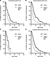 Figure 1
Incorporation of the adjunct triad manifestations of APECED rash, enamel hypoplasia, and autoimmune enteritis into the classic triad manifestations results in accelerated diagnosis across independent cohorts of American and European APECED patients. Shown in A–D are Kaplan-Meier curves comparing the time to APECED diagnosis between developing any diagnostic dyad among the classic triad criteria (CMC, adrenal insufficiency, hypoparathyroidism) or the expanded diagnostic criteria (classic triad plus adjunct triad manifestations of APECED rash, autoimmune enteritis, and enamel hypoplasia), as shown in the first 35 American patients (previously reported in and modified from Ferre et al., ref. 12) (A), a second independent cohort of 57 American patients (current study) (B), a cohort of 12 European patients (current study) (C), and in the entire cohort of all 104 APECED patients combined (D). Vertical hash marks on Kaplan-Meier curves represent individuals who have not yet met the designated clinical diagnostic criteria at the specified age during the time of evaluation at the NIH Clinical Center.
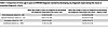 Table 3
Comparison of mean age in years of APECED diagnosis reached by developing any diagnostic dyad among the classic or expanded diagnostic criteria
When evaluating the European cohort by the classic diagnostic criteria, we found 9 (75%) patients reached a classic diagnostic dyad by a mean age of 4.8 (range, 1.5–8) years, earlier than the American cohort (Tables 2 and 3). The other 3 genetically confirmed patients with a mean age of 9 (range, 3–12) years had not yet developed a classic diagnostic dyad at the time of NIH evaluation. Similar to the American patients, 10 (83.3%) European patients developed an average of 2 (range, 1–7) non-classic triad manifestations before reaching a classic diagnostic dyad, among which the 3 most common were our proposed adjunct triad manifestations of APECED rash (7 patients, 58%), enamel hypoplasia (5 patients, 42%), and autoimmune enteritis (3 patients, 25%) (Supplemental Table 5). Implementing our expanded diagnostic criteria would have reduced the time to diagnosis by half (mean, 2.8 years; range, 0.75–7 years) relative to the diagnosis reached by the classic diagnostic criteria (Figure 1C and Table 3). In 5 patients (41.7%) who manifested hypocalcemic seizures and/or adrenal crises before reaching a classic diagnostic dyad, these events could have been prevented with earlier diagnosis by the expanded diagnostic criteria. Among these patients, 2 could also have benefited from earlier recognition and treatment of autoimmune pneumonitis and autoimmune hepatitis, which occurred before reaching a classic diagnostic dyad.
When we combined the 69 patients from this study with our 35 previously reported American patients, implementing the expanded diagnostic criteria in the entire cohort (n = 104) also decreased the time to diagnosis by half relative to the time needed with the classic criteria (Figure 1D and Table 3); this beneficial effect may be underestimated because 13 patients (12.5%) with a current mean age of 9.1 years have not yet reached a classic diagnostic dyad but 11 of them have reached an adjunct diagnostic dyad at a mean age of 2.6 years. Two genetically confirmed patients, one 20-year-old Salvadoran male with CMC, autoimmune keratoconjunctivitis, growth hormone deficiency, and B12 deficiency and one 12-year-old Irish girl with CMC, alopecia areata, and autoimmune pneumonitis have not yet met a diagnostic dyad by either the classic or expanded diagnostic criteria.
We next aimed to validate our previous observation (12) that APECED rash, enamel hypoplasia, and autoimmune enteritis are indeed the 3 most appropriate manifestations, being prevalent early on, for inclusion in the expanded diagnostic criteria. For that, we analyzed the patients’ first 2 consecutive manifestations. Among the 57 American patients, the most common diseases occurring among their first 2 manifestations were CMC (70.2%) and APECED rash (33.3%), followed by autoimmune enteritis (21.1%), enamel hypoplasia and hypoparathyroidism (19.3% each), and autoimmune pneumonitis (12.3%) (Supplemental Table 6). As previously described, adrenal insufficiency occurred late (7, 12). Hence, the 3 most common non-classic triad diseases occurring among the first 2 manifestations were APECED rash, autoimmune enteritis, and enamel hypoplasia. Strikingly, in 39 patients (68.4%), their first 2 manifestations were among the expanded diagnostic criteria, whereas in only 5 patients (8.8%) were their first 2 manifestations among the classic diagnostic triad (P < 0.0001) (Table 4). European patients developed the same 6 manifestations as the most common among the first 2 that developed and the adjunct triad manifestations were the 3 most prevalent non-classic triad manifestations observed early on (Supplemental Table 6). As with American patients, in 10 European patients (83.3%) their first 2 manifestations were among the expanded diagnostic criteria manifestations, whereas in only 2 patients (16.7%) were their first 2 manifestations among the classic diagnostic triad (P = 0.003) (Table 4).
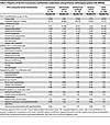 Table 4
Frequency of the first 2 consecutive manifestation combinations among American and European patients with APECED
When considering the entire cohort of 104 patients, the first 2 consecutive manifestations were approximately 5 times more likely to be within the expanded diagnostic criteria (74 patients, 71.2%) than being within the classic diagnostic triad (14 patients, 13.5%; P < 0.0001) (Table 4). CMC and APECED rash were the most common first 2 manifestations, as previously reported (12), with CMC being most prevalent within the entire cohort. Importantly, CMC combined with any manifestation is most commonly seen (68 patients, 65.4%) within the first 2 consecutive manifestations in the entire cohort (Table 4), including in the aforementioned 12-year-old Irish girl with biallelic AIRE variants who developed CMC, alopecia areata, and autoimmune pneumonitis and has not yet met the classic or expanded diagnostic criteria (censored patient, Figure 1C). Notably, 28.8% of all patients developed combinations of the first 2 consecutive manifestations that were not both captured within either the classic or expanded diagnostic criteria. Therefore, a high index of suspicion for APECED is required in patients who develop serial autoimmune manifestations and particularly when these include any of the expanded diagnostic criteria manifestations of CMC, adrenal insufficiency, hypoparathyroidism, APECED rash, enamel hypoplasia, and autoimmune enteritis.
Uncommon APECED manifestations in the entire cohort. Evaluation of all 104 patients in our prospective NIH natural history study revealed several uncommon and/or previously under-recognized manifestations of APECED. Neurologic manifestations included episodic migraines in 12.5% of patients, a frequency similar to the average rate in population prevalence studies (24, 25), and seizure disorders in 5% diagnosed by EEG in the absence of hypocalcemia, hypoglycemia, or other metabolic abnormalities; most were well controlled, although one patient developed progressive cognitive decline, movement disorder, and intractable hiccups. Rare central nervous system autoimmune or structural findings included acute noninfectious encephalitis (n = 2), and one patient each with progressive encephalomyelitis with rigidity and myoclonus (PERM) (26), Arnold-Chiari malformation type 1, and hippocampal inversion, further expanding the previously reported central nervous system manifestations in APECED (27–30). Uncommon ocular diseases included recurrent blepharitis and chalazia (n = 5), amblyopia with esotropia in one individual with concurrent trisomy 21, and single cases of macular coloboma and retinopathy with nystagmus, optic nerve atrophy, and retinal dystrophy. Cardiac abnormalities were infrequent, including patent foramen ovale (n = 2) and one each of atrial septal defect (31) and tetralogy of Fallot. Pituitary adenomas were incidentally identified on MRI in 6 individuals (6%). Hepatobiliary disease included cholelithiasis in 14 patients (13.5%), consistent with prior reports, with a variable age of onset (range, 5–52 years) (32); 12 patients underwent cholecystectomy. Additional rare gastrointestinal manifestations included rectal prolapse due to severe autoimmune enteritis-associated diarrhea or constipation (n = 2), abdominal cocoon (33) in the context of autoimmune enteritis (n = 1), and postpartum sigmoid rupture (n = 1). Musculoskeletal manifestations comprised polyarthropathy with associated fever and myalgias in one 2-year-old child (n = 1) that responded to corticosteroids, and metaphyseal dysplasia in 2 individuals, one resolving spontaneously and another associated with genu varum of both lower extremities. Hematologic abnormalities were frequent, with iron deficiency anemia in 15 patients (14.5%) (34) and unprovoked hypercoagulability in 3 presenting as pulmonary emboli (n = 2) or recurrent deep vein thromboses (n = 1).
Malignancy was infrequent. We observed 2 gastric carcinomas (one each signet-ring and gastroesophageal junction adenocarcinoma) requiring total gastrectomy. Interestingly, both patients had intestinal metaplasia, prompting ongoing endoscopic surveillance in additional patients with premalignant gastric histological changes (35, 36). Oral or esophageal squamous carcinomas, described in other APECED cohorts (37–40), were not observed in our cohort, although 2 patients have had oral leukoplakia and remain under clinical surveillance.
Collectively, these findings further expand the recognized clinical spectrum of APECED, revealing diverse autoimmune, inflammatory, and structural complications that extend beyond the classic and adjunct triad manifestations. This underscores the importance of continued longitudinal evaluation to define disease penetrance, natural history, and mechanisms of organ-specific autoimmunity in APECED.
Causes of mortality. Among the 104 APECED patients, 10 (9.6%) died at a mean age of 32 years (range, 14–61 years). The principal causes of death were respiratory failure and/or infections associated with APECED-associated pneumonitis (n = 5), other infection-related complications (n = 2), endocrine failure (n = 2), and acute hepatic failure (n = 1).
Five deaths were attributed to advanced APECED pneumonitis complicated by irreversible bronchiectasis and secondary multidrug-resistant (MDR) bacterial infections. These included a 14-year-old boy with severe bronchiectasis who succumbed to acute respiratory failure and pneumonia; a 56-year-old man with a 40-year history of APECED pneumonitis who developed MDR Pseudomonas aeruginosa sepsis (41); a 61-year-old woman with longstanding pneumonitis and bronchiectasis who died of postoperative sepsis due to carbapenem-resistant Pseudomonas aeruginosa and Klebsiella pneumoniae; a 22-year-old woman who suffered from multiple recurrent catheter-associated bloodstream infections and bronchiectasis-associated bacterial pneumonias; and a 20-year-old woman who required extracorporeal membrane oxygenation for acute respiratory failure. In each case, chronic untreated autoimmune pneumonitis led to recurrent pulmonary infections and antibiotic exposure, promoting antimicrobial resistance and progressive respiratory decline.
Two patients died from complications of endocrine failure. A 28-year-old woman with known hypoparathyroidism was found deceased at home, possibly due to cardiac arrhythmia from severe hypocalcemia. Another patient, a man in his early 20s with adrenal insufficiency and type 1 diabetes died from combined adrenal crisis and hypoglycemia. A 42-year-old woman died in inpatient hospice care after developing encephalitis in the context of polyomavirus reactivation following immunosuppression with alemtuzumab and corticosteroids. A 24-year-old woman developed fulminant hepatic failure following a brief prodrome of elevated transaminases, suspected to be triggered by a viral infection on a background of unrecognized APECED-associated hepatitis. Finally, a 30-year-old woman hospitalized for septic shock developed fatal disseminated mucormycosis (42).
Taken together, these findings underscore that preventable complications — most commonly pulmonary sequelae of autoimmune pneumonitis, followed by endocrine crises, hepatic failure, and severe systemic infections (21, 41, 43, 44) — remain the leading causes of mortality in APECED. These outcomes highlight the critical importance of early diagnosis, structured endocrine management, comprehensive surveillance and screening (Figure 2 and Table 5), and timely immunomodulatory therapy to prevent irreversible organ damage while not promoting immune compromise. Together, these approaches will improve long-term survival in APECED.
 Figure 2
Proposed screening of prospective APECED patients. A proposed schema for screening practices to identify patients with APECED. AIRE, autoimmune regulator; PHD, plant homeodomain. AAbs, autoantibodies. *Note that pathogenic deep intronic variants may be missed by conventional genetic panels and whole exome sequencing. Whole genome sequencing, when available, may facilitate identification of such variants and may also reveal alternative genetic etiologies when AIRE variants are not detected.
Immunophenotyping of patient lymphocytes. We had previously demonstrated an expansion of CD4+ naive T cells, autoreactive CD21loCD38lo B cells, and plasmablasts, and a contraction of NK cells in the peripheral blood of APECED patients (12). We performed immunophenotyping of peripheral blood lymphocytes in the entire cohort of 104 APECED patients and again found similar perturbations of these 4 lymphocyte populations (Supplemental Tables 7 and 8). In addition, we observed an expansion of CD8+ naive T cells and NKT cells in peripheral blood (Supplemental Tables 7 and 8). Together, these data define a characteristic immunophenotypic signature in APECED, marked by expansion of naive T cell subsets, dysregulated B cell compartments, and altered innate lymphocyte populations, reflecting the broad immune activation and loss of tolerance that underlie the disease.
Serum CXCL9 levels are elevated across patient cohorts compared with healthy donors. Given our previous observation that APECED is characterized by IFN-γ–driven inflammation with elevated serum and tissue CXCL9 (5, 6), we assessed serum CXCL9 levels across the initial American cohort, the newly enrolled cohort, and healthy donors. Both the initial and newly enrolled cohorts exhibited higher serum CXCL9 levels than healthy donors, consistent with a shared IFN-γ–driven inflammatory signature across cohorts (Supplemental Figure 3).


