
Research ArticleImmunologyOncology
Open Access |  10.1172/jci.insight.205984
T cell–mediated antibody-dependent cellular cytotoxicity provides a therapeutic approach for solid cancers
Alon Richter,1 Shahar Dotan,2 Petr Shendrik,3 Diana Rasoulouniriana,1 Amichai Zarfin,1 Nadine Santana-Magal,1 Aseel Khateeb,1 Adi Frisch,1 Gal Verbin,1 Ariel Galor,1 Eran Sadot,4 Oran Zlotnik,4 Avishai Maliah,1 Ori Moskowitz,1 Tom Ben-Dov,1,5 Maya Halpern,1,5 Chen Luxenburg,1 Alexander Leithner,6 Yu Zhang,6 Natalia T. Freund,1 Merav Cohen,1 Carmit Levy,1 Matthew H. Spitzer,7 Raya Sorkin,3 Michael L. Dustin,6 Peleg Rider,1 and Yaron Carmi1
1Gray School of Medicine, Tel Aviv University, Tel Aviv, Israel.
2Gilboa Therapeutics Ltd., Rehovot, Israel.
3School of Chemistry Tel Aviv University, Tel Aviv, Israel.
4Department of General Surgery, Rabin Medical Center- Beilinson Campus, Petach Tikva, Israel.
5Department of Otolaryngology, Head and Neck Surgery-Meir Medical Center, Kfar-Saba, Israel.
6The Kennedy Institute of Rheumatology, Oxford University, Oxford, England.
7Gladstone Institutes and Departments of Otolaryngology–Head and Neck Surgery, UCSF, San Francisco, California, USA.
Address for correspondence. Yaron Carmi, Tel-Aviv University, Klatzkin St. 35, Tel Aviv-Yafo, 6997801, Israel. Email: yaron.carmi@gmail.com.
Find articles by Richter, A. in: PubMed | Google Scholar
1Gray School of Medicine, Tel Aviv University, Tel Aviv, Israel.
2Gilboa Therapeutics Ltd., Rehovot, Israel.
3School of Chemistry Tel Aviv University, Tel Aviv, Israel.
4Department of General Surgery, Rabin Medical Center- Beilinson Campus, Petach Tikva, Israel.
5Department of Otolaryngology, Head and Neck Surgery-Meir Medical Center, Kfar-Saba, Israel.
6The Kennedy Institute of Rheumatology, Oxford University, Oxford, England.
7Gladstone Institutes and Departments of Otolaryngology–Head and Neck Surgery, UCSF, San Francisco, California, USA.
Address for correspondence. Yaron Carmi, Tel-Aviv University, Klatzkin St. 35, Tel Aviv-Yafo, 6997801, Israel. Email: yaron.carmi@gmail.com.
Find articles by
Dotan, S.
in:
PubMed
|
Google Scholar
|

1Gray School of Medicine, Tel Aviv University, Tel Aviv, Israel.
2Gilboa Therapeutics Ltd., Rehovot, Israel.
3School of Chemistry Tel Aviv University, Tel Aviv, Israel.
4Department of General Surgery, Rabin Medical Center- Beilinson Campus, Petach Tikva, Israel.
5Department of Otolaryngology, Head and Neck Surgery-Meir Medical Center, Kfar-Saba, Israel.
6The Kennedy Institute of Rheumatology, Oxford University, Oxford, England.
7Gladstone Institutes and Departments of Otolaryngology–Head and Neck Surgery, UCSF, San Francisco, California, USA.
Address for correspondence. Yaron Carmi, Tel-Aviv University, Klatzkin St. 35, Tel Aviv-Yafo, 6997801, Israel. Email: yaron.carmi@gmail.com.
Find articles by Shendrik, P. in: PubMed | Google Scholar
1Gray School of Medicine, Tel Aviv University, Tel Aviv, Israel.
2Gilboa Therapeutics Ltd., Rehovot, Israel.
3School of Chemistry Tel Aviv University, Tel Aviv, Israel.
4Department of General Surgery, Rabin Medical Center- Beilinson Campus, Petach Tikva, Israel.
5Department of Otolaryngology, Head and Neck Surgery-Meir Medical Center, Kfar-Saba, Israel.
6The Kennedy Institute of Rheumatology, Oxford University, Oxford, England.
7Gladstone Institutes and Departments of Otolaryngology–Head and Neck Surgery, UCSF, San Francisco, California, USA.
Address for correspondence. Yaron Carmi, Tel-Aviv University, Klatzkin St. 35, Tel Aviv-Yafo, 6997801, Israel. Email: yaron.carmi@gmail.com.
Find articles by Rasoulouniriana, D. in: PubMed | Google Scholar
1Gray School of Medicine, Tel Aviv University, Tel Aviv, Israel.
2Gilboa Therapeutics Ltd., Rehovot, Israel.
3School of Chemistry Tel Aviv University, Tel Aviv, Israel.
4Department of General Surgery, Rabin Medical Center- Beilinson Campus, Petach Tikva, Israel.
5Department of Otolaryngology, Head and Neck Surgery-Meir Medical Center, Kfar-Saba, Israel.
6The Kennedy Institute of Rheumatology, Oxford University, Oxford, England.
7Gladstone Institutes and Departments of Otolaryngology–Head and Neck Surgery, UCSF, San Francisco, California, USA.
Address for correspondence. Yaron Carmi, Tel-Aviv University, Klatzkin St. 35, Tel Aviv-Yafo, 6997801, Israel. Email: yaron.carmi@gmail.com.
Find articles by Zarfin, A. in: PubMed | Google Scholar
1Gray School of Medicine, Tel Aviv University, Tel Aviv, Israel.
2Gilboa Therapeutics Ltd., Rehovot, Israel.
3School of Chemistry Tel Aviv University, Tel Aviv, Israel.
4Department of General Surgery, Rabin Medical Center- Beilinson Campus, Petach Tikva, Israel.
5Department of Otolaryngology, Head and Neck Surgery-Meir Medical Center, Kfar-Saba, Israel.
6The Kennedy Institute of Rheumatology, Oxford University, Oxford, England.
7Gladstone Institutes and Departments of Otolaryngology–Head and Neck Surgery, UCSF, San Francisco, California, USA.
Address for correspondence. Yaron Carmi, Tel-Aviv University, Klatzkin St. 35, Tel Aviv-Yafo, 6997801, Israel. Email: yaron.carmi@gmail.com.
Find articles by Santana-Magal, N. in: PubMed | Google Scholar
1Gray School of Medicine, Tel Aviv University, Tel Aviv, Israel.
2Gilboa Therapeutics Ltd., Rehovot, Israel.
3School of Chemistry Tel Aviv University, Tel Aviv, Israel.
4Department of General Surgery, Rabin Medical Center- Beilinson Campus, Petach Tikva, Israel.
5Department of Otolaryngology, Head and Neck Surgery-Meir Medical Center, Kfar-Saba, Israel.
6The Kennedy Institute of Rheumatology, Oxford University, Oxford, England.
7Gladstone Institutes and Departments of Otolaryngology–Head and Neck Surgery, UCSF, San Francisco, California, USA.
Address for correspondence. Yaron Carmi, Tel-Aviv University, Klatzkin St. 35, Tel Aviv-Yafo, 6997801, Israel. Email: yaron.carmi@gmail.com.
Find articles by Khateeb, A. in: PubMed | Google Scholar
1Gray School of Medicine, Tel Aviv University, Tel Aviv, Israel.
2Gilboa Therapeutics Ltd., Rehovot, Israel.
3School of Chemistry Tel Aviv University, Tel Aviv, Israel.
4Department of General Surgery, Rabin Medical Center- Beilinson Campus, Petach Tikva, Israel.
5Department of Otolaryngology, Head and Neck Surgery-Meir Medical Center, Kfar-Saba, Israel.
6The Kennedy Institute of Rheumatology, Oxford University, Oxford, England.
7Gladstone Institutes and Departments of Otolaryngology–Head and Neck Surgery, UCSF, San Francisco, California, USA.
Address for correspondence. Yaron Carmi, Tel-Aviv University, Klatzkin St. 35, Tel Aviv-Yafo, 6997801, Israel. Email: yaron.carmi@gmail.com.
Find articles by Frisch, A. in: PubMed | Google Scholar
1Gray School of Medicine, Tel Aviv University, Tel Aviv, Israel.
2Gilboa Therapeutics Ltd., Rehovot, Israel.
3School of Chemistry Tel Aviv University, Tel Aviv, Israel.
4Department of General Surgery, Rabin Medical Center- Beilinson Campus, Petach Tikva, Israel.
5Department of Otolaryngology, Head and Neck Surgery-Meir Medical Center, Kfar-Saba, Israel.
6The Kennedy Institute of Rheumatology, Oxford University, Oxford, England.
7Gladstone Institutes and Departments of Otolaryngology–Head and Neck Surgery, UCSF, San Francisco, California, USA.
Address for correspondence. Yaron Carmi, Tel-Aviv University, Klatzkin St. 35, Tel Aviv-Yafo, 6997801, Israel. Email: yaron.carmi@gmail.com.
Find articles by Verbin, G. in: PubMed | Google Scholar
1Gray School of Medicine, Tel Aviv University, Tel Aviv, Israel.
2Gilboa Therapeutics Ltd., Rehovot, Israel.
3School of Chemistry Tel Aviv University, Tel Aviv, Israel.
4Department of General Surgery, Rabin Medical Center- Beilinson Campus, Petach Tikva, Israel.
5Department of Otolaryngology, Head and Neck Surgery-Meir Medical Center, Kfar-Saba, Israel.
6The Kennedy Institute of Rheumatology, Oxford University, Oxford, England.
7Gladstone Institutes and Departments of Otolaryngology–Head and Neck Surgery, UCSF, San Francisco, California, USA.
Address for correspondence. Yaron Carmi, Tel-Aviv University, Klatzkin St. 35, Tel Aviv-Yafo, 6997801, Israel. Email: yaron.carmi@gmail.com.
Find articles by Galor, A. in: PubMed | Google Scholar
1Gray School of Medicine, Tel Aviv University, Tel Aviv, Israel.
2Gilboa Therapeutics Ltd., Rehovot, Israel.
3School of Chemistry Tel Aviv University, Tel Aviv, Israel.
4Department of General Surgery, Rabin Medical Center- Beilinson Campus, Petach Tikva, Israel.
5Department of Otolaryngology, Head and Neck Surgery-Meir Medical Center, Kfar-Saba, Israel.
6The Kennedy Institute of Rheumatology, Oxford University, Oxford, England.
7Gladstone Institutes and Departments of Otolaryngology–Head and Neck Surgery, UCSF, San Francisco, California, USA.
Address for correspondence. Yaron Carmi, Tel-Aviv University, Klatzkin St. 35, Tel Aviv-Yafo, 6997801, Israel. Email: yaron.carmi@gmail.com.
Find articles by Sadot, E. in: PubMed | Google Scholar
1Gray School of Medicine, Tel Aviv University, Tel Aviv, Israel.
2Gilboa Therapeutics Ltd., Rehovot, Israel.
3School of Chemistry Tel Aviv University, Tel Aviv, Israel.
4Department of General Surgery, Rabin Medical Center- Beilinson Campus, Petach Tikva, Israel.
5Department of Otolaryngology, Head and Neck Surgery-Meir Medical Center, Kfar-Saba, Israel.
6The Kennedy Institute of Rheumatology, Oxford University, Oxford, England.
7Gladstone Institutes and Departments of Otolaryngology–Head and Neck Surgery, UCSF, San Francisco, California, USA.
Address for correspondence. Yaron Carmi, Tel-Aviv University, Klatzkin St. 35, Tel Aviv-Yafo, 6997801, Israel. Email: yaron.carmi@gmail.com.
Find articles by Zlotnik, O. in: PubMed | Google Scholar
1Gray School of Medicine, Tel Aviv University, Tel Aviv, Israel.
2Gilboa Therapeutics Ltd., Rehovot, Israel.
3School of Chemistry Tel Aviv University, Tel Aviv, Israel.
4Department of General Surgery, Rabin Medical Center- Beilinson Campus, Petach Tikva, Israel.
5Department of Otolaryngology, Head and Neck Surgery-Meir Medical Center, Kfar-Saba, Israel.
6The Kennedy Institute of Rheumatology, Oxford University, Oxford, England.
7Gladstone Institutes and Departments of Otolaryngology–Head and Neck Surgery, UCSF, San Francisco, California, USA.
Address for correspondence. Yaron Carmi, Tel-Aviv University, Klatzkin St. 35, Tel Aviv-Yafo, 6997801, Israel. Email: yaron.carmi@gmail.com.
Find articles by Maliah, A. in: PubMed | Google Scholar
1Gray School of Medicine, Tel Aviv University, Tel Aviv, Israel.
2Gilboa Therapeutics Ltd., Rehovot, Israel.
3School of Chemistry Tel Aviv University, Tel Aviv, Israel.
4Department of General Surgery, Rabin Medical Center- Beilinson Campus, Petach Tikva, Israel.
5Department of Otolaryngology, Head and Neck Surgery-Meir Medical Center, Kfar-Saba, Israel.
6The Kennedy Institute of Rheumatology, Oxford University, Oxford, England.
7Gladstone Institutes and Departments of Otolaryngology–Head and Neck Surgery, UCSF, San Francisco, California, USA.
Address for correspondence. Yaron Carmi, Tel-Aviv University, Klatzkin St. 35, Tel Aviv-Yafo, 6997801, Israel. Email: yaron.carmi@gmail.com.
Find articles by Moskowitz, O. in: PubMed | Google Scholar
1Gray School of Medicine, Tel Aviv University, Tel Aviv, Israel.
2Gilboa Therapeutics Ltd., Rehovot, Israel.
3School of Chemistry Tel Aviv University, Tel Aviv, Israel.
4Department of General Surgery, Rabin Medical Center- Beilinson Campus, Petach Tikva, Israel.
5Department of Otolaryngology, Head and Neck Surgery-Meir Medical Center, Kfar-Saba, Israel.
6The Kennedy Institute of Rheumatology, Oxford University, Oxford, England.
7Gladstone Institutes and Departments of Otolaryngology–Head and Neck Surgery, UCSF, San Francisco, California, USA.
Address for correspondence. Yaron Carmi, Tel-Aviv University, Klatzkin St. 35, Tel Aviv-Yafo, 6997801, Israel. Email: yaron.carmi@gmail.com.
Find articles by Ben-Dov, T. in: PubMed | Google Scholar
1Gray School of Medicine, Tel Aviv University, Tel Aviv, Israel.
2Gilboa Therapeutics Ltd., Rehovot, Israel.
3School of Chemistry Tel Aviv University, Tel Aviv, Israel.
4Department of General Surgery, Rabin Medical Center- Beilinson Campus, Petach Tikva, Israel.
5Department of Otolaryngology, Head and Neck Surgery-Meir Medical Center, Kfar-Saba, Israel.
6The Kennedy Institute of Rheumatology, Oxford University, Oxford, England.
7Gladstone Institutes and Departments of Otolaryngology–Head and Neck Surgery, UCSF, San Francisco, California, USA.
Address for correspondence. Yaron Carmi, Tel-Aviv University, Klatzkin St. 35, Tel Aviv-Yafo, 6997801, Israel. Email: yaron.carmi@gmail.com.
Find articles by Halpern, M. in: PubMed | Google Scholar
1Gray School of Medicine, Tel Aviv University, Tel Aviv, Israel.
2Gilboa Therapeutics Ltd., Rehovot, Israel.
3School of Chemistry Tel Aviv University, Tel Aviv, Israel.
4Department of General Surgery, Rabin Medical Center- Beilinson Campus, Petach Tikva, Israel.
5Department of Otolaryngology, Head and Neck Surgery-Meir Medical Center, Kfar-Saba, Israel.
6The Kennedy Institute of Rheumatology, Oxford University, Oxford, England.
7Gladstone Institutes and Departments of Otolaryngology–Head and Neck Surgery, UCSF, San Francisco, California, USA.
Address for correspondence. Yaron Carmi, Tel-Aviv University, Klatzkin St. 35, Tel Aviv-Yafo, 6997801, Israel. Email: yaron.carmi@gmail.com.
Find articles by Luxenburg, C. in: PubMed | Google Scholar
1Gray School of Medicine, Tel Aviv University, Tel Aviv, Israel.
2Gilboa Therapeutics Ltd., Rehovot, Israel.
3School of Chemistry Tel Aviv University, Tel Aviv, Israel.
4Department of General Surgery, Rabin Medical Center- Beilinson Campus, Petach Tikva, Israel.
5Department of Otolaryngology, Head and Neck Surgery-Meir Medical Center, Kfar-Saba, Israel.
6The Kennedy Institute of Rheumatology, Oxford University, Oxford, England.
7Gladstone Institutes and Departments of Otolaryngology–Head and Neck Surgery, UCSF, San Francisco, California, USA.
Address for correspondence. Yaron Carmi, Tel-Aviv University, Klatzkin St. 35, Tel Aviv-Yafo, 6997801, Israel. Email: yaron.carmi@gmail.com.
Find articles by Leithner, A. in: PubMed | Google Scholar
1Gray School of Medicine, Tel Aviv University, Tel Aviv, Israel.
2Gilboa Therapeutics Ltd., Rehovot, Israel.
3School of Chemistry Tel Aviv University, Tel Aviv, Israel.
4Department of General Surgery, Rabin Medical Center- Beilinson Campus, Petach Tikva, Israel.
5Department of Otolaryngology, Head and Neck Surgery-Meir Medical Center, Kfar-Saba, Israel.
6The Kennedy Institute of Rheumatology, Oxford University, Oxford, England.
7Gladstone Institutes and Departments of Otolaryngology–Head and Neck Surgery, UCSF, San Francisco, California, USA.
Address for correspondence. Yaron Carmi, Tel-Aviv University, Klatzkin St. 35, Tel Aviv-Yafo, 6997801, Israel. Email: yaron.carmi@gmail.com.
Find articles by Zhang, Y. in: PubMed | Google Scholar
1Gray School of Medicine, Tel Aviv University, Tel Aviv, Israel.
2Gilboa Therapeutics Ltd., Rehovot, Israel.
3School of Chemistry Tel Aviv University, Tel Aviv, Israel.
4Department of General Surgery, Rabin Medical Center- Beilinson Campus, Petach Tikva, Israel.
5Department of Otolaryngology, Head and Neck Surgery-Meir Medical Center, Kfar-Saba, Israel.
6The Kennedy Institute of Rheumatology, Oxford University, Oxford, England.
7Gladstone Institutes and Departments of Otolaryngology–Head and Neck Surgery, UCSF, San Francisco, California, USA.
Address for correspondence. Yaron Carmi, Tel-Aviv University, Klatzkin St. 35, Tel Aviv-Yafo, 6997801, Israel. Email: yaron.carmi@gmail.com.
Find articles by
Freund, N.
in:
PubMed
|
Google Scholar
|
1Gray School of Medicine, Tel Aviv University, Tel Aviv, Israel.
2Gilboa Therapeutics Ltd., Rehovot, Israel.
3School of Chemistry Tel Aviv University, Tel Aviv, Israel.
4Department of General Surgery, Rabin Medical Center- Beilinson Campus, Petach Tikva, Israel.
5Department of Otolaryngology, Head and Neck Surgery-Meir Medical Center, Kfar-Saba, Israel.
6The Kennedy Institute of Rheumatology, Oxford University, Oxford, England.
7Gladstone Institutes and Departments of Otolaryngology–Head and Neck Surgery, UCSF, San Francisco, California, USA.
Address for correspondence. Yaron Carmi, Tel-Aviv University, Klatzkin St. 35, Tel Aviv-Yafo, 6997801, Israel. Email: yaron.carmi@gmail.com.
Find articles by Cohen, M. in: PubMed | Google Scholar
1Gray School of Medicine, Tel Aviv University, Tel Aviv, Israel.
2Gilboa Therapeutics Ltd., Rehovot, Israel.
3School of Chemistry Tel Aviv University, Tel Aviv, Israel.
4Department of General Surgery, Rabin Medical Center- Beilinson Campus, Petach Tikva, Israel.
5Department of Otolaryngology, Head and Neck Surgery-Meir Medical Center, Kfar-Saba, Israel.
6The Kennedy Institute of Rheumatology, Oxford University, Oxford, England.
7Gladstone Institutes and Departments of Otolaryngology–Head and Neck Surgery, UCSF, San Francisco, California, USA.
Address for correspondence. Yaron Carmi, Tel-Aviv University, Klatzkin St. 35, Tel Aviv-Yafo, 6997801, Israel. Email: yaron.carmi@gmail.com.
Find articles by Levy, C. in: PubMed | Google Scholar
1Gray School of Medicine, Tel Aviv University, Tel Aviv, Israel.
2Gilboa Therapeutics Ltd., Rehovot, Israel.
3School of Chemistry Tel Aviv University, Tel Aviv, Israel.
4Department of General Surgery, Rabin Medical Center- Beilinson Campus, Petach Tikva, Israel.
5Department of Otolaryngology, Head and Neck Surgery-Meir Medical Center, Kfar-Saba, Israel.
6The Kennedy Institute of Rheumatology, Oxford University, Oxford, England.
7Gladstone Institutes and Departments of Otolaryngology–Head and Neck Surgery, UCSF, San Francisco, California, USA.
Address for correspondence. Yaron Carmi, Tel-Aviv University, Klatzkin St. 35, Tel Aviv-Yafo, 6997801, Israel. Email: yaron.carmi@gmail.com.
Find articles by
Spitzer, M.
in:
PubMed
|
Google Scholar
|
1Gray School of Medicine, Tel Aviv University, Tel Aviv, Israel.
2Gilboa Therapeutics Ltd., Rehovot, Israel.
3School of Chemistry Tel Aviv University, Tel Aviv, Israel.
4Department of General Surgery, Rabin Medical Center- Beilinson Campus, Petach Tikva, Israel.
5Department of Otolaryngology, Head and Neck Surgery-Meir Medical Center, Kfar-Saba, Israel.
6The Kennedy Institute of Rheumatology, Oxford University, Oxford, England.
7Gladstone Institutes and Departments of Otolaryngology–Head and Neck Surgery, UCSF, San Francisco, California, USA.
Address for correspondence. Yaron Carmi, Tel-Aviv University, Klatzkin St. 35, Tel Aviv-Yafo, 6997801, Israel. Email: yaron.carmi@gmail.com.
Find articles by Sorkin, R. in: PubMed | Google Scholar
1Gray School of Medicine, Tel Aviv University, Tel Aviv, Israel.
2Gilboa Therapeutics Ltd., Rehovot, Israel.
3School of Chemistry Tel Aviv University, Tel Aviv, Israel.
4Department of General Surgery, Rabin Medical Center- Beilinson Campus, Petach Tikva, Israel.
5Department of Otolaryngology, Head and Neck Surgery-Meir Medical Center, Kfar-Saba, Israel.
6The Kennedy Institute of Rheumatology, Oxford University, Oxford, England.
7Gladstone Institutes and Departments of Otolaryngology–Head and Neck Surgery, UCSF, San Francisco, California, USA.
Address for correspondence. Yaron Carmi, Tel-Aviv University, Klatzkin St. 35, Tel Aviv-Yafo, 6997801, Israel. Email: yaron.carmi@gmail.com.
Find articles by
Dustin, M.
in:
PubMed
|
Google Scholar
|
1Gray School of Medicine, Tel Aviv University, Tel Aviv, Israel.
2Gilboa Therapeutics Ltd., Rehovot, Israel.
3School of Chemistry Tel Aviv University, Tel Aviv, Israel.
4Department of General Surgery, Rabin Medical Center- Beilinson Campus, Petach Tikva, Israel.
5Department of Otolaryngology, Head and Neck Surgery-Meir Medical Center, Kfar-Saba, Israel.
6The Kennedy Institute of Rheumatology, Oxford University, Oxford, England.
7Gladstone Institutes and Departments of Otolaryngology–Head and Neck Surgery, UCSF, San Francisco, California, USA.
Address for correspondence. Yaron Carmi, Tel-Aviv University, Klatzkin St. 35, Tel Aviv-Yafo, 6997801, Israel. Email: yaron.carmi@gmail.com.
Find articles by Rider, P. in: PubMed | Google Scholar
1Gray School of Medicine, Tel Aviv University, Tel Aviv, Israel.
2Gilboa Therapeutics Ltd., Rehovot, Israel.
3School of Chemistry Tel Aviv University, Tel Aviv, Israel.
4Department of General Surgery, Rabin Medical Center- Beilinson Campus, Petach Tikva, Israel.
5Department of Otolaryngology, Head and Neck Surgery-Meir Medical Center, Kfar-Saba, Israel.
6The Kennedy Institute of Rheumatology, Oxford University, Oxford, England.
7Gladstone Institutes and Departments of Otolaryngology–Head and Neck Surgery, UCSF, San Francisco, California, USA.
Address for correspondence. Yaron Carmi, Tel-Aviv University, Klatzkin St. 35, Tel Aviv-Yafo, 6997801, Israel. Email: yaron.carmi@gmail.com.
Find articles by Carmi, Y. in: PubMed | Google Scholar
Published November 9, 2026 - More info
JCI Insight. 2026;11(21):e205984. https://doi.org/10.1172/jci.insight.205984.
© 2026 Richter et al. This work is licensed under the Creative Commons Attribution 4.0 International License. To view a copy of this license, visit http://creativecommons.org/licenses/by/4.0/.
Received: February 17, 2026; Accepted: September 21, 2026


-
Results
A rare population of FcγRI+CD8+ T cells is present in mouse tumors. In past projects, our group identified a mixed population of exhausted infiltrating CD4+ T cells that express FcγRI in mouse and human tumors (18). To determine whether a similar population exists among CD8+ T cells, we analyzed CD8+ T cells from draining lymph nodes (DLNs) and tumors of mice bearing day 10 B16F10 melanomas by cytometry by time-of-flight (CyTOF). After excluding non-T cell lineages, unbiased uniform manifold approximation and projection (UMAP) analysis identified a small population of TCRβ+CD8+ T cells expressing FcγRI. These cells co-expressed CD44, T-bet, perforin, Sca-1, CD40, CCR7, and CX3CR1, while lacking CD27 and CD62L. Unlike naive, central memory, and early activated CD8+ T cells, FcγRI+CD8+ T cells were detected exclusively in tumors, together with perforinhi effector cells (Figure 1, A–C, and Supplemental Figure 1A; supplemental material available online with this article; https://doi.org/10.1172/jci.insight.205984DS1).
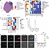 Figure 1
A rare population of FcγRI+CD8+ T cells is present in mouse tumors. (A) UMAP plot of CD8+ T cells from tumors and DLNs of mice bearing day 10 B16F10 tumors (n = 3). (B) Heatmap of median marker intensities in CD8+ T cell clusters. (C) Differential abundance test results and normalized proportions for CD8+ T cell clusters obtained using the diffcyt package. (D) Mean percentages of FcγRI expression on T cells in mice bearing day 10 B16F10 (cold) tumors (n = 5) and YUMMER1.7 (hot) tumors (n = 8). (E) Immunofluorescence of FcγRI in histological sections from day 10 B16F10 (cold) tumor–bearing mouse and YUMMER1.7 (hot) tumor–bearing mouse. Original magnification, ×400. Arrowheads indicate positively stained cells. (F) Percentages of apoptotic B16F10 melanoma 24 hours after incubation with T cells from tumor-bearing mice (n = 4). CM, central memory; EM, effector memory; TRM, tissue-resident memory; GZB, granzyme B. Shown is the summary of 1 experiment out of at least 3 independent experiments. ****P < 0.00001 by ANOVA with Tukey’s correction for multiple comparisons. Error bars represent SEM.
FcγRI+CD8+ T cells were more frequent in immunogenic YUMMER1.7 tumors than in B16F10 tumors (~2.5% vs. ~0.5%) and were not detected in the DLNs or blood (Figure 1D and Supplemental Figure 1B). Confocal microscopy confirmed FcγRI expression on the CD8+ T cell membrane (Figure 1E). To determine whether the FcγRI was functional, infiltrating FcγRI+CD8+ T cells were isolated from OT-I mice bearing B16F10 tumors and selected for an irrelevant OVA-specific T cell receptor (TCR) to exclude TCR-mediated tumor recognition. These cells efficiently killed B16F10 cells only in the presence of the tumor-binding anti-gp75 antibody TA99, demonstrating functional FcγRI-mediated ADCC (Figure 1F).
FcγRI+CD8+ T cells are present at sites of inflammation in humans. We next examined whether FcγRI+CD8+ T cells exist in human tumors. Reanalysis of CyTOF data from patients with head and neck squamous cell carcinoma identified FcγRI expression almost exclusively among activated CD8+ T cells characterized by CD69 and CD38 expression and reduced TCF-1 and CD45RA. FcγRI+ cells also expressed FcγRIII (Figure 2, A and B, and Supplemental Figure 1, C and D). Flow cytometric analysis of freshly resected tumors detected FcγRI+CD8+ T cells in all 5 melanoma patients examined, but not in 9 patients with pancreatic ductal adenocarcinoma (PDAC). Confocal microscopy confirmed FcγRI+CD8+ T cells within melanoma tumors, frequently adjacent to FcγRI+ non-T cells (Figure 2, C and D, and Supplemental Figure 1E). In addition, we checked for the existence of FcγRI+CD8+ T cells in inflammatory settings. FcγRI+CD8+ T cells were also detected in tonsils from 4 of 10 patients with recurrent tonsillitis, including 2 patients with circulating FcγRI+CD8+ T cells, and in the blood of all 6 patients who had recovered from acute COVID-19 (Figure 2E and Supplemental Figure 1, F and G).
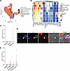 Figure 2
FcγRI+CD8+ T cells are present at sites of inflammation in humans. (A) UMAP plot of CD8+ T cells from tumors, metastatic DLNs, and normal LNs of patients with head and neck squamous cell carcinoma (n = 10). (B) Heatmap of median marker intensities in CD8+ T cell clusters. CM, central memory; EM, effector memory; TRM, tissue-resident memory; GZB, granzyme B. (C) Mean percentages of T cells expressing FcγRI, out of total T cells (n = 5 for healthy patients’ blood, n = 10 for PDAC patients, n = 5 for melanoma patients). (D) Immunofluorescence of FcγRI in histological sections from patient with breast cancer. Original magnification, ×400. Arrowheads indicate positively stained cells. (E) Mean percentages of FcγRI-expressing T cells from freshly resected tonsils of patients suffering from chronic tonsillitis, blood of COVID-19–recovered patients, and blood of healthy patients (n = 5 for healthy blood, n = 5 for COVID-19 blood, n = 10 for tonsillitis patients). Shown is the summary of 1 experiment out of at least 3 independent experiments.
CD8+ T cells acquire FcγRI through trogocytosis from myeloid cells. To investigate the origin of FcγRI on CD8+ T cells, we reanalyzed published single-cell RNA-seq (scRNA-seq) datasets of tumor-infiltrating T cells. Although FcγRI protein was readily detected, FCGR1A transcripts were absent from CD8+ T cell clusters, suggesting a non-transcriptional mechanism (Figure 3, A–C, and Supplemental Figure 2, A and B).
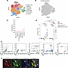 Figure 3
CD8+ T cells acquire FcγRI through trogocytosis from myeloid cells. (A) UMAP plot of tumor-infiltrating CD8+ T cell subclusters (n = 316). (B) Relative intensity of FCGR1A superimposed on UMAP projection. (C) Percentage of FCGR1A-expressing CD8+ T cells across T cell clusters (n = 316). (D) Mean percentages of FcγRI expression on CD8+ T cells co-cultured with monocytes and various cytokines after 1 and 6 days (n = 4). BF, bright-field. (E) Representative FACS analysis of FcγRI-expressing CD8+ T cells co-cultured with monocytes. (F) Representative structured illumination microscopy (SIM) of CD8+ T cells co-cultured with monocytes. Scale bars: 2 μm. Original magnification, ×630. Shown is the summary of 1 experiment out of at least 3 independent experiments. *P < 0.05; **P < 0.001; ****P < 0.00001 by ANOVA with Tukey’s correction for multiple comparisons. Error bars represent SEM.
Because FcγRI+CD8+ T cells were consistently observed near FcγRI+TCR– cells, we tested whether FcγRI could be acquired through trogocytosis. Activated human CD8+ T cells were co-cultured with autologous CD14+ monocytes. FcγRI became detectable on approximately 1% of T cells after 1 day and increased to approximately 2% after 6 days or following IFN-γ exposure, whereas cytokines alone did not induce FcγRI expression (Figure 3, D and E, and Supplemental Figure 2, C and D). Lattice structured illumination microscopy (SIM) imaging demonstrated acquisition of large membrane fragments from monocytes by CD8+ T cells (Figure 3F and Supplemental Figure 2E). These findings indicate that FcγRI is acquired by CD8+ T cells through trogocytosis.
FcγRI acquired by CD8+ T cells remains functional for several hours. We next tested whether trogocytosed FcγRI remains functional. To this end, CD8+ T cells were co-cultured with autologous CD14+ cells and later exposed to HER2-containing lipid bilayers in the presence of trastuzumab. Approximately one-third of FcγRI+ cells formed immune synapses within 20 minutes and exhibited phosphorylated ERK1/2 (Figure 4, A–C). FcγRI accumulation at the synapse was also observed upon interaction with HER2-expressing tumor cells (Figure 4D and Supplemental Figure 2F). Importantly, FcγRI expression was lost within 4–6 hours after sorting, indicating rapid receptor turnover rather than a stable FcγRI+ T cell subset. Thus, CD8+ T cells can transiently acquire functional FcγRI from myeloid cells through trogocytosis.
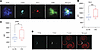 Figure 4
FcγRI acquired by CD8+ T cells remains functional for several hours. (A) Representative TIRF imaging of sorted FcγRI-expressing CD8+ T cells interaction through intermediate antibodies with Her2 on SLB. (B and C) Mean fluorescence intensity of HER2 (B) and p-ERK (C) during interaction of FcγRI-expressing CD8+ T cells with HER2 on SLBs with and without a relevant antibody intermediate (n = 35). (D) Representative SIM-sorted FcγRI-expressing CD8+ T cells forming immune synapses with NCI-N87 cancer cells in the presence of trastuzumab. Shown is the summary of 1 experiment out of at least 3 independent experiments. Original magnification, ×1000 (A and D). **P < 0.001, ***P < 0.0001 by ANOVA with Tukey’s correction for multiple comparisons. Error bars represent SEM.
FcγRI signaling is controlled by antigen density. To define the signaling requirements of FcγRI-mediated ADCC, human CD8+ T cells were engineered to express FcγRI and its ITAM-containing FcRγ chain (AG2G) (Figure 5A). On HER2-containing lipid bilayers, FcγRI formed dense clusters with HER2-trastuzumab complexes while excluding TCRβ and CD45. FcγRI clustering required a minimum HER2 density of approximately 50 molecules/μm2, whereas TCR signaling occurred at substantially lower densities (10–20 molecules/μm2) (Figure 5, B and C, and Supplemental Figure 3A).
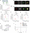 Figure 5
FcγRI signaling is controlled by antigen density. (A) Illustration of FcγRI (A2G) and AG2G constructs. (B) Representative SIM-TIRF imaging of transduced T cell interaction through intermediate antibodies with HER2 on SLBs. (C) Integrated HER2 density on SLBs during interaction with transduced T cells (3 donors, n = 9–11 each). (D) Representative TIRF imaging of transduced T cell interaction through intermediate antibodies with HER2 on SLBs.Original magnification, ×1000 (B and D). (E–G). Integrated density of p-Syk (E), p-ERK (F), and p-LAT (G) during interaction of transduced T cells with HER2 on SLBs (n = 10). (H) Mean levels of IFN-γ in the supernatants of transduced human T cells incubated 48 hours with fixed antibody (n = 4). (I) Principal components analysis of RNA-seq from transduced T cells activated with human antigens (n = 4). Experiments were repeated at least 3 independent times. *P < 0.05; **P < 0.001; ***P < 0.0001; ****P < 0.00001 by ANOVA with Tukey’s correction for multiple comparisons. Error bars represent SEM.
FcγRI signaling depended on Syk rather than ZAP70 and induced phosphorylation of LAT and ERK1/2. Addition of the FcRγ chain enhanced phosphorylation of Syk, LAT, and ERK1/2 but did not reduce the antigen-density threshold, with AG2G activation also beginning at approximately 50 HER2 molecules/μm² (Figure 5, B and C, and Supplemental Figure 3B). Increasing ITAM copy number further resulted in ligand-independent tonic signaling (Supplemental Figure 3, C and D). Consistent with enhanced signaling, AG2G T cells exhibited greater tumor-cell killing and IFN-γ production than cells expressing FcγRI alone (Figure 5, D–H).
Transcriptomic analysis further demonstrated that AG2G T cells remained minimally activated below the antigen-density threshold but underwent marked activation above that threshold, characterized by increased TNF-α– and IFN-γ–associated programs and reduced TGF-β signaling (Figure 5I and Supplemental Figure 3, E–G).
T cell ADCC selectively targets cells exceeding a defined antigen-density threshold. We next tested whether antigen-density-dependent signaling enables selective tumor targeting. AG2G T cells were exposed to tumor cells expressing above-threshold EGFR, borderline HER2, or below-threshold MUC16. Cetuximab induced robust tumor-cell killing, whereas trastuzumab produced limited killing and anti-MUC16 antibody produced no detectable cytotoxicity (Figure 6, A and B). FcγRI and cortical actin accumulated at the tumor-cell interface only in the presence of the activating antibody, forming stable synapses lasting 30 minutes or longer (Figure 6, C and D, and Supplemental Figure 3H).
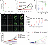 Figure 6
T cell ADCC selectively targets cells exceeding a defined antigen-density threshold. (A) FACS absolute quantification of antigen expression on HT29 tumor cells (n = 6). (B) Normalized HT29 cell confluence following incubation with transduced human T cells in combination with cetuximab or trastuzumab or sofituzumab (n = 4 for each combination). (C) Mean synapse length between AG2G-transduced T cells and tumor cells in the presence of relevant antibody (cetuximab), irrelevant antibody (trastuzumab), and no antibody (n = 18). (D) Representative SIM of HT29 tumor cells incubated overnight with AG2G-transduced T cells. Original magnification, ×630. (E) Integrated density of HER2 on SLBs during interaction with transduced T cells (3 donors, n = 8 cells each). (F) Normalized epithelial cell count following incubation with AG2G-, CAR HER2–, or sham-transduced T cells in combination with trastuzumab (n = 4). (G) Tumor size following treatment with AG2G and relevant antibody (anti–mouse HER2), irrelevant antibody (rituximab), or sham and relevant antibody (n = 4). Experiments were repeated at least 3 independent times. *P < 0.05; ***P < 0.0001; ****P < 0.00001 by ANOVA with Tukey’s correction for multiple comparisons. Error bars represent SEM.
Compared with donor-matched HER2 CAR T cells, which responded to HER2 densities as low as 10 molecules/μm2, AG2G T cells displayed a substantially higher activation threshold (Figure 6E and Supplemental Figure 3I). Consequently, HER2 CAR T cells efficiently killed both normal epithelial cells and HER2-overexpressing tumor cells, whereas AG2G T cells selectively killed tumor cells while sparing normal cells (Figure 6F, Supplemental Figure 3J, and Supplemental Videos 1–6). In vivo, AG2G T cells significantly reduced established HER2-overexpressing tumors only when administered with a tumor-binding antibody and caused no detectable inflammation in major organs (Figure 6G). These findings demonstrate that FcγRI-mediated T cell activation is strongly dependent on antigen density and can discriminate between malignant and normal tissues expressing the same antigen.
Soluble IgG does not activate AG2G T cells. We next compared AG2G with a conventional FcR-based CAR containing the FcγRI extracellular domain linked to CD28 and CD3ζ signaling domains. Unlike AG2G, the FcR-based CAR induced cytotoxicity and cytokine production in response to both tumor-bound and irrelevant soluble antibodies (Figure 7, A–C, Supplemental Figure 4A, and Supplemental Videos 7 and 8). In contrast, AG2G T cells remained inactive in the presence of soluble trastuzumab unless antibody concentrations exceeded approximately 1.375 mg/mL (Figure 7D and Supplemental Figure 4, B and C). Fluorescence analysis demonstrated rapid removal of surface-bound IgG from AG2G T cells, with clearance within approximately 2 hours. High-resolution imaging showed that soluble IgG was internalized and degraded in endosomal compartments (Figure 7, E and F, Supplemental Figure 4D, and Supplemental Videos 9 and 10). Thus, soluble antibody is efficiently cleared without inducing productive FcγRI signaling.
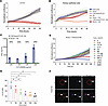 Figure 7
Soluble IgG does not activate AG2G T cells. (A) Normalized H1975 cancer cell confluence following incubation with AG2G- or CAR-based FcγR-transduced T cells in combination with trastuzumab (n = 4). (B) Normalized kidney epithelial cell confluence following incubation with AG2G- or CAR-based FcγR-transduced T cells in combination with trastuzumab (n = 4). (C) Mean levels of IFN-γ in the supernatants of AG2G- and CAR-based FcγR-transduced T cells incubated with various concentrations of free antibody (n = 3). (D) Normalized HT29 cell confluence following incubation with AG2G-transduced T cells in combination with various concentrations of trastuzumab (n = 4) (E) Mean percentages of stained transduced human T cells following wash of FITC-IgG1 (n = 3). (F) Representative SIM of AG2G-transduced human T cells following addition of FITC-IgG1. Scale bars: 1 μm. Original magnification, ×630. Arrowheads indicate positively stained organelles. Experiments were repeated at least 3 independent times. *P < 0.05; ****P < 0.00001 by ANOVA with Tukey’s correction for multiple comparisons. Error bars represent SEM.
Soluble IgG does not interfere with FcγRI-mediated tumor-cell killing. To define the mechanism underlying this selectivity, we compared FcγRI signaling following exposure to immobilized versus soluble IgG. Immobilized trastuzumab induced FcγRI and phosphorylated-Syk clustering, and cortical actin accumulation, whereas soluble IgG induced none of these responses (Figure 8, A and B, and Supplemental Figure 4E). These findings indicate that productive AG2G signaling requires spatially restricted receptor clustering at an antibody-coated target–T cell interface.
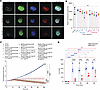 Figure 8
Soluble IgG does not interfere with FcγRI-mediated tumor-cell killing. (A) Representative SIM of AG2G-transduced T cells incubated for 20 minutes with trastuzumab. Scale bars: 1 μm. Original magnification, ×630. (B) Mean percentages of p-Syk in AG2G-transduced T cells incubated for 20 minutes with trastuzumab. (C) Mean counts of HER2+ HT29 tumor cells following incubation with AG2G-transduced human T cells and in the presence of trastuzumab and IVIg (n = 4). (D) Mean percentages of stained transduced human T cells blocked with 10 mg/mL trastuzumab and incubated with FITC-IgG1 (n = 3). Experiments were repeated at least 3 independent times. *P < 0.05; **P < 0.001; ***P < 0.0001; ****P < 0.00001 by ANOVA with Tukey’s correction for multiple comparisons. Error bars represent SEM.
Importantly, high concentrations of irrelevant human IgG (IVIg) did not substantially inhibit AG2G-mediated killing of HER2+ tumor cells in the presence of trastuzumab (Figure 8C). Although soluble IgG rapidly occupied most FcγRI molecules, subsequent antibody exchange demonstrated continuous receptor turnover (Figure 8D, Supplemental Figure 4F, and Supplemental Videos 11 and 12). Thus, soluble IgG neither activates AG2G T cells nor prevents their response to antibody-coated tumor cells.
Repeated AG2G T cell administration promotes tumor eradication and effector memory T cell formation. Next we tested the in vivo effect of AG2G therapy. A single infusion of 5 × 10⁶ AG2G T cells combined with trastuzumab controlled but did not eradicate established NCI-N87 gastric tumors in NSG mice. Yet, residual tumors retained HER2 expression, arguing against antigen loss as a mechanism of resistance (Figure 9A). AG2G T cells preferentially accumulated in tumors, peaking approximately 14 days after transfer and declining substantially by day 30 (Figure 9B). Weekly trastuzumab injection combined with 2 infusions of 5 × 10⁶ AG2G T cells, administered 21 days apart, resulted in complete tumor eradication, whereas a single infusion produced only partial control (Figure 9, C–E, and Supplemental Figure 5, A–D). Most persisting AG2G T cells in the bone marrow and spleen of tumor-free mice displayed an effector memory phenotype (Figure 9, F and G).
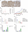 Figure 9
Repeated AG2G T cell administration promotes tumor eradication and effector memory formation. (A) Representative histology of HER2 in NCI-N87 tumor core from NSG mice treated with AG2G- or sham-transduced T cells in combination with trastuzumab. Scale bar: 100 μm. (B) AG2G-transduced T cell relative quantification (RQ) in various organs of NCI-N87 tumor–bearing NSG mice (n = 6). (C) NCI-N87 tumor volume in NSG mice following treatment with AG2G- and sham-transduced T cells in combination with trastuzumab (n = 6). (D) NCI-N87 tumor necrosis score in NSG mice following treatment with AG2G- and sham-transduced T cells in combination with trastuzumab (n = 6). (E) NCI-N87 tumor weight in NSG mice following treatment with AG2G- and sham-transduced T cells in combination with trastuzumab (n = 6). (F and G) Mean percentages of central memory (CM) and effector memory T cells from bone marrow (F) and spleen (G) in NSG mice challenged with NCI-N87 tumors following treatment with AG2G-transduced T cells in combination with trastuzumab (n = 4). Experiments were repeated at least 3 independent times. Statistical significance assessed by ANOVA with Tukey’s correction for multiple comparisons. Error bars represent SEM.
AG2G T cells produce memory subsets in vivo and provide long-term protection. To determine whether tumor eradication reflected cumulative T cell dose, mice received escalating numbers of AG2G T cells. Doses of 1 × 10⁷ or 2 × 10⁷ cells induced complete tumor regression, whereas 1 × 10⁶ cells had limited activity (Figure 10A). Persisting cells again predominantly displayed an effector memory phenotype (Figure 10, B and C).
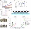 Figure 10
AG2G T cells produce memory subsets in vivo and provide long-term protection. (A) NCI-N87 tumor volume in NSG mice following treatment with 250 μg trastuzumab and i.v. injection of AG2G-transduced T cells (n = 6). (B and C) Representative FACS analysis (B) and mean percentages (C) of memory T cells in mice challenged with NCI-N87 cells on day 60 (n = 6). (D) Illustration of experimental design. (E) Image of NCI-N87 tumor after tumor rechallenge with and without treatment with trastuzumab (n = 4). (F) NCI-N87 tumor volume in NSG mice following treatment with AG2G-transduced T cells and 250 μg trastuzumab. (n = 6). Experiments were repeated at least 3 independent times. Statistical significance assessed by ANOVA with Tukey’s correction for multiple comparisons. Error bars represent SEM.
To test the functionality of this memory subset, mice were rechallenged with NCI-N87 cells 2 months after tumor eradication. Following trastuzumab administration, all previously cured mice rapidly eliminated the rechallenged tumors, whereas untreated mice developed progressive disease (Figure 10, D–F). These results indicate that AG2G therapy can generate persistent effector memory T cells capable of antibody-dependent tumor control upon rechallenge.
AG2G T cells display enhanced tumor infiltration compared with CAR T cells. AG2G T cells’ ability to eradicate tumors suggested overcoming the infiltration limitation posed by solid tumors. To test that, we compared AG2G T cell tumor-infiltrating capacities to that of HER2 CAR– and sham-transfected T cells, all from the same donors. In all 3 tested donors, the levels of infiltrating AG2G-transduced T cells were significantly higher compared with CAR- and sham-transduced T cells, composing up to 7% of the total lymphocytes, and AG2G T cells could be found in the tumor core (Figure 11, A and B).
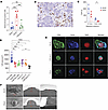 Figure 11
AG2G T cells display enhanced tumor infiltration compared with CAR T cells. (A) Mean percentage of transduced T cells out of non-tumor cells in NCI-N87 tumor core taken from NSG mice after treatment (n = 6) (B) Representative histology of core tumor from NSG mice treated with AG2G-transduced T cells in combination with trastuzumab. (C) Mean migration distance of transduced T cells on Matrigel (n = 3). (D) Mean cell area of transduced T cells on glass and Matrigel (n = 18). (E) Representative SIM of AG2G-, HER2 CAR–, and sham-transduced T cells on Matrigel. (F) Representative SEM of AG2G- and sham-transduced T cells on Matrigel. Experiments were repeated at least 3 independent times. Scale bars: 50 μm (B), 1 μm (E) (original magnification, ×630), and 7 μm (F). Statistical significance was calculated using ANOVA with Tukey’s correction for multiple comparisons. For cumulative distribution function statistical analysis, a Kolmogorov-Smirnov test was used. For interaction classification analysis, a Mann-Whitney U test was used. *P < 0.05; **P < 0.001; ***P < 0.0001; ****P < 0.00001. Error bars represent SEM.
Since there are no relevant suppressive cues from the tumor microenvironment of NSG mice, the main factor limiting T cell infiltration is the basement membrane that forms a mechanical barrier. Hence, we coated migration slides with artificial basement membrane (Matrigel) and recorded T cell motility and migration distance towards CCL5 by confocal microscopy. Consistent with their in vivo infiltration capacities, AG2G-transduced T cells moved rapidly on the slides and for a longer distance compared with CAR- and sham-transfected T cells from matched donors (Figure 11C and Supplemental Videos 13 and 14). Interestingly, we noticed that when AG2G-transduced T cells are placed on Matrigel, they acquire polarity characterized by actin polymerization at the moving front and a spreading morphology (Figure 11, D and E, and Supplemental Figure 6A). Analysis by electron microscopy further corroborated the flattened spreading morphology that AG2G T cells acquire once placed on Matrigel (Figure 11F).
Reduced basement membrane adhesion promotes AG2G T cell infiltration. Reanalysis of tumor-infiltrating T cell datasets identified a distinct integrin profile associated with FcγRIII+CD8+ T cells, characterized by increased CD11b, CD11a, and CD11c and reduced CD29 and CD49a, the latter being prominent mediators of collagen IV interactions (Figure 12A and Supplemental Figure 6B). AG2G T cells similarly displayed reduced CD49a and increased CD11b expression (Figure 12, B and C).
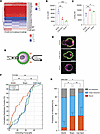 Figure 12
Reduced basement membrane adhesion promotes AG2G T cell infiltration. (A) Pearson correlation scores between the mean log-normalized expression of various integrin and FcR genes across T cell clusters from human single-cell published data (n = 316). (B) Mean fluorescence intensity (MFI) of CD11b on transduced T cells (n = 3). (C) MFI of CD49a on transduced T cells (n = 3). (D) Force spectroscopy setup illustration. Pasp, aspiration pressure. (E) Representative fluorescence images of AG2G, CAR HER2, and sham cells aspirated with a micropipette (green, CD8; red, CD64\HER2; magenta, F-actin). Scale bars: 2 μm. (F) Cumulative distribution function (CDF) of transduced T cell interaction with Matrigel-coated beads. (G) Interaction classification for transduced T cells. The data represent 5 independent experiments, with up to 3 measured interactions per cell (AG2G – 20 cells, n = 59; CAR HER2 – 21 cells, n = 61; sham – 22 cells, n = 62). Boxes represent the fraction of each interaction type, and whiskers indicate 5%–95% CI, shown only for “bound.” Supplemental Figure 7J shows CI’s for all interaction types. Experiments were repeated at least 3 independent times. Statistical significance was calculated using ANOVA with Tukey’s correction for multiple comparisons. For CDF statistical analysis, a Kolmogorov-Smirnov (K-S) test was used. For interaction classification analysis, a Mann-Whitney U (MWU) test was used.*P < 0.05; **P < 0.001; ****P < 0.00001. Error bars represent SEM.
To determine whether this profile alters T cell–ECM interactions, we developed an optical tweezers/micropipette force spectroscopy assay using Matrigel-coated beads (Figure 12D and Supplemental Figure 6, C–G). Integrin-dependent binding was confirmed by EDTA-mediated reduction of unbinding forces (Supplemental Figure 6, H and I). Although AG2G, sham, and HER2 CAR T cells exhibited similar probabilities of forming initial interactions with Matrigel, AG2G T cells showed substantially fewer strong interactions (~7% vs. ~35% for sham and CAR T cells). Consistent with this finding, most AG2G interactions were weak, with 52% occurring between 10 and 20 pN, whereas sham and CAR T cells displayed distributions shifted toward stronger interactions (>28 pN) (Figure 12, E–G, Supplemental Figure 6J, and Supplemental Videos 15–18). Jointly, our data suggest that the integrin profile expressed on AG2G-transduced T cells results in lower interactions with the basement membrane and subsequently improves their capacity to infiltrate cancers.


