Issue published October 22, 2026 Previous issue

- Volume 11, Issue 20
On the cover:
Transcriptomic profiling of immune cells in malignant pleural effusions identifies macrophage reprogramming associated with survival
Khatri et al. report that macrophage reprogramming is associated with survival outcomes in lung cancer metastasis. The cover image shows tumor-associated macrophages (CD163, red) surrounding an invading lung cancer metastatic lesion (pan-cytokeratin, green). Credit: Aaditya Khatri.
Khatri et al. report that macrophage reprogramming is associated with survival outcomes in lung cancer metastasis. The cover image shows tumor-associated macrophages (CD163, red) surrounding an invading lung cancer metastatic lesion (pan-cytokeratin, green). Credit: Aaditya Khatri.
Go to section:
-
Research Letter
×
Abstract
Authors
Jianfei Ji, Wenjing You, Xiaoli Sun, Peng Zhao

-
Research Articles
×
Abstract
Ubiquitination is an important posttranslational modification associated with essential cellular processes. Here we show that deletion of E3 ubiquitin ligase HECTD3 — germline or in hematopoietic compartment, including in Cd11c+ cells — causes severe disease and increased production of proinflammatory cytokines in a murine model of ulcerative colitis (UC). HECTD3 mRNA levels were found reduced in colonic tissues of patients with UC, which highlights a potential role for HECTD3 in regulating inflammatory responses in UC. We identified MyD88, a central adaptor in the TLR/IL-1R signaling and inflammatory response, as a target for HECTD3 ubiquitination. We demonstrate that HECTD3 ubiquitinates MyD88 through K27-linked Poly-Ub chains in a nondegradative manner. HECTD3-KO GM-CSF–bone marrow–derived cells treated with the TLR4 ligand LPS produced more proinflammatory cytokines, showed elevated phosphorylation of NF-κB and IRAK4, as well as increased association of MyD88 with IRAK4, demonstrating that HECTD3 controls MyD88/IRAK4/NF-κB axis. Inhibition of MyD88 activity rescued colitis severity in the HECTD3-KO mice, including the elevated proinflammatory cytokine production. Thus, our results establish MyD88 as a target for HECTD3 nondegradative polyubiquitination and restriction of innate immune colonic inflammation.
Authors
Shamima Islam, Shahnewaj Mannan, Ashley Zuniga, Upasana Parthasarathy, Valeriu B. Cismasiu, Leonardo Silvane, Sayan Chakraborty, Divya Priyanka Talada, Raghwendra Pratap Singh, Tomas Zelenka, Amreen Naveen, Sephra Chyanne Vickers, Michael G.M. Grant, Jonathan J. Cho, Theodore Drashansky, Alexander J. Kwiatkowski, Xintong Liu, Ross Tomaino, Olga A. Guryanova, Mariola J. Ferraro, Sang Yong Kim, Christian Jobin, Benjamin G. Keselowsky, Hongmin Li, Paulo C. Rodriguez, Martina Molgora, Amer A. Beg, Timothy I. Shaw, Zheng Ruan, Lixin Wan, Dorina Avram
×Abstract
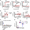
Tregs play an essential role in immune tolerance, and Treg-promoting therapies are in development for the treatment of many inflammatory disorders. IL-2–based therapies increase Treg frequency, but little is known about impacts on Treg heterogeneity and function. We extended analyses of an IL-2 mutein (MK-6194) single ascending dose trial in healthy human participants by comprehensively defining Treg subsets and gene expression changes in vitro and in vivo. We found highly specific and dose-dependent activation and expansion of Tregs in clinical and preclinical studies. Following a single subcutaneous dose in humans, thymus-derived Tregs were selectively activated and expanded, while peripherally induced Tregs were unaffected. Expanded Tregs had increased expression of genes and proteins consistent with activation, suppressor function, and homing to nonlymphoid tissue, as well as increased transendocytotic activity, as measured by CTLA-4–dependent capture of CD80 and CD86 from non-Tregs. These results shed light onto underlying mechanisms by which Treg-targeted therapy may promote immune tolerance.
Authors
Laura A. Cooney, Mitch Fahning, Liliane Khoryati, Anna Kus, Sheila Scheiding, Lori Blanchfield, Matt Lawrance, Basilin Benson, Kristina M. Harris, Gretchen A. Baltus, Shiuli Agarwal, Richard Wnek, Johannes F. Scheid, Kiki Cunningham-Bussel, Nancy D. Kim, S. Aubrey Stoch, Jyothsna Visweswaraiah, Nathan Higginson-Scott, Katalin Kis-Toth, Joanne L. Viney, Kevin L. Otipoby, Erik Sampson, Bridget Larkin, Daniel J. Campbell, S. Alice Long
×Abstract
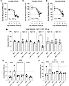
Genome-targeting strategies to address NaV1.7-mediated signaling in nociceptive afferents are of great clinical interest given its high selectivity and persistent analgesic outcomes. In the present work, we analyze concentration-dependent effects of suppressing primary afferent NaV1.7 using the intrathecal delivery of an antisense oligonucleotide (ASO) on pain behaviors and demonstrate that NaV1.7 ASOs are likely to modulate pain traffic by blocking afferent signaling at the dorsal root ganglion (DRG) level. Intrathecal NaV1.7 ASOs induced a concentration-dependent gradient of knockdown in DRGs (lumbar to cervical) of Scn9a mRNA and ATTO488-ProTx-II binding in small DRG neurons, and a suppression of pain behaviors initiated by mechanical compression, inflammation, and the NaV1.7 agonist OD-1. The rostro-caudal reduction in mRNA correlated with ASO distribution, with implications for dose-related analgesic profiles. Marked reduction in DRG NaV1.7 binding was seen after intrathecal delivery of an ASO optimal dose, with high selectivity for small DRG neurons. Changes in sciatic nerve NaV1.7 binding were not detected, indicating a pain traffic modulation likely happening due to a block of afferent signaling at the DRG level.
Authors
Kaue Malange, Julia Borges Paes Lemes, Briana Noble, Yuvicza Anchondo, Carlos E. Morado-Urbina, Saee Jadhav, Saurabh Yadav, Jingrui Ma, Vineet Augustine, Sara A. Dochnal, Chiraag Kambalimath, Marie Duong, Pedro Alvarez, Bethany Fitzsimmons, Curt Mazur, Holly B. Kordasiewicz, Kim Dore, Hien T. Zhao, Tony Yaksh
×Abstract
DNA ligase IV (LIGIV or LIG4) is essential for DNA double-strand break (DSB) repair. Hypomorphic LIG4 variants cause LIG4 syndrome, characterized by growth disturbance, increased radiosensitivity, predisposition to malignancies, adaptive immunodeficiency, and inflammatory conditions. Most of these manifestations are recapitulated in hypomorphic Lig4 mutant mice. However, no model mice with defective DSB repair have consistently exhibited inflammation. Here, we generated mutant mice carrying the LIG4 missense variant, p.W447C, found in a patient with LIG4 syndrome. Lig4W447C/W447C mice showed functional defects of LIGIV and manifested growth retardation, increased radiosensitivity, and life-threatening intestinal inflammation under severe adaptive immunodeficiency. The inflammation was dependent on lymphocytes and characterized by marked infiltration of Th1 cells and macrophages, along with elevated expression of IFN-γ–inducible genes. When Ifng was deleted, Th2 and Th17 instead of Th1 cells drove the inflammation. Single-cell RNA-seq analyses with TCR repertoire revealed that T cells from Lig4W447C/W447C mice preferentially used proximal Vα and Jα segments in V regions of TCRα chains and exhibited expansion of several clonotypes, a substantial portion of which were CD4+ T cells expressing IFN-γ. Thus, our hypomorphic Lig4 mutant mice represent a unique model for studying Th1-skewed intestinal inflammation under severe adaptive immunodeficiency.
Authors
Yusuke Yamashita, Hideki Kosako, Takashi Kato, Izumi Sasaki, Sadahiro Iwabuchi, Yuri Fukuda-Ohta, Tadashi Okamura, Misato Tane, Shotaro Tabata, Kazutaka Nakashima, Ken Tanaka, Kazunori Shiraishi, Yuki Uchihara, Daisuke Okuzaki, Kyoichi Isono, Atsushi Shibata, Tsunehiro Mizushima, Hiroaki Hemmi, Nobuo Kanazawa, Seiji Kodama, Hiroaki Miyoshi, Koichi Ohshima, Shinichi Hashimoto, Yoshio Fujitani, Takashi Sonoki, Shinobu Tamura, Tsuneyasu Kaisho
×Abstract
Clinical immunity to malaria develops after repeated malaria episodes. In this process, the inflammatory response is modulated to respond less vigorously upon reinfection. Monocytes are a major source of proinflammatory mediators during blood-stage infection and are known to adapt to repeated pathogen exposure. Here, we investigated the effect of previous malaria exposure on monocytes during blood-stage malaria by comparing the response in previously exposed and primary infected individuals. We observed reduced levels of several proinflammatory chemokines in previously exposed individuals, linked to changes in monocytes. Similarly, BAFF levels were lower in these individuals and associated with modulation of monocyte and dendritic cells. This affected the BAFF/BAFF-R axis, crucial for B cell responses, correlating with increasing parasite-specific antibody levels. Collectively, we present insights into how previous malaria exposure shapes monocyte responses during acute malaria and how these, in turn, correlate with modulation of the B cell compartment and humoral immune response.
Authors
Maximilian Julius Lautenbach, Pengjun Xi, Linn Kleberg, Alan-Dine Courey-Ghaouzi, Maia Serene Gower, Carolina Sousa Silva, Felicia Chammas, Anna Färnert, Christopher Sundling
×Abstract
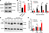
Autophagy is a critical host defense mechanism that restricts intracellular pathogens such as Mycobacterium tuberculosis. A key step in this process is the ubiquitination of M. tuberculosis or M. tuberculosis–associated structures. The E3 ligase Smad-specific E3 ubiquitin protein ligase 1 (SMURF1) catalyzes K48-linked ubiquitination, promoting bacterial clearance. However, the function of its homolog, SMURF2, in host defense remains undefined. Here, we demonstrate that Smurf2 deletion in murine macrophages increases SMURF1 levels, enhances LC3B lipidation, augments K48 ubiquitination of M. tuberculosis–associated structures, and reduces intracellular M. tuberculosis replication. These effects are reversed by Smurf1 deletion, supporting a role for SMURF1 in SMURF2-dependent control of M. tuberculosis. Mice with myeloid-specific Smurf2 deletion exhibit modestly prolonged survival after aerosol M. tuberculosis infection. In human macrophages, SMURF2 knockdown or its pharmacological inhibition with the HECT E3-ligase inhibitor Heclin reduces M. tuberculosis replication. Together, our findings identify SMURF2 as a negative regulator of macrophage control of M. tuberculosis and support further investigation of SMURF2 as a potential target for host-directed therapy in tuberculosis.
Authors
Priscila C. Campos, Kathryn C. Rahlwes, Victoria A. Ektnitphong, Beatriz R.S. Dias, Kubra F. Naqvi, Samuel Alvarez-Arguedas, Michael U. Shiloh
×Abstract
The adult mammalian heart has a limited ability to regenerate lost myocardium following myocardial infarction (MI), largely due to the poor proliferative capacity of cardiomyocytes (CMs). Dual-specificity tyrosine phosphorylation–regulated kinase 1A (DYRK1A) is a known regulator of cell quiescence, though the mechanisms underlying its function remain unclear. Previous studies have shown that pharmacological inhibition of DYRK1A using harmine induces CM cell cycle reentry after ischemia/reperfusion (I/R) MI. Here, we developed a computational network model of DYRK1A-mediated regulation of the cell cycle, which predicts how DYRK1A inhibition promotes CM reentry. To validate these predictions, we tested selective DYRK1A inhibitors and observed robust induction of cell cycle activity in neonatal rat cardiomyocytes (NRCMs). Integrating our network model with bulk RNA sequencing data from DYRK1A inhibitor–treated NRCMs, we identified E2F1 as a key transcriptional driver of cell cycle gene expression. Finally, we demonstrate that both pharmacological and post-developmental inhibition of DYRK1A enhances heart function and increases CM cycling following I/R MI. Our findings suggest that functional recovery induced by small-molecule inhibitors of DYRK1A is mediated by the induction of cycling CMs.
Authors
Bryce C. Murillo, Alexander Young, Kaitlyn L. Wintruba, Alexander J. Eichert, Klara Siejda, Dennon Hoernig, Leigh A. Bradley, Bryana N. Harris, Catherine Zhao, Michelle Wu, Emmanuel Deau, Mattias F. Lindberg, Laurent Meijer, Jeffrey J. Saucerman, Matthew J. Wolf
×Abstract
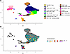
Despite advances in treatment approaches for lung cancer, the morbidity and survival of patients with lung cancer with malignant pleural effusions (MPE) remain poor. This is in part due to gaps in understanding the role of immune cells in the pleural fluid microenvironment. We performed single-cell analysis with flow cytometry validation of CD45+ cells in 8 MPE and 5 benign pleural fluid effusion (BPE) specimens to identify changes in the transcriptomic landscape of immune cells across disease states. We found upregulation of proinflammatory signaling pathways, including IFN and TNF signaling, in T cells, B cells, and macrophages in benign compared with malignant pleural effusions. Proinflammatory HLA-DR+ macrophages were associated with good survival outcomes while protumorigenic HLA-DR– macrophages with upregulation of angiogenesis, TGF-β, and fibronectin signaling were associated with poor survival outcomes in patients with MPE. We also validated these findings with macrophage cell surface expression markers using flow cytometry in 14 MPE and 7 BPE specimens. Finally, we performed multiplex cytokine analysis, which showed enrichment of the type 3 inflammatory cytokine, IL-17A, in MPE as a putative mechanism for macrophage reprogramming. These data provide a rich resource for interrogating the immune cell types and states present across the spectrum of pleural disease. They offer not only prognostic value for patient outcomes at the time of pleural fluid collection but also insights into novel immunotherapy targets.
Authors
Aaditya Khatri, Huimin Wang, Zhicheng Ji, Prekshaben Patel, Smita K. Nair, Javid P. Mohammed, Beth H. Shaz, Andrew B. Nixon, Scott M. Palmer, Kamran Mahmood
×Abstract
Ebola virus disease is a severe viral hemorrhagic illness with high maternal and perinatal mortality. Although the replication-competent vesicular stomatitis virus–based Ebola vaccine (VSV-EBOV) has been deployed successfully in outbreak settings, pregnant women were excluded from clinical trials, leaving critical safety gaps. To address this, we developed a murine model to assess pregnancy outcomes following VSV-EBOV exposure at different gestational stages. Pregnant C57BL/6 mice vaccinated at embryonic day 7 (E7), E10, or E15 mounted robust anti–Ebola virus (anti-EBOV) antibody responses comparable to those of non-pregnant controls and efficiently cleared the vaccine virus without evidence of vertical transmission. However, peri-implantation vaccination modestly increased fetal resorption and early postnatal mortality, accompanied by placental pathology characterized by vascular remodeling defects and inflammation. Later gestational vaccination had minimal impact on survival or offspring development. Mechanistic studies revealed that these adverse effects were mediated by type I interferon signaling rather than direct viral infection, as IFN-β–deficient mice exhibited normal pregnancy outcomes and blocking interferon receptor activity ameliorated placental pathology despite higher viral loads. To our knowledge, these findings represent the first comprehensive preclinical safety evaluation of VSV-EBOV vaccination during pregnancy and identify a critical window of vulnerability early in gestation. More broadly, this work underscores the central role of interferons in shaping placental immune responses and provides an experimental framework to guide evaluation of live-virus vaccines in pregnancy.
Authors
Aaron P. Lewkowicz, Yiru Wang, Mirian I. Mendoza, Kaliroi Engel, Logan Kelley-Baker, Iara Rattner, Amber Lopez, Ian L. McWilliams, Derek D.C. Ireland, Mohanraj Manangeeswaran, Ha-Na Lee, Daniela Verthelyi, Sandra M. Blois
×Abstract
TGF-β is a central driver of kidney fibrosis, a common pathological hallmark of chronic kidney disease (CKD). Initiation of TGF-β signaling requires not only its synthesis but also the conversion of latent TGF-β to its bioactive form. However, the mechanisms governing TGF-β activation in the kidney and their contribution to kidney fibrosis remain poorly understood. Glycoprotein A repetitions predominant (GARP) anchors latent TGF-β on the cell surface and facilitates its bioactive release. Here, we show that GARP-mediated TGF-β activation promotes kidney fibrosis. GARP was upregulated in both human and mouse CKD kidneys, predominantly in fibroblasts, and was induced by TNF in an NF-κB–dependent fashion. In multiple mouse models of kidney fibrosis, either global or fibroblast-specific deletion of GARP markedly reduced fibrosis. Mechanistically, GARP enables sustained production of active TGF-β, thereby amplifying fibroblast stimulation. Deletion of GARP in kidney fibroblasts lowered active TGF-β levels and attenuated fibroblast activation, whereas GARP overexpression enhanced TGF-β signaling. Notably, tamoxifen-induced deletion of GARP after fibrosis onset attenuated kidney fibrosis. Together, our findings identify GARP-mediated release of active TGF-β as a critical step in sustaining fibroblast activation during kidney fibrosis and highlight GARP as a promising therapeutic target for CKD.
Authors
Yintong Chen, Weiwei Xu, Jieli Yu, Pei Deng, Nianping Liu, Yinyin Li, Hui Zhou, Hong Zhou, Jianchuan Wang, Bo Zhao, Florian Winau, Fan Fan Hou, Yu Hu
×Abstract
Identifying and monitoring autoreactive T cells that drive β cell destruction remains a major obstacle to developing effective immunotherapies for type 1 diabetes (T1D). These cells are extremely rare in peripheral blood and cannot be accessed directly from the pancreas. We used intradermal injection of glutamic acid decarboxylase (GAD)-alum to recruit GAD-specific T cells to accessible sites in the skin and skin-draining lymph nodes (LNs), sampled by skin suction blisters and ultrasound-guided LN aspiration. Peripheral blood samples obtained before GAD injection were restimulated with GAD in vitro to detect reactive CD4+ T cells. Single-cell RNA sequencing (scRNA-seq) followed by re-expression of selected T cell receptors (TCRs) confirmed antigen specificity. Up to 70% of T cells at the skin injection site were clonally expanded and 4 of 14 (28%) re-expressed TCRs were GAD reactive. In LNs, 1 of 14 (4%) clonally expanded TCRs was GAD reactive, representing approximately 0.08% of all T cells. GAD-reactive cells across compartments displayed Th1- and Th17-associated transcription signatures. These results demonstrate that intradermal autoantigen challenge and scRNA-seq enable direct identification and molecular profiling of autoreactive T cells in vivo. This minimally invasive approach provides a powerful platform for tracking antigen-specific T cells to monitor disease activity and evaluate immune interventions in T1D.
Authors
Stephanie J. Hanna, Emma J.S. Robinson, Terri C. Thayer, Maki Nakayama, Laurie Landry, Robert Andrews, Garry Dolton, Joanne Davies, Evangelia Williams, James A. Pearson, Andrew K. Sewell, Parth Narendran, David C. Wraith, Alexandra Howell, Philippa Young, Mary Hart, Anton Lindqvist, F. Susan Wong, Tim I.M. Tree, Colin M. Dayan, Danijela Tatovic
×Abstract
Inflammatory cytokines reprogram keratinocyte metabolism, but the metabolic pathways that couple immune signals to pathological epidermal growth remain incompletely defined. Here, we identify glutaminase 1–mediated (GLS1-mediated) glutaminolysis as a metabolic program preferentially induced in keratinocytes under type 3 inflammatory conditions. Integrated transcriptomic, metabolomic, genetic, and functional analyses showed that IL-17A induced GLS1 expression and glutaminolysis in keratinocytes. Keratinocyte-specific Gls1 deletion reduced the intracellular availability of arginine, proline, and methionine, impaired amino acid–dependent mTORC1 activation, disrupted redox homeostasis, and limited keratinocyte proliferation. Amino acid or antioxidant supplementation partially rescued these defects, whereas rapamycin blocked the amino acid–mediated proliferative rescue. Gls1 deletion did not impair steady-state skin development or homeostasis and did not alter MC903-induced type 2 dermatitis, but it delayed wound re-epithelialization and attenuated imiquimod-induced psoriasiform inflammation. Loss of keratinocyte GLS1 also reduced epidermal chemokine expression and the accumulation of neutrophils and IL-17A–producing γδ T cells, revealing a role for glutaminolysis in amplifying epithelial-immune crosstalk. These findings define GLS1-mediated glutaminolysis as a context-specific metabolic checkpoint linking type 3 inflammation to keratinocyte proliferation and cutaneous immune amplification, and support locally or temporally controlled GLS1 inhibition as a potential therapeutic strategy for psoriasis.
Authors
Yongfei Hu, Hai Yu, Kaiyu Liang, Liyan Yuan, Wenjun Zhang, Zhe Zhuang, Suyun Ji, Xichun Xia
×Endothelial cell cycle inhibition enables blood vessel maturation to normalize the tumor vasculature
Abstract
Dysfunctional tumor vessels promote disease progression, whereas improved function enhances therapeutic delivery. However, current approaches to normalize tumor vasculature have limited efficacy. In vascular malformations, vessels are similarly dysfunctional, with endothelial cell (EC) hyperproliferation impairing arterial-venous specification. These defects are corrected with palbociclib, a cyclin-dependent kinase 4/6 inhibitor (CDK4/6i) that has beneficial effects on tumor and immune cells, but the effects on tumor vasculature are not well characterized. In our studies, murine mammary tumor ECs (TECs) exhibited disrupted cell cycle and specification, and CDK4/6i promoted TEC cycle control, enabling improved tumor vascular function. To investigate transcriptomic changes, we performed single-cell RNA sequencing (scRNA-seq) of treated and untreated tumors, and healthy tissues. CDK4/6i-mediated TEC cycle arrest promoted arterial-venous specification, cellular junctions, and pericyte association, and suppressed glycolytic and immunosuppressive gene expression. These effects were associated with increased vessel perfusion, decreased tumor hypoxia, and a more favorable immune landscape with immunotherapy. In scRNA-seq datasets from patients treated long-term with CDK4/6i, TECs exhibited similar transcriptomic changes associated with arterial-venous specification, pericyte recruitment, and immune signaling. Thus, in contrast to current strategies, CDK4/6i-mediated vascular changes may be maintained with continued treatment, highlighting the relevance of modulating TEC cycle to improve vessel maturation/function.
Authors
Shelby R. Cain, Gael Genet, Nafiisha Genet, Jordon W. Aragon, Madeline G. Jackson, Victoria M. Milosek, Mark R. Schwartz, Umadevi Paila, Aleksandra Cwiek, Zaneta Markowska, Nicholas W. Chavkin, Richard J. Price, Andrew C. Dudley, Karen K. Hirschi
×Abstract
Cell metabolic rewiring is associated with resistance to venetoclax-azacitidine (Ven-Aza) combination therapy and relapse in patients with acute myeloid leukemia (AML). Drug-resistant cells exhibit an enhanced reliance on oxidative phosphorylation (OXPHOS) for energy production. Therefore, impairing mitochondrial metabolism represents an exciting strategy to face this unmet clinical need. We recently demonstrated that the specific activation of the phosphatase PP2A-B56α enhances the proapoptotic efficacy of venetoclax in AML. Here, through leveraging unbiased multi-omics–based approaches and using both genetic and pharmacological tools, we define key roles for the tumor suppressor PP2A-B56α complex in OXPHOS regulation and treatment response in disease-relevant AML models. From a translational perspective, the specific stabilization of PP2A-B56α heterocomplex with the potentially novel PP2A molecular glue activator, RPT04402, reduces OXPHOS levels in treatment-resistant AML cells and improves treatment response in both Ven-Aza–sensitive and –resistant AML cell lines, primary cells, and in vivo models. Together, our work supports further research on targeted combination therapy approaches based on PP2A-B56α stabilization to counteract OXPHOS-related treatment resistance and improve AML responses in a patient population with historically poor outcomes.
Authors
Silvia Romero-Murillo, Irene Peris, Anna Maria Lucianò, Nerea Marcotegui, Carmen Vicente, Brian Tran, Kelsey Barrie, Caitlin M. O’Connor, Andrea Torres-López, Maria C. Mateos, Maria L. Cayuela, Victoriano Mulero, Joaquín Fernández-Irigoyen, Enrique Santamaría, Maria D. Odero, Goutham Narla
×Abstract
Purine metabolism dysregulation has been implicated in Alzheimer’s disease (AD), but its temporal dynamics and mechanistic links to core pathologies remain unclear. This translational study integrated a longitudinal preclinical AD mouse model and clinical cohort investigations to define the role of purine metabolism in AD progression. In APPswe/PSEN1dE9 (APP/PS1) mice, serum-untargeted metabolomics identified purine metabolism as the most notably altered pathway, with metabolite levels (e.g., adenine, adenosine) shifting dynamically across disease stages alongside increasing amyloid-β (Aβ) burden. In the pathology-defined clinical cohort, targeted analysis revealed that serum uric acid and allantoin were specifically depleted in the Aβ+ cognitively impaired (CI) group and correlated strongly with higher brain Aβ load, increased neuroinflammation, and poorer cognitive scores. Formal mediation analysis elucidated a plausible mechanistic cascade: the association of uric acid/allantoin with cognitive impairment was primarily mediated by Aβ pathology. Two-year longitudinal data further suggest that changes in specific purine metabolites might predict the rate of cognitive decline. Allantoin supplementation reduced Aβ pathology through promoting microglial phagocytosis of Aβ in APP/PS1 mice. Our findings identified purine metabolism as a key hub connecting Aβ pathology, neuroinflammation, and clinical progression in AD.
Authors
Xiaohang Qian, Zhongxuan Wang, Siyue Chen, Yang Xue, Yawen Yang, Peijing Cui, Miao Zhang, Huidong Tang
×Abstract
Bone metastases are a frequent complication of breast cancer, leading to reduced quality of life and increased morbidity. By intratibially injecting PyMT breast cancer cells in mice, we showed that the presence in the gut microbiome of segmented filamentous bacteria (SFB), a taxon that induces IL-17–producing Th17 cells and TNF-α (TNF)–producing T cells (TNF+ T cells), accelerated intraosseous tumor growth. SFB increased the activity of parathyroid hormone–related protein (PTHrP), a known driver of breast cancer bone metastasis produced by breast cancer cells. SFB promoted the PTHrP-induced expansion of intestinal Th17 cells and TNF+ T cells, their migration to bone marrow (BM) in tumor-bearing bones, and the BM expression of TNF, IL-17, and RANKL, cytokines that promote bone resorption. Demonstrating the functional relevance of IL-17 and TNF, silencing of either IL-17 or TNF blunted the growth of bone lesions in SFB+ mice. Thus, the abundance of SFB in the microbiome of mice and of SFB-like taxa in humans may determine the potency of PTHrP and the rate of growth of bone metastasis in breast cancer. Targeting the gut microbiota may represent a novel therapeutic strategy for breast cancer bone metastasis.
Authors
Nand K. Roy, Subhashis Pal, Hamid Y. Dar, Sanchiti Patil, M. Neale Weitzmann, Roberto Pacifici
×Abstract
Septins are RAS-like GTP-binding proteins involved in cytoskeletal organization and cellular division. We previously described 2 pediatric patients harboring SEPT6 germline mutations (GM1: SEPT6 c.1282T>C and GM2: SEPT6 c.1282T>A) presenting with congenital neutropenia associated with the development of myelodysplastic syndrome. In one patient, an additional somatic mutation (SM: SEPT6 c.43C>T) was detected at a low allelic fraction in the BM. Here, we describe the consequences of these mutations for hematopoiesis. Transgenic expression or gene editing of GM1 or GM2 in human CD34+ cells led to enlarged, multinucleated cells associated with accelerated exhaustion and premature senescence of hematopoietic stem and progenitor cells (HSPCs). Senescence defects were associated with the upregulation of mTOR phosphorylation, and pharmacologic inhibition of the mTOR pathway resulted in rescue of senescence. In parallel, HSPCs exhibited marked mitochondrial abnormalities, including increased expression of OPA-1, a key regulator of mitochondrial fusion, and enhanced oxygen consumption during mitochondrial respiration. Introduction of the SM in cis with GM1 mitigated all observed defects. Collectively, our findings demonstrate a gain-of-function mechanism underlying marrow failure in patients carrying germline SEPT6 mutations and suggest that acquisition of a secondary somatic SEPT6 variant may confer a compensatory effect within hematopoietic cells.
Authors
Safa F. Mohamad, Meaghan McGuinness, Yu Zhou, Xin Jiang, Gabriele Casirati, Adele Mucci, Sara Canovas Nunes, Akiko Shimamura, Pietro Genovese, David A. Williams
×Abstract
Activation of the mechanistic target of rapamycin (mTOR) complex1 (mTORC1) promotes muscle protein synthesis, mass, and function. Muscle mTORC1 can be activated by feeding and contraction. Here, muscle mTORC1 signaling, protein synthesis, mass, and function are characterized in a genetic mouse model that separates these 2 major modes of muscle mTORC1 regulation. AKT signaling is required for feeding-induced muscle mTORC1 signaling and protein synthesis, and mice expressing a mutant of tuberous sclerosis complex 2 (TSC2) that cannot be phosphorylated by AKT specifically in skeletal muscle (SkM-TSC2-5A) attenuate these effects of feeding. Despite this loss of postprandial protein synthesis, SkM-TSC2-5A mice have similar muscle and myofiber size compared with SkM-TSC2-WT mice. SkM-TSC2-5A mice maintain normal muscle mTORC1 activation in response to contraction and exhibit no differences in atrophy-related gene expression or ribosomal content. SkM-TSC2-5A mice exhibit improved maximal endurance capacity without changes in muscle contractile function. This phenotype occurs without alterations in muscle glycogen content or myofiber type, but it does coincide with a modest increase in muscle mitochondrial content. Therefore, AKT-mediated phosphorylation of TSC2 is required for postprandial mTORC1 activation and the induction of protein synthesis; however, these are dispensable for the development and maintenance of muscle mass in sedentary mice.
Authors
Samuel C. Lapp, Krystle C. Kalafut, Madi Y. Cissé, Khaled Tighanimine, Dean M. Rosenthal, Will Doxsey, Sheng Hui, Karen E. Inouye, Claire E. Morrow, Yann Cormerais, Brendan D. Manning