
Research ArticleImmunology
Open Access |  10.1172/jci.insight.203119
Tracking GAD-specific T cell expansions in type 1 diabetes by intradermal GAD-alum challenge
Stephanie J. Hanna,1,2 Emma J.S. Robinson,1 Terri C. Thayer,1,3 Maki Nakayama,4 Laurie Landry,4 Robert Andrews,1 Garry Dolton,1 Joanne Davies,1 Evangelia Williams,5 James A. Pearson,2 Andrew K. Sewell,1 Parth Narendran,2 David C. Wraith,2 Alexandra Howell,1 Philippa Young,6 Mary Hart,7 Anton Lindqvist,8 F. Susan Wong,1 Tim I.M. Tree,5 Colin M. Dayan,2 and Danijela Tatovic2
1Division of Infection and Immunity, Cardiff University School of Medicine, Cardiff, United Kingdom.
2Division of Immunology and Immunotherapy, University of Birmingham, Birmingham, United Kingdom.
3Center for Infectious Diseases and Immunology, Rochester General Hospital Research Institute, Rochester, New York, USA.
4Barbara Davis Center for Childhood Diabetes, University of Colorado, Denver, USA.
5Department of Immunobiology, School of Immunology & Microbial Sciences, King’s College London, London, United Kingdom.
6Breast Test Wales, Public Health Wales, Cardiff, United Kingdom.
7Department of Radiology, University Hospital of Wales, Cardiff, United Kingdom.
8Diamyd Medical AB, Stockholm, Sweden.
Address correspondence to: Colin M. Dayan, Department of Immunology and Immunotherapy, 4th Floor IBR, Wolfson Drive, Medical School, University of Birmingham, Edgbaston, Birmingham B15 2TT, United Kingdom. Email: c.m.dayan@bham.ac.uk.
Find articles by Hanna, S. in: PubMed | Google Scholar
1Division of Infection and Immunity, Cardiff University School of Medicine, Cardiff, United Kingdom.
2Division of Immunology and Immunotherapy, University of Birmingham, Birmingham, United Kingdom.
3Center for Infectious Diseases and Immunology, Rochester General Hospital Research Institute, Rochester, New York, USA.
4Barbara Davis Center for Childhood Diabetes, University of Colorado, Denver, USA.
5Department of Immunobiology, School of Immunology & Microbial Sciences, King’s College London, London, United Kingdom.
6Breast Test Wales, Public Health Wales, Cardiff, United Kingdom.
7Department of Radiology, University Hospital of Wales, Cardiff, United Kingdom.
8Diamyd Medical AB, Stockholm, Sweden.
Address correspondence to: Colin M. Dayan, Department of Immunology and Immunotherapy, 4th Floor IBR, Wolfson Drive, Medical School, University of Birmingham, Edgbaston, Birmingham B15 2TT, United Kingdom. Email: c.m.dayan@bham.ac.uk.
Find articles by Robinson, E. in: PubMed | Google Scholar
1Division of Infection and Immunity, Cardiff University School of Medicine, Cardiff, United Kingdom.
2Division of Immunology and Immunotherapy, University of Birmingham, Birmingham, United Kingdom.
3Center for Infectious Diseases and Immunology, Rochester General Hospital Research Institute, Rochester, New York, USA.
4Barbara Davis Center for Childhood Diabetes, University of Colorado, Denver, USA.
5Department of Immunobiology, School of Immunology & Microbial Sciences, King’s College London, London, United Kingdom.
6Breast Test Wales, Public Health Wales, Cardiff, United Kingdom.
7Department of Radiology, University Hospital of Wales, Cardiff, United Kingdom.
8Diamyd Medical AB, Stockholm, Sweden.
Address correspondence to: Colin M. Dayan, Department of Immunology and Immunotherapy, 4th Floor IBR, Wolfson Drive, Medical School, University of Birmingham, Edgbaston, Birmingham B15 2TT, United Kingdom. Email: c.m.dayan@bham.ac.uk.
Find articles by Thayer, T. in: PubMed | Google Scholar
1Division of Infection and Immunity, Cardiff University School of Medicine, Cardiff, United Kingdom.
2Division of Immunology and Immunotherapy, University of Birmingham, Birmingham, United Kingdom.
3Center for Infectious Diseases and Immunology, Rochester General Hospital Research Institute, Rochester, New York, USA.
4Barbara Davis Center for Childhood Diabetes, University of Colorado, Denver, USA.
5Department of Immunobiology, School of Immunology & Microbial Sciences, King’s College London, London, United Kingdom.
6Breast Test Wales, Public Health Wales, Cardiff, United Kingdom.
7Department of Radiology, University Hospital of Wales, Cardiff, United Kingdom.
8Diamyd Medical AB, Stockholm, Sweden.
Address correspondence to: Colin M. Dayan, Department of Immunology and Immunotherapy, 4th Floor IBR, Wolfson Drive, Medical School, University of Birmingham, Edgbaston, Birmingham B15 2TT, United Kingdom. Email: c.m.dayan@bham.ac.uk.
Find articles by Nakayama, M. in: PubMed | Google Scholar
1Division of Infection and Immunity, Cardiff University School of Medicine, Cardiff, United Kingdom.
2Division of Immunology and Immunotherapy, University of Birmingham, Birmingham, United Kingdom.
3Center for Infectious Diseases and Immunology, Rochester General Hospital Research Institute, Rochester, New York, USA.
4Barbara Davis Center for Childhood Diabetes, University of Colorado, Denver, USA.
5Department of Immunobiology, School of Immunology & Microbial Sciences, King’s College London, London, United Kingdom.
6Breast Test Wales, Public Health Wales, Cardiff, United Kingdom.
7Department of Radiology, University Hospital of Wales, Cardiff, United Kingdom.
8Diamyd Medical AB, Stockholm, Sweden.
Address correspondence to: Colin M. Dayan, Department of Immunology and Immunotherapy, 4th Floor IBR, Wolfson Drive, Medical School, University of Birmingham, Edgbaston, Birmingham B15 2TT, United Kingdom. Email: c.m.dayan@bham.ac.uk.
Find articles by Landry, L. in: PubMed | Google Scholar
1Division of Infection and Immunity, Cardiff University School of Medicine, Cardiff, United Kingdom.
2Division of Immunology and Immunotherapy, University of Birmingham, Birmingham, United Kingdom.
3Center for Infectious Diseases and Immunology, Rochester General Hospital Research Institute, Rochester, New York, USA.
4Barbara Davis Center for Childhood Diabetes, University of Colorado, Denver, USA.
5Department of Immunobiology, School of Immunology & Microbial Sciences, King’s College London, London, United Kingdom.
6Breast Test Wales, Public Health Wales, Cardiff, United Kingdom.
7Department of Radiology, University Hospital of Wales, Cardiff, United Kingdom.
8Diamyd Medical AB, Stockholm, Sweden.
Address correspondence to: Colin M. Dayan, Department of Immunology and Immunotherapy, 4th Floor IBR, Wolfson Drive, Medical School, University of Birmingham, Edgbaston, Birmingham B15 2TT, United Kingdom. Email: c.m.dayan@bham.ac.uk.
Find articles by Andrews, R. in: PubMed | Google Scholar
1Division of Infection and Immunity, Cardiff University School of Medicine, Cardiff, United Kingdom.
2Division of Immunology and Immunotherapy, University of Birmingham, Birmingham, United Kingdom.
3Center for Infectious Diseases and Immunology, Rochester General Hospital Research Institute, Rochester, New York, USA.
4Barbara Davis Center for Childhood Diabetes, University of Colorado, Denver, USA.
5Department of Immunobiology, School of Immunology & Microbial Sciences, King’s College London, London, United Kingdom.
6Breast Test Wales, Public Health Wales, Cardiff, United Kingdom.
7Department of Radiology, University Hospital of Wales, Cardiff, United Kingdom.
8Diamyd Medical AB, Stockholm, Sweden.
Address correspondence to: Colin M. Dayan, Department of Immunology and Immunotherapy, 4th Floor IBR, Wolfson Drive, Medical School, University of Birmingham, Edgbaston, Birmingham B15 2TT, United Kingdom. Email: c.m.dayan@bham.ac.uk.
Find articles by Dolton, G. in: PubMed | Google Scholar
1Division of Infection and Immunity, Cardiff University School of Medicine, Cardiff, United Kingdom.
2Division of Immunology and Immunotherapy, University of Birmingham, Birmingham, United Kingdom.
3Center for Infectious Diseases and Immunology, Rochester General Hospital Research Institute, Rochester, New York, USA.
4Barbara Davis Center for Childhood Diabetes, University of Colorado, Denver, USA.
5Department of Immunobiology, School of Immunology & Microbial Sciences, King’s College London, London, United Kingdom.
6Breast Test Wales, Public Health Wales, Cardiff, United Kingdom.
7Department of Radiology, University Hospital of Wales, Cardiff, United Kingdom.
8Diamyd Medical AB, Stockholm, Sweden.
Address correspondence to: Colin M. Dayan, Department of Immunology and Immunotherapy, 4th Floor IBR, Wolfson Drive, Medical School, University of Birmingham, Edgbaston, Birmingham B15 2TT, United Kingdom. Email: c.m.dayan@bham.ac.uk.
Find articles by Davies, J. in: PubMed | Google Scholar
1Division of Infection and Immunity, Cardiff University School of Medicine, Cardiff, United Kingdom.
2Division of Immunology and Immunotherapy, University of Birmingham, Birmingham, United Kingdom.
3Center for Infectious Diseases and Immunology, Rochester General Hospital Research Institute, Rochester, New York, USA.
4Barbara Davis Center for Childhood Diabetes, University of Colorado, Denver, USA.
5Department of Immunobiology, School of Immunology & Microbial Sciences, King’s College London, London, United Kingdom.
6Breast Test Wales, Public Health Wales, Cardiff, United Kingdom.
7Department of Radiology, University Hospital of Wales, Cardiff, United Kingdom.
8Diamyd Medical AB, Stockholm, Sweden.
Address correspondence to: Colin M. Dayan, Department of Immunology and Immunotherapy, 4th Floor IBR, Wolfson Drive, Medical School, University of Birmingham, Edgbaston, Birmingham B15 2TT, United Kingdom. Email: c.m.dayan@bham.ac.uk.
Find articles by Williams, E. in: PubMed | Google Scholar
1Division of Infection and Immunity, Cardiff University School of Medicine, Cardiff, United Kingdom.
2Division of Immunology and Immunotherapy, University of Birmingham, Birmingham, United Kingdom.
3Center for Infectious Diseases and Immunology, Rochester General Hospital Research Institute, Rochester, New York, USA.
4Barbara Davis Center for Childhood Diabetes, University of Colorado, Denver, USA.
5Department of Immunobiology, School of Immunology & Microbial Sciences, King’s College London, London, United Kingdom.
6Breast Test Wales, Public Health Wales, Cardiff, United Kingdom.
7Department of Radiology, University Hospital of Wales, Cardiff, United Kingdom.
8Diamyd Medical AB, Stockholm, Sweden.
Address correspondence to: Colin M. Dayan, Department of Immunology and Immunotherapy, 4th Floor IBR, Wolfson Drive, Medical School, University of Birmingham, Edgbaston, Birmingham B15 2TT, United Kingdom. Email: c.m.dayan@bham.ac.uk.
Find articles by Pearson, J. in: PubMed | Google Scholar
1Division of Infection and Immunity, Cardiff University School of Medicine, Cardiff, United Kingdom.
2Division of Immunology and Immunotherapy, University of Birmingham, Birmingham, United Kingdom.
3Center for Infectious Diseases and Immunology, Rochester General Hospital Research Institute, Rochester, New York, USA.
4Barbara Davis Center for Childhood Diabetes, University of Colorado, Denver, USA.
5Department of Immunobiology, School of Immunology & Microbial Sciences, King’s College London, London, United Kingdom.
6Breast Test Wales, Public Health Wales, Cardiff, United Kingdom.
7Department of Radiology, University Hospital of Wales, Cardiff, United Kingdom.
8Diamyd Medical AB, Stockholm, Sweden.
Address correspondence to: Colin M. Dayan, Department of Immunology and Immunotherapy, 4th Floor IBR, Wolfson Drive, Medical School, University of Birmingham, Edgbaston, Birmingham B15 2TT, United Kingdom. Email: c.m.dayan@bham.ac.uk.
Find articles by
Sewell, A.
in:
PubMed
|
Google Scholar
|

1Division of Infection and Immunity, Cardiff University School of Medicine, Cardiff, United Kingdom.
2Division of Immunology and Immunotherapy, University of Birmingham, Birmingham, United Kingdom.
3Center for Infectious Diseases and Immunology, Rochester General Hospital Research Institute, Rochester, New York, USA.
4Barbara Davis Center for Childhood Diabetes, University of Colorado, Denver, USA.
5Department of Immunobiology, School of Immunology & Microbial Sciences, King’s College London, London, United Kingdom.
6Breast Test Wales, Public Health Wales, Cardiff, United Kingdom.
7Department of Radiology, University Hospital of Wales, Cardiff, United Kingdom.
8Diamyd Medical AB, Stockholm, Sweden.
Address correspondence to: Colin M. Dayan, Department of Immunology and Immunotherapy, 4th Floor IBR, Wolfson Drive, Medical School, University of Birmingham, Edgbaston, Birmingham B15 2TT, United Kingdom. Email: c.m.dayan@bham.ac.uk.
Find articles by Narendran, P. in: PubMed | Google Scholar
1Division of Infection and Immunity, Cardiff University School of Medicine, Cardiff, United Kingdom.
2Division of Immunology and Immunotherapy, University of Birmingham, Birmingham, United Kingdom.
3Center for Infectious Diseases and Immunology, Rochester General Hospital Research Institute, Rochester, New York, USA.
4Barbara Davis Center for Childhood Diabetes, University of Colorado, Denver, USA.
5Department of Immunobiology, School of Immunology & Microbial Sciences, King’s College London, London, United Kingdom.
6Breast Test Wales, Public Health Wales, Cardiff, United Kingdom.
7Department of Radiology, University Hospital of Wales, Cardiff, United Kingdom.
8Diamyd Medical AB, Stockholm, Sweden.
Address correspondence to: Colin M. Dayan, Department of Immunology and Immunotherapy, 4th Floor IBR, Wolfson Drive, Medical School, University of Birmingham, Edgbaston, Birmingham B15 2TT, United Kingdom. Email: c.m.dayan@bham.ac.uk.
Find articles by Wraith, D. in: PubMed | Google Scholar
1Division of Infection and Immunity, Cardiff University School of Medicine, Cardiff, United Kingdom.
2Division of Immunology and Immunotherapy, University of Birmingham, Birmingham, United Kingdom.
3Center for Infectious Diseases and Immunology, Rochester General Hospital Research Institute, Rochester, New York, USA.
4Barbara Davis Center for Childhood Diabetes, University of Colorado, Denver, USA.
5Department of Immunobiology, School of Immunology & Microbial Sciences, King’s College London, London, United Kingdom.
6Breast Test Wales, Public Health Wales, Cardiff, United Kingdom.
7Department of Radiology, University Hospital of Wales, Cardiff, United Kingdom.
8Diamyd Medical AB, Stockholm, Sweden.
Address correspondence to: Colin M. Dayan, Department of Immunology and Immunotherapy, 4th Floor IBR, Wolfson Drive, Medical School, University of Birmingham, Edgbaston, Birmingham B15 2TT, United Kingdom. Email: c.m.dayan@bham.ac.uk.
Find articles by Howell, A. in: PubMed | Google Scholar
1Division of Infection and Immunity, Cardiff University School of Medicine, Cardiff, United Kingdom.
2Division of Immunology and Immunotherapy, University of Birmingham, Birmingham, United Kingdom.
3Center for Infectious Diseases and Immunology, Rochester General Hospital Research Institute, Rochester, New York, USA.
4Barbara Davis Center for Childhood Diabetes, University of Colorado, Denver, USA.
5Department of Immunobiology, School of Immunology & Microbial Sciences, King’s College London, London, United Kingdom.
6Breast Test Wales, Public Health Wales, Cardiff, United Kingdom.
7Department of Radiology, University Hospital of Wales, Cardiff, United Kingdom.
8Diamyd Medical AB, Stockholm, Sweden.
Address correspondence to: Colin M. Dayan, Department of Immunology and Immunotherapy, 4th Floor IBR, Wolfson Drive, Medical School, University of Birmingham, Edgbaston, Birmingham B15 2TT, United Kingdom. Email: c.m.dayan@bham.ac.uk.
Find articles by Young, P. in: PubMed | Google Scholar
1Division of Infection and Immunity, Cardiff University School of Medicine, Cardiff, United Kingdom.
2Division of Immunology and Immunotherapy, University of Birmingham, Birmingham, United Kingdom.
3Center for Infectious Diseases and Immunology, Rochester General Hospital Research Institute, Rochester, New York, USA.
4Barbara Davis Center for Childhood Diabetes, University of Colorado, Denver, USA.
5Department of Immunobiology, School of Immunology & Microbial Sciences, King’s College London, London, United Kingdom.
6Breast Test Wales, Public Health Wales, Cardiff, United Kingdom.
7Department of Radiology, University Hospital of Wales, Cardiff, United Kingdom.
8Diamyd Medical AB, Stockholm, Sweden.
Address correspondence to: Colin M. Dayan, Department of Immunology and Immunotherapy, 4th Floor IBR, Wolfson Drive, Medical School, University of Birmingham, Edgbaston, Birmingham B15 2TT, United Kingdom. Email: c.m.dayan@bham.ac.uk.
Find articles by Hart, M. in: PubMed | Google Scholar
1Division of Infection and Immunity, Cardiff University School of Medicine, Cardiff, United Kingdom.
2Division of Immunology and Immunotherapy, University of Birmingham, Birmingham, United Kingdom.
3Center for Infectious Diseases and Immunology, Rochester General Hospital Research Institute, Rochester, New York, USA.
4Barbara Davis Center for Childhood Diabetes, University of Colorado, Denver, USA.
5Department of Immunobiology, School of Immunology & Microbial Sciences, King’s College London, London, United Kingdom.
6Breast Test Wales, Public Health Wales, Cardiff, United Kingdom.
7Department of Radiology, University Hospital of Wales, Cardiff, United Kingdom.
8Diamyd Medical AB, Stockholm, Sweden.
Address correspondence to: Colin M. Dayan, Department of Immunology and Immunotherapy, 4th Floor IBR, Wolfson Drive, Medical School, University of Birmingham, Edgbaston, Birmingham B15 2TT, United Kingdom. Email: c.m.dayan@bham.ac.uk.
Find articles by Lindqvist, A. in: PubMed | Google Scholar
1Division of Infection and Immunity, Cardiff University School of Medicine, Cardiff, United Kingdom.
2Division of Immunology and Immunotherapy, University of Birmingham, Birmingham, United Kingdom.
3Center for Infectious Diseases and Immunology, Rochester General Hospital Research Institute, Rochester, New York, USA.
4Barbara Davis Center for Childhood Diabetes, University of Colorado, Denver, USA.
5Department of Immunobiology, School of Immunology & Microbial Sciences, King’s College London, London, United Kingdom.
6Breast Test Wales, Public Health Wales, Cardiff, United Kingdom.
7Department of Radiology, University Hospital of Wales, Cardiff, United Kingdom.
8Diamyd Medical AB, Stockholm, Sweden.
Address correspondence to: Colin M. Dayan, Department of Immunology and Immunotherapy, 4th Floor IBR, Wolfson Drive, Medical School, University of Birmingham, Edgbaston, Birmingham B15 2TT, United Kingdom. Email: c.m.dayan@bham.ac.uk.
Find articles by
Wong, F.
in:
PubMed
|
Google Scholar
|
1Division of Infection and Immunity, Cardiff University School of Medicine, Cardiff, United Kingdom.
2Division of Immunology and Immunotherapy, University of Birmingham, Birmingham, United Kingdom.
3Center for Infectious Diseases and Immunology, Rochester General Hospital Research Institute, Rochester, New York, USA.
4Barbara Davis Center for Childhood Diabetes, University of Colorado, Denver, USA.
5Department of Immunobiology, School of Immunology & Microbial Sciences, King’s College London, London, United Kingdom.
6Breast Test Wales, Public Health Wales, Cardiff, United Kingdom.
7Department of Radiology, University Hospital of Wales, Cardiff, United Kingdom.
8Diamyd Medical AB, Stockholm, Sweden.
Address correspondence to: Colin M. Dayan, Department of Immunology and Immunotherapy, 4th Floor IBR, Wolfson Drive, Medical School, University of Birmingham, Edgbaston, Birmingham B15 2TT, United Kingdom. Email: c.m.dayan@bham.ac.uk.
Find articles by Tree, T. in: PubMed | Google Scholar
1Division of Infection and Immunity, Cardiff University School of Medicine, Cardiff, United Kingdom.
2Division of Immunology and Immunotherapy, University of Birmingham, Birmingham, United Kingdom.
3Center for Infectious Diseases and Immunology, Rochester General Hospital Research Institute, Rochester, New York, USA.
4Barbara Davis Center for Childhood Diabetes, University of Colorado, Denver, USA.
5Department of Immunobiology, School of Immunology & Microbial Sciences, King’s College London, London, United Kingdom.
6Breast Test Wales, Public Health Wales, Cardiff, United Kingdom.
7Department of Radiology, University Hospital of Wales, Cardiff, United Kingdom.
8Diamyd Medical AB, Stockholm, Sweden.
Address correspondence to: Colin M. Dayan, Department of Immunology and Immunotherapy, 4th Floor IBR, Wolfson Drive, Medical School, University of Birmingham, Edgbaston, Birmingham B15 2TT, United Kingdom. Email: c.m.dayan@bham.ac.uk.
Find articles by
Dayan, C.
in:
PubMed
|
Google Scholar
|
1Division of Infection and Immunity, Cardiff University School of Medicine, Cardiff, United Kingdom.
2Division of Immunology and Immunotherapy, University of Birmingham, Birmingham, United Kingdom.
3Center for Infectious Diseases and Immunology, Rochester General Hospital Research Institute, Rochester, New York, USA.
4Barbara Davis Center for Childhood Diabetes, University of Colorado, Denver, USA.
5Department of Immunobiology, School of Immunology & Microbial Sciences, King’s College London, London, United Kingdom.
6Breast Test Wales, Public Health Wales, Cardiff, United Kingdom.
7Department of Radiology, University Hospital of Wales, Cardiff, United Kingdom.
8Diamyd Medical AB, Stockholm, Sweden.
Address correspondence to: Colin M. Dayan, Department of Immunology and Immunotherapy, 4th Floor IBR, Wolfson Drive, Medical School, University of Birmingham, Edgbaston, Birmingham B15 2TT, United Kingdom. Email: c.m.dayan@bham.ac.uk.
Find articles by Tatovic, D. in: PubMed | Google Scholar
Published September 1, 2026 - More info
JCI Insight. 2026;11(20):e203119. https://doi.org/10.1172/jci.insight.203119.
© 2026 Hanna et al. This work is licensed under the Creative Commons Attribution 4.0 International License. To view a copy of this license, visit http://creativecommons.org/licenses/by/4.0/.
Received: December 2, 2025; Accepted: August 21, 2026

-
Results
Response to intradermal GAD-alum injection. In order to recruit GAD-specific T cells to the skin and skin-draining LNs, we enrolled participants with T1D and positive for GAD autoantibodies and gave them an intradermal injection of GAD-alum in the deltoid region (Supplemental Tables 1–3; supplemental material available online with this article; https://doi.org/10.1172/jci.insight.203119DS1). All participants who received intradermal GAD-alum developed an immediate local skin reaction characterized by a circular erythematous area approximately 2–3 cm in diameter. The erythema resolved within 45–60 minutes under observation. A delayed asymptomatic erythematous skin induration of similar size then appeared at the injection site 2 days later and resolved completely within 15 days (Supplemental Figure 1).
Response to intradermal GAD-alum in the skin and skin-draining LNs. Based on our previous work, we hypothesized that intradermal injection of GAD-alum would recruit clonally expanded T cells to the skin and skin-draining LNs (18). The rapid onset of local skin responses suggested recruitment of pre-existing antigen-experienced T cells, rather than de novo clonal expansion following antigen exposure and we hypothesized that they might also traffic to the draining LNs. By examining cells in the skin and draining LNs after intradermal GAD challenge, we aimed to enrich for GAD-specific cells by recirculation into LNs and be able to capture their in vivo phenotype without requiring in vivo (or ex vivo) expansion. Such clonal expansions would have occurred prior to intervention, in response to endogenous GAD.
Skin suction blisters were obtained from the injection sites of 3 participants with T1D (Donors C, D, and E), 5–13 days after GAD-alum administration. The fluid from the suction blisters was aspirated and the cells in the fluid were subjected to scRNA-seq. scRNA-seq–derived TCR sequences were obtained from all 3 donors and gene expression (GEX) data were generated from blisters from Donors D and E (Figure 1A and Supplemental Tables 4–6). LN aspirates were collected before and after GAD-alum injection from 5 donors. We refer to these samples as LN_Pre and LN_Post. A total of 67,780 LN cells and 2620 blister cells passed quality control (QC) after sequencing and were used in this analysis (Supplemental Tables 7–9). We annotated cell clusters based on expression of curated lists of genes defining canonical T cell subsets (Supplemental Figure 2). All blisters with GEX information demonstrated T cell infiltrations (Figure 1A and Supplemental Figures 3 and 4). LNs contained abundant CD4+ T cells, smaller CD8+ T cell proportions, and substantial B cell fractions (Figure 1A and Supplemental Figure 4). Clonally expanded T cells, defined by identical paired TCRα and TCRβ sequences, were mapped onto Uniform Manifold Approximation and Projection (UMAP) plots (Figure 1B) (TCRs were compared at the amino acid level. To be defined as a clone, CDR3 sequences were identical, as were V and J gene usage. Amino acid changes outside of the CDR3 were permissible. Morisita-Horn indexes were calculated from the Seurat objects after QC).
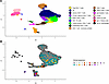 Figure 1
scRNA-seq of lymph node and skin blister samples following intradermal GAD-alum injection. (A) UMAP plot showing all pre- and post-GAD lymph node (LN) cells and post-GAD blister cells, clustered in Seurat and colored by cell type. (B) UMAP plot of T cells expressing paired TCRs, colored by clonal expansion number. Data are pooled from 5 donors. TCRs were compared at the amino acid level. To be defined as a clone, CDR3 sequences were identical, as were V and J gene usage. Amino acid changes outside of the CDR3 were permissible. Morisita-Horn indexes were calculated from the Seurat objects after QC. Total cells in this analysis were 67,780 LN cells and 2620 blister cells.
Consistent with our previous findings (18, 20), we expected GAD-specific T cells to be enriched among clonally expanded populations in the LNs. Clonal expansions were present both before and after intradermal GAD-alum injection, representing approximately 0.1%–2.5% of all T cells, with no significant change in abundance (Figure 2, A and B, and Supplemental Figure 5).
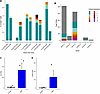 Figure 2
TCRs in LN and skin blister samples following intradermal GAD-alum injection. (A) Bar graph showing the proportion of clonally expanded T cells in the LNs, expressed as a percentage of all T cells with sequenced TCRs and colored by expansion size. (B) Total number of TCRs sequenced from each of the skin blisters, colored by clonal expansion number. Clonal expansion legend colors refer to A and B. (C) Total number of TCRs in blisters. (D) Number of clonal TCRs in blisters. C and D show pooled data from 3 donors in each group. *P < 0.05 by 1-tailed t test. TCRs were compared at the amino acid level. To be defined as a clone, CDR3 sequences were required to be identical, as were V and J gene usage. Amino acid changes outside of the CDR3 were permissible. Morisita-Horn indexes were calculated from the Seurat objects after QC. A total of 52,959 TCRs were analyzed across all samples.
Donors C, D, and E all demonstrated substantial clonal expansions in skin blisters (~20%, 5%, and 45% of all T cells, respectively, exceeding those observed in LNs) (Figure 2B). Donor E also showed some evidence of antigen-presenting cells clustering with T cells, possibly reflecting ongoing physical interactions and antigen recognition in situ (21).
Uninjected control blisters were collected from 3 donors with T1D who did not receive GAD-alum (Supplemental Tables 10–12). The total number of TCR-bearing cells was significantly higher in GAD-injected compared with control blisters (P < 0.05, 1-tailed t test), although the increase in clonally expanded TCRs did not reach statistical significance (Figure 2, C and D). The total number of cells recovered from the GAD-injected blisters was also increased compared with controls (data not shown).
Re-expression of TCRs to confirm GAD specificity. The large clonal expansions observed in the skin strongly suggested an antigen-specific response (18), which we sought to confirm by re-expressing the TCRs in a cell-based system. We selected TCRs with paired α and β chains representing the highest clonal expansion, including both CD4+ and CD8+ clonotypes where possible (Tables 1 and 2) and expressed them in the 5KC reporter line (22). TCR transductants were first screened for activation by GAD-alum presented by autologous lymphoblastoid cell lines (LCLs) and those showing a significant increase in NFAT reporter signal (data not shown) were subsequently tested against GAD-expressing LCLs. This analysis demonstrated that 4 of 14 (29%) of the blister-derived TCRs were GAD specific, including at least one GAD-reactive TCR from each donor as indicated by increased fluorescence in the NFAT reporter assay (Figure 3A, Tables 1 and 2, and Supplemental Tables 13–22).
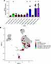 Figure 3
GAD specificity of re-expressed TCRs. (A) TCRs identified by scRNA-seq were re-expressed in 5KC reporter cells. TCRs showing positive responses when screened with GAD-alum were retested using autologous GAD-expressing LCL lines. **P < 0.01 by 1-tailed t test. Data are pooled from duplicate observations in 3 independent experiments. (B) UMAP plot showing the distribution of all re-expressed TCRs mapped back onto the single-cell dataset, illustrating phenotypes of GAD-specific T cells compared with those of other specificities, colored by clone ID. Data are pooled from 5 donors. A total of 38 TCR sequences were analyzed (Tables 1 and 2).
Re-expression of LN TCRs identified GAD-specific T cells. From the LN clonal expansions, we re-expressed the most abundant postinjection TCRs and assessed their GAD reactivity in vitro using the 5KC reporter system. TCR transductants were initially screened with GAD-alum, and those showing significant NFAT reporter response (not shown) were subsequently screened using GAD-expressing LCLs. Of the 24 TCRs re-expressed from 22 T cell clones studied (2 T cell clones each expressing 2 TCRα chains), one clonotype (~4% of those tested) demonstrated GAD specificity in postinjection LN samples (Figure 3A and Tables 1 and 2) and was designated TCR B_post_GAD_LN-5.
Shared clonally expanded TCRs detected in both LN_Post and LN_Pre samples from the same donor were not GAD specific (Tables 1 and 2). All confirmed GAD-specific TCRs derived from CD4+ T cells, while none of the 11 CD8+ T cell TCRs tested exhibited GAD reactivity (Tables 1 and 2). Alum alone did not induce NFAT reporter activation in 5KCs expressing these TCRs (data not shown). When mapped back onto the UMAP plot, cells expressing the same GAD-specific TCR clonotype exhibited highly similar gene expression profiles, while expression patterns differed between distinct GAD-specific clonotypes (Figure 3B).
T cell phenotypes in the LN and blister. LN T cells had phenotypes consistent with recirculating T cell populations and across all samples, clonally expanded cells were enriched within 2 major clusters: CD8+ T cells expressing IFNG, and CD4+ T cells expressing high levels of the Th1- and Th17-associated chemokine receptors CCR6 and CXCR3 (Figure 1 and Supplemental Figure 2). We focused particularly on Th17.1 cells, given our previous observations that reductions in this subset predict response to ustekinumab in T1D (20). However, in the absence of ex vivo stimulation, these CD4+ T cells did not express IL17A (Figure 4).
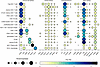 Figure 4
Gene expression profiles of cell subsets. Dot plot showing expression of major subset markers across scRNA-seq clusters from combined LNs and blister samples. Data are pooled from 5 donors. Total cells in this analysis were 67,780 LN cells and 2620 blister cells.
Phenotypes of GAD-specific cells in the skin. Because a high proportion of the re-expressed clonally expanded TCRs from the blisters were GAD specific, we compared all blister T cells (with a defined TCR and not associated with antigen-presenting cells) from the GAD-injected participants to those from control donors. Following GAD injection, T cells showed upregulation of mitochondrial and ribosomal genes, consistent with increased cellular activation. TRRUST analysis indicated that these genes were regulated by transcription factors from the RFX family (Figure 5A and Supplemental Table 23). We next examined canonical T cell subset markers and found substantial interdonor variability. Donor D exhibited expression of IL22, CXCR3, CCR6, and LTB, genes associated with Th17 cells and autoimmune responses (23), whereas this pattern was not observed in Donor E (Supplemental Figure 6).
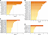 Figure 5
Gene expression profiles of T cells following in vivo GAD. (A) Transcription factors identified by TRRUST analysis of all genes upregulated in blister T cells following GAD injection compared with control blisters. (B) Transcription factors identified by TRRUST analysis of genes upregulated in conventional CD4+ T cells from blisters compared with pre-GAD LN samples. (C) Transcription factors identified by TRRUST analysis of all upregulated genes in clonally expanded CD4+ T cells post-GAD vs. pre-GAD in the LNs. (D) Transcription factors identified by TRRUST analysis of all genes upregulated in clonally expanded CD8+ T cells post-GAD vs. pre-GAD in the LNs. Data are pooled from 5 donors in LN groups and 2 donors in blister groups. A total of 740 clonally expanded T cells were analyzed. DEG pathway analysis was conducted using Metascape standard parameters on all DEGs with an uncorrected P value of less than 0.05 and Metascape parameters using Benjamini-Hochberg P value correction are reported.
The above analysis was limited by the small number of cells in the control blisters. To further characterize phenotypes associated with GAD-specific T cells, we compared all post-GAD Alum blister T cells within conventional CD4+ T cell clusters (i.e., excluding those associated with monocytes) to CD4+ T cells from pre-GAD LNs. The top DEGs included IL22, CXCR6, and CD26 as well as activation markers such as TNFRSF18 (GITR) (Supplemental Table 24). These changes were primarily driven by high expression in Donor D, who contributed more conventional CD4+ T cells than Donor E (Supplemental Figure 7). TRRUST analysis identified NF-κB, RelA, and SP1 (which regulates TBET expression; ref. 24) as the top 3 transcription factors driving the observed gene expression profile in blister T cells (Figure 5B)
Phenotypes of T cells in the LNs following intradermal GAD-alum. In addition to sampling suction blister fluid, we also performed ultrasound-guided fine-needle aspiration (FNA) of the skin-draining LNs before and after intradermal injection of GAD-alum. We examined changes in clonally expanded T cells following GAD-alum injection, hypothesizing that recruitment of GAD-specific T cells into the clonally expanded T cell clusters would generate a detectable transcriptional signature. Focussing first on CD4+ clonally expanded T cells, differential expression analysis of post-GAD samples revealed upregulation of ribosomal genes consistent with increased transcriptional activity, and of TXNIP. TRRUST analysis performed using Metascape (25) on upregulated genes (uncorrected P < 0.05) identified NF-κB, RelA, and STAT3 as the top 3 transcription factors driving the post-GAD signature (Figure 5C and Supplemental Table 25). Genes regulated by these factors included TXNIP and members of the TNF superfamily. When examined by donor, TXNIP upregulation was particularly consistent across individuals (Supplemental Figure 8). Similar results were observed when a pseudobulk analysis was performed (Supplemental Figures 9–11, and Supplemental Table 26).
We next analyzed clonally expanded cells in the IFNG+ CD8+ T cell cluster. Again, the most significantly upregulated genes were associated with increased activation and protein translation. TRRUST analysis identified RBMX, NF1, and FOXO1 as key transcriptional regulators of this signature (Figure 5D and Supplemental Table 27). Similar results were observed when a pseudobulk analysis was performed (Supplemental Figures 9–11, and Supplemental Table 28), although the small number of donors limited the statistical power of this approach.
Comparison of gene expression in GAD-reactive peripheral blood T cells reveals distinctive autoantigenic phenotypes. All participants were positive for anti-GAD autoantibodies and had T1D. We therefore hypothesized that GAD-specific T cells could be detected in peripheral blood prior to GAD challenge. To test this, we used an in vitro activation-induced maker (AIM) assay to enrich and identify GAD-reactive CD4+ T cells in PBMC samples collected before antigen challenge.
To this end, we performed an in vitro AIM assay to detect GAD-reactive CD4+ T cells prior to GAD injection. Unstimulated cells served as a negative control for unactivated cells, staphylococcal enterotoxin B–stimulated (SEB-stimulated) cells as a positive control, and buffer-stimulated control (PBMC_Ctrl) as a control for non-specific activation, or preactivated responses. A total of 693 cells passed QC from the Ctrl and GAD-stimulated wells and were included in the analysis (Supplemental Tables 29 and 30). Sorted cells formed distinct clusters, including resting cells, Tregs, 3 populations of activated central memory (TCM) cells, and 2 clusters coexpressing IL17A and IFNG, designated Th17.1s (Figure 6, A and B). As expected, cluster frequencies differed substantially between SEB-stimulated and unstimulated conditions (data not shown). The mean stimulation index of GAD versus control stimulation, corrected for initial PBMC input, was 2.2 (range 1.2–4.9). Th17.1 clusters were enriched in IFNG, IL17A, IL17F, and CSF2. Subsets designated Th17.1 IL2+ and Th17.1 IL22+ coexpressed IL2 or IL22, respectively (Figure 6B). These populations were of particular interest given our previous finding that reduction in Th17.1 cells predicts response to ustekinumab in T1D (20). Compared with PBMC_Ctrl cells, PBMC_GAD cells exhibited increased expression of IFNG, IL17A, IL17F, CSF2, IL2, and IL22 as well as the Th1- and Th17-associated transcription factors STAT1 and STAT3 (Figure 6B). Pairwise comparisons between control and GAD stimulation for each donor showed this pattern of particularly elevated IL17A, IL17F, and IL22 expression, was consistent across all donors except Donor B, who displayed the lowest stimulation index and an insufficient number of surviving cells after scRNA-seq QC (Supplemental Figure 12). Despite these transcriptional changes, the frequency of cells within individual clusters did not increase. Differential gene expression analysis across all cells revealed PBMC_GAD cells were defined by upregulation of IL2RA, IL2RB, LTB, DPP4 (CD26), and IL22 (Figure 6B and Supplemental Table 31). TRRUST analysis identified STAT1 and STAT3 in the top transcription factors driving the gene expression program in GAD-reactive cells (Figure 7A). Pseudobulk analysis demonstrated similar findings, with IL17F and IL22 in particular consistently upregulated in the GAD-stimulated samples (Supplemental Figures 9–11, and Supplemental Table 32).
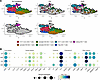 Figure 6
In vitro stimulation of CD4+ T cells from peripheral blood. (A) UMAP plots of all flow-sorted cells separated by in vitro restimulation condition. (B) Dot plots showing expression of key markers across each cluster. Data are pooled from 6 donors and 3486 total cells.
 Figure 7
GAD-reactive T cells from peripheral blood have Th17.1 phenotypes. (A) Transcription factors identified by TRRUST analysis of genes upregulated in GAD-activated compared with control-activated cells. (B) UMAP plot highlighting a clonally expanded population of cells (“PBMC-1”) detected only in the GAD-activated sample. Data are pooled from 6 donors. (C) PBMC-1 TCR re-expressed in 5KC reporter cells and stimulated with control or GAD-expressing LCLs. *P < 0.05 by unpaired 1-tailed t test from 3 independent experiments. A total of 693 T cells from Ctrl and GAD-stimulated conditions were analyzed in this figure. DEG pathway analysis was conducted using Metascape standard parameters on all DEGs with an uncorrected P value of less than 0.05 and Metascape parameters using Benjamini-Hochberg P value correction are reported.
One large clonal expansion of GAD-reactive cells was detected in Donor D (15 cells, Figure 7B). These cells predominantly exhibited a Th17.1 IL2+ phenotype and re-expression of their TCR in the 5KC reporter line confirmed GAD specificity (Tables 1 and 2, and Figure 7C). Interestingly, despite being from a different donor (Donor B), the TCRα chain of the LN-derived clone B_post_GAD_LN-5 (CAENGAGGTSYGKLTF) differed from that of the PBMC clone PBMC-1 (Donor D, CAENNAGGTSYGKLTF) by only a single amino acid. These donors shared several HLA alleles, including HLA-DRB1*04:01:01.
In addition, we also examined the phenotype of GAD-reactive cells in the peripheral blood after the injection of GAD-alum using the AIM assay, comparing their frequency and phenotype to those sampled prior to in vivo GAD-alum (Supplemental Figure 13). The percentage of GAD-reactive T cells in the peripheral blood was not altered (data not shown).
PBMC-1 GAD-specific clones were also present in the post-GAD sample, stimulated with in vitro GAD (i.e., only in the in vitro GAD-stimulated samples before and after in vivo GAD and not at all in the Ctrl stimulated wells at either time point, Supplemental Figure 13B), and predominantly retained a Th17.1 IL2+ phenotype.
Comparing abundance of clusters (again excluding Donor B who had insufficient cells), there was a significant (P < 0.05, paired t test) increase in GAD-reactive cells with a Th17.1 IL2+ phenotype and a decrease in GAD-reactive cells with a Th17.1 IL22+ phenotype (this significance did not remain after correction for multiple testing). In contrast, no changes in the Th17.1 populations were noted in the Ctrl stimulations, although the proportion of Ctrl Tregs did increase after in vivo GAD-alum.
Sharing of paired TCRs was predominantly observed between samples from the same donor (Supplemental Figure 14 and Supplemental Table 33). Overlap was detected between peripheral blood, LN, and blister samples within donors, including persistence of clonotypes across LN time points and sharing between LN and blister compartments. In contrast, sharing between donors was limited. A notably high Morisita-Horn index was observed for Donor D due to the high clonal expansion of PBMC_1 in peripheral blood samples. Higher sharing was also noted in the LNs where clones persisted between the 2 sampling time points in all 5 donors. Both blister samples with GEX information showed evidence of repertoire sharing with the same donor’s LN samples. Sharing between Ctrl PBMC samples was skewed by very low total cell numbers (cells that were activated in response to buffer components or cells that were already activated in vivo, i.e., a persistent immune response to pathogens between the sampling time points and compartments). Sharing of clones (paired TCRs) between donors was more limited (Supplemental Table 33).
Together, these findings demonstrate that GAD-specific CD4+ T cells can be isolated and characterized from peripheral blood, skin, and LN samples following intradermal GAD-alum administration. Across compartments, these cells displayed transcriptional features consistent with Th1 and Th17.1 phenotypes, including activation of NF-κB–, RelA-, and STAT-driven pathways. The identification of clonally expanded, GAD-reactive TCRs in both peripheral and lymphoid compartments provides new opportunities to track autoantigen-specific T cells longitudinally and to evaluate their modulation during immunotherapeutic intervention.


