
Research ArticleAgingImmunologyInfectious disease
Open Access |  10.1172/jci.insight.195618
Age-related differences in immune responses to inactivated influenza and adjuvanted recombinant herpes zoster vaccines
Gizem Kilic,1 Esther J.M. Taks,1 Leonie S. Helder,1 Elisabeth A. Dulfer,1 Büsra Geckin,1 Liesbeth van Emst,1 Heidi Lemmers,1 Stefano Berrè,2 Adhidev Biswas,3 Mumin Ozturk,4,5 Yutaka Negishi,4,5 Wivine Burny,2 Sofia M. Buonocore,2 Jaap ten Oever,1 Musa M. Mhlanga,4,5 and Mihai G. Netea1,6
1Department of Internal Medicine and Radboud Community for Infectious Diseases, Radboud University Medical Center, Nijmegen, Netherlands.
2GSK, Rixensart, Belgium.
3GSK, Bengaluru, India.
4Department of Cell Biology, Faculty of Science, Radboud University, Nijmegen, Netherlands.
5Department of Human Genetics, Radboud University Medical Center, Nijmegen, Netherlands.
6Department of Immunology and Metabolism, Life and Medical Sciences Institute, University of Bonn, Bonn, Germany.
Address correspondence to: Mihai G. Netea, Department of Internal Medicine, Radboud University Nijmegen Medical Center, Geert Grooteplein Zuid 8, 6525 GA Nijmegen, Netherlands. Phone: 31.0.24.361.4652; Email: mihai.netea@radboudumc.nl.
Find articles by
Kilic, G.
in:
PubMed
|
Google Scholar
|

1Department of Internal Medicine and Radboud Community for Infectious Diseases, Radboud University Medical Center, Nijmegen, Netherlands.
2GSK, Rixensart, Belgium.
3GSK, Bengaluru, India.
4Department of Cell Biology, Faculty of Science, Radboud University, Nijmegen, Netherlands.
5Department of Human Genetics, Radboud University Medical Center, Nijmegen, Netherlands.
6Department of Immunology and Metabolism, Life and Medical Sciences Institute, University of Bonn, Bonn, Germany.
Address correspondence to: Mihai G. Netea, Department of Internal Medicine, Radboud University Nijmegen Medical Center, Geert Grooteplein Zuid 8, 6525 GA Nijmegen, Netherlands. Phone: 31.0.24.361.4652; Email: mihai.netea@radboudumc.nl.
Find articles by Taks, E. in: PubMed | Google Scholar
1Department of Internal Medicine and Radboud Community for Infectious Diseases, Radboud University Medical Center, Nijmegen, Netherlands.
2GSK, Rixensart, Belgium.
3GSK, Bengaluru, India.
4Department of Cell Biology, Faculty of Science, Radboud University, Nijmegen, Netherlands.
5Department of Human Genetics, Radboud University Medical Center, Nijmegen, Netherlands.
6Department of Immunology and Metabolism, Life and Medical Sciences Institute, University of Bonn, Bonn, Germany.
Address correspondence to: Mihai G. Netea, Department of Internal Medicine, Radboud University Nijmegen Medical Center, Geert Grooteplein Zuid 8, 6525 GA Nijmegen, Netherlands. Phone: 31.0.24.361.4652; Email: mihai.netea@radboudumc.nl.
Find articles by Helder, L. in: PubMed | Google Scholar
1Department of Internal Medicine and Radboud Community for Infectious Diseases, Radboud University Medical Center, Nijmegen, Netherlands.
2GSK, Rixensart, Belgium.
3GSK, Bengaluru, India.
4Department of Cell Biology, Faculty of Science, Radboud University, Nijmegen, Netherlands.
5Department of Human Genetics, Radboud University Medical Center, Nijmegen, Netherlands.
6Department of Immunology and Metabolism, Life and Medical Sciences Institute, University of Bonn, Bonn, Germany.
Address correspondence to: Mihai G. Netea, Department of Internal Medicine, Radboud University Nijmegen Medical Center, Geert Grooteplein Zuid 8, 6525 GA Nijmegen, Netherlands. Phone: 31.0.24.361.4652; Email: mihai.netea@radboudumc.nl.
Find articles by Dulfer, E. in: PubMed | Google Scholar
1Department of Internal Medicine and Radboud Community for Infectious Diseases, Radboud University Medical Center, Nijmegen, Netherlands.
2GSK, Rixensart, Belgium.
3GSK, Bengaluru, India.
4Department of Cell Biology, Faculty of Science, Radboud University, Nijmegen, Netherlands.
5Department of Human Genetics, Radboud University Medical Center, Nijmegen, Netherlands.
6Department of Immunology and Metabolism, Life and Medical Sciences Institute, University of Bonn, Bonn, Germany.
Address correspondence to: Mihai G. Netea, Department of Internal Medicine, Radboud University Nijmegen Medical Center, Geert Grooteplein Zuid 8, 6525 GA Nijmegen, Netherlands. Phone: 31.0.24.361.4652; Email: mihai.netea@radboudumc.nl.
Find articles by Geckin, B. in: PubMed | Google Scholar
1Department of Internal Medicine and Radboud Community for Infectious Diseases, Radboud University Medical Center, Nijmegen, Netherlands.
2GSK, Rixensart, Belgium.
3GSK, Bengaluru, India.
4Department of Cell Biology, Faculty of Science, Radboud University, Nijmegen, Netherlands.
5Department of Human Genetics, Radboud University Medical Center, Nijmegen, Netherlands.
6Department of Immunology and Metabolism, Life and Medical Sciences Institute, University of Bonn, Bonn, Germany.
Address correspondence to: Mihai G. Netea, Department of Internal Medicine, Radboud University Nijmegen Medical Center, Geert Grooteplein Zuid 8, 6525 GA Nijmegen, Netherlands. Phone: 31.0.24.361.4652; Email: mihai.netea@radboudumc.nl.
Find articles by van Emst, L. in: PubMed | Google Scholar
1Department of Internal Medicine and Radboud Community for Infectious Diseases, Radboud University Medical Center, Nijmegen, Netherlands.
2GSK, Rixensart, Belgium.
3GSK, Bengaluru, India.
4Department of Cell Biology, Faculty of Science, Radboud University, Nijmegen, Netherlands.
5Department of Human Genetics, Radboud University Medical Center, Nijmegen, Netherlands.
6Department of Immunology and Metabolism, Life and Medical Sciences Institute, University of Bonn, Bonn, Germany.
Address correspondence to: Mihai G. Netea, Department of Internal Medicine, Radboud University Nijmegen Medical Center, Geert Grooteplein Zuid 8, 6525 GA Nijmegen, Netherlands. Phone: 31.0.24.361.4652; Email: mihai.netea@radboudumc.nl.
Find articles by
Lemmers, H.
in:
PubMed
|
Google Scholar
|
1Department of Internal Medicine and Radboud Community for Infectious Diseases, Radboud University Medical Center, Nijmegen, Netherlands.
2GSK, Rixensart, Belgium.
3GSK, Bengaluru, India.
4Department of Cell Biology, Faculty of Science, Radboud University, Nijmegen, Netherlands.
5Department of Human Genetics, Radboud University Medical Center, Nijmegen, Netherlands.
6Department of Immunology and Metabolism, Life and Medical Sciences Institute, University of Bonn, Bonn, Germany.
Address correspondence to: Mihai G. Netea, Department of Internal Medicine, Radboud University Nijmegen Medical Center, Geert Grooteplein Zuid 8, 6525 GA Nijmegen, Netherlands. Phone: 31.0.24.361.4652; Email: mihai.netea@radboudumc.nl.
Find articles by Berrè, S. in: PubMed | Google Scholar
1Department of Internal Medicine and Radboud Community for Infectious Diseases, Radboud University Medical Center, Nijmegen, Netherlands.
2GSK, Rixensart, Belgium.
3GSK, Bengaluru, India.
4Department of Cell Biology, Faculty of Science, Radboud University, Nijmegen, Netherlands.
5Department of Human Genetics, Radboud University Medical Center, Nijmegen, Netherlands.
6Department of Immunology and Metabolism, Life and Medical Sciences Institute, University of Bonn, Bonn, Germany.
Address correspondence to: Mihai G. Netea, Department of Internal Medicine, Radboud University Nijmegen Medical Center, Geert Grooteplein Zuid 8, 6525 GA Nijmegen, Netherlands. Phone: 31.0.24.361.4652; Email: mihai.netea@radboudumc.nl.
Find articles by Biswas, A. in: PubMed | Google Scholar
1Department of Internal Medicine and Radboud Community for Infectious Diseases, Radboud University Medical Center, Nijmegen, Netherlands.
2GSK, Rixensart, Belgium.
3GSK, Bengaluru, India.
4Department of Cell Biology, Faculty of Science, Radboud University, Nijmegen, Netherlands.
5Department of Human Genetics, Radboud University Medical Center, Nijmegen, Netherlands.
6Department of Immunology and Metabolism, Life and Medical Sciences Institute, University of Bonn, Bonn, Germany.
Address correspondence to: Mihai G. Netea, Department of Internal Medicine, Radboud University Nijmegen Medical Center, Geert Grooteplein Zuid 8, 6525 GA Nijmegen, Netherlands. Phone: 31.0.24.361.4652; Email: mihai.netea@radboudumc.nl.
Find articles by Ozturk, M. in: PubMed | Google Scholar
1Department of Internal Medicine and Radboud Community for Infectious Diseases, Radboud University Medical Center, Nijmegen, Netherlands.
2GSK, Rixensart, Belgium.
3GSK, Bengaluru, India.
4Department of Cell Biology, Faculty of Science, Radboud University, Nijmegen, Netherlands.
5Department of Human Genetics, Radboud University Medical Center, Nijmegen, Netherlands.
6Department of Immunology and Metabolism, Life and Medical Sciences Institute, University of Bonn, Bonn, Germany.
Address correspondence to: Mihai G. Netea, Department of Internal Medicine, Radboud University Nijmegen Medical Center, Geert Grooteplein Zuid 8, 6525 GA Nijmegen, Netherlands. Phone: 31.0.24.361.4652; Email: mihai.netea@radboudumc.nl.
Find articles by Negishi, Y. in: PubMed | Google Scholar
1Department of Internal Medicine and Radboud Community for Infectious Diseases, Radboud University Medical Center, Nijmegen, Netherlands.
2GSK, Rixensart, Belgium.
3GSK, Bengaluru, India.
4Department of Cell Biology, Faculty of Science, Radboud University, Nijmegen, Netherlands.
5Department of Human Genetics, Radboud University Medical Center, Nijmegen, Netherlands.
6Department of Immunology and Metabolism, Life and Medical Sciences Institute, University of Bonn, Bonn, Germany.
Address correspondence to: Mihai G. Netea, Department of Internal Medicine, Radboud University Nijmegen Medical Center, Geert Grooteplein Zuid 8, 6525 GA Nijmegen, Netherlands. Phone: 31.0.24.361.4652; Email: mihai.netea@radboudumc.nl.
Find articles by Burny, W. in: PubMed | Google Scholar
1Department of Internal Medicine and Radboud Community for Infectious Diseases, Radboud University Medical Center, Nijmegen, Netherlands.
2GSK, Rixensart, Belgium.
3GSK, Bengaluru, India.
4Department of Cell Biology, Faculty of Science, Radboud University, Nijmegen, Netherlands.
5Department of Human Genetics, Radboud University Medical Center, Nijmegen, Netherlands.
6Department of Immunology and Metabolism, Life and Medical Sciences Institute, University of Bonn, Bonn, Germany.
Address correspondence to: Mihai G. Netea, Department of Internal Medicine, Radboud University Nijmegen Medical Center, Geert Grooteplein Zuid 8, 6525 GA Nijmegen, Netherlands. Phone: 31.0.24.361.4652; Email: mihai.netea@radboudumc.nl.
Find articles by Buonocore, S. in: PubMed | Google Scholar
1Department of Internal Medicine and Radboud Community for Infectious Diseases, Radboud University Medical Center, Nijmegen, Netherlands.
2GSK, Rixensart, Belgium.
3GSK, Bengaluru, India.
4Department of Cell Biology, Faculty of Science, Radboud University, Nijmegen, Netherlands.
5Department of Human Genetics, Radboud University Medical Center, Nijmegen, Netherlands.
6Department of Immunology and Metabolism, Life and Medical Sciences Institute, University of Bonn, Bonn, Germany.
Address correspondence to: Mihai G. Netea, Department of Internal Medicine, Radboud University Nijmegen Medical Center, Geert Grooteplein Zuid 8, 6525 GA Nijmegen, Netherlands. Phone: 31.0.24.361.4652; Email: mihai.netea@radboudumc.nl.
Find articles by ten Oever, J. in: PubMed | Google Scholar
1Department of Internal Medicine and Radboud Community for Infectious Diseases, Radboud University Medical Center, Nijmegen, Netherlands.
2GSK, Rixensart, Belgium.
3GSK, Bengaluru, India.
4Department of Cell Biology, Faculty of Science, Radboud University, Nijmegen, Netherlands.
5Department of Human Genetics, Radboud University Medical Center, Nijmegen, Netherlands.
6Department of Immunology and Metabolism, Life and Medical Sciences Institute, University of Bonn, Bonn, Germany.
Address correspondence to: Mihai G. Netea, Department of Internal Medicine, Radboud University Nijmegen Medical Center, Geert Grooteplein Zuid 8, 6525 GA Nijmegen, Netherlands. Phone: 31.0.24.361.4652; Email: mihai.netea@radboudumc.nl.
Find articles by Mhlanga, M. in: PubMed | Google Scholar
1Department of Internal Medicine and Radboud Community for Infectious Diseases, Radboud University Medical Center, Nijmegen, Netherlands.
2GSK, Rixensart, Belgium.
3GSK, Bengaluru, India.
4Department of Cell Biology, Faculty of Science, Radboud University, Nijmegen, Netherlands.
5Department of Human Genetics, Radboud University Medical Center, Nijmegen, Netherlands.
6Department of Immunology and Metabolism, Life and Medical Sciences Institute, University of Bonn, Bonn, Germany.
Address correspondence to: Mihai G. Netea, Department of Internal Medicine, Radboud University Nijmegen Medical Center, Geert Grooteplein Zuid 8, 6525 GA Nijmegen, Netherlands. Phone: 31.0.24.361.4652; Email: mihai.netea@radboudumc.nl.
Find articles by Netea, M. in: PubMed | Google Scholar
Published June 2, 2026 - More info
JCI Insight. 2026;11(14):e195618. https://doi.org/10.1172/jci.insight.195618.
© 2026 Kilic et al. This work is licensed under the Creative Commons Attribution 4.0 International License. To view a copy of this license, visit http://creativecommons.org/licenses/by/4.0/.
Received: May 21, 2025; Accepted: May 28, 2026

-
Results
Safety data and participant demographics. For the study, 84 young and 63 older adults were recruited. After the initial screening and informed consent process, participants were randomized into different study arms based on age and vaccine type, as illustrated in Figure 1A. Because of the extended period between the screening/randomization and vaccination visits (D0), some participants dropped out or withdrew their consent. Ultimately, 22 young and 31 older participants were immunized with RZV, and 30 young and 27 older adults received IIV4. Additionally, 11 young adults in the placebo-IIV4 group and 11 adults in the placebo-RZV group were vaccinated with a placebo. The timeline, sample collection, and performed assays are summarized in Figure 1B. The demographics, comorbidities, and risk factors of the study cohort are presented in Table 1. As expected, older adults had more comorbidities and risk factors, such as hypertension and chronic cardiovascular disease.
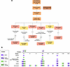 Figure 1
Summary of the clinical study and the time points of each assay. (A) The flow diagram depicts participant recruitment, randomization, and the number of people who started and finished the study. (B) Vaccination groups in the study and the time points for each injection and experiment.
Overall, both vaccines were well tolerated, showing acceptable safety profiles in young and older participants. The most common local adverse reaction within the first week after vaccination was pain at the injection site, and the most common systemic adverse reactions were myalgia and fatigue (Supplemental Tables 1–3; supplemental material available online with this article; https://doi.org/10.1172/jci.insight.195618DS1). Most of the adverse reactions were rated mild or moderate by the study participants, whereas some severe (grade 3) adverse reactions were also reported (Supplemental Tables 4–6). These adverse reactions typically resolved spontaneously within a week. Three serious adverse events (SAEs) were recorded during the study, which were a road traffic accident (RZV-young group), atrial fibrillation (RZV-old), and acute heart failure (RZV-old), the last SAE being fatal (Supplemental Table 7). Given that the participants with atrial fibrillation and acute heart failure both had a prior history of similar conditions and were receiving related medications, and the participant involved in the traffic accident did not develop any condition that contributed to the accident, the SAEs were not considered related to the study vaccines.
Baseline differences in immune parameters between young and older adults. We first evaluated the immune system parameters in young and older adults before they received any study vaccination. The numbers of WBCs, neutrophils, monocytes, and lymphocytes were similar between the 2 age groups (Supplemental Figure 1A). Additionally, the baseline concentrations of anti-VZV gE antibodies and the HA inhibition (HAI) titers against the A/Victoria/H1N1 and B/Phuket virus strains were also comparable in young and older adults (Supplemental Figure 1, B and C). In contrast, the IFN-γ production of PBMCs from young adults was significantly higher after a 7-day incubation with the IIV4 vaccines and gE antigen compared with that from older counterparts (Supplemental Figure 1D). Lastly, many inflammatory proteins were significantly more abundant in the circulation of older individuals than in young volunteers (Supplemental Figure 1E).
IIV4-induced modulation of immune cell numbers, HAI titers, antigen-specific CD4+ T cells, and IFN-γ production in young and older adults. Next, we analyzed changes in circulating immune cells after IIV4 vaccination compared with baseline (D0). In young adults, immune cell counts, except lymphocytes, increased 1 day after vaccination (Figure 2A). Although lymphocyte numbers decreased on D1, they remained significantly elevated for the remainder of the study compared with the baseline. WBC counts remained higher on day 180 compared with the baseline, possibly due to the higher numbers of lymphocytes and monocytes. In older adults, changes were minimal (Figure 2B). Absolute cell counts after IIV4 and placebo vaccination are presented in Supplemental Figure 2, A–C.
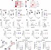 Figure 2
IIV4-induced changes in immune cell counts and adaptive immune responses in young and older adults. Heatmaps showing the fold changes in immune cell counts after IIV4 vaccination versus before vaccination in the (A) young and (B) older groups. Fold changes at D1, D7, D60, and D180 were compared with D0, after correction for multiple testing using the Benjamini-Hochberg method. The scale displays fold change values; stars on the heatmap represent FDR values. (C) HAI titers and (D) comparison of fold changes in antibody production against the A/Victoria/H1N1 strain. (E) HAI titers and (F) comparison of fold changes (D60/D0) in antibody production against the B/Phuket strain. Spearman’s correlation between baseline HAI titers against the A/Victoria/H1N1 and B/Phuket strains and fold changes after IIV4 vaccination in (G) young and (H) older adults. (I) IFN-γ production from PBMCs after 7-day stimulation with 1 μg/mL of IIV4 2021/2022 and 2022/2023 season vaccines. PBMCs from each individual were stimulated with the same season of the vaccine they received. (J) Spearman’s correlation of fold IFN-γ response (D60/D0) and fold HAI titers (D60/D0) for the A/Victoria/H1N1 and B/Phuket strains. The scale indicates the correlation coefficient (r). (K) The frequency of activated (4-1BB+) CD4+ T cells per 106 CD4+ T cells that were positive for CD40L, IL-2, TNF, and IFN-γ after stimulation with A/H1N1/Victoria and B/Phuket. The y axis values in C–F are displayed on a log2 scale; values in I are shown on a log10 scale. The dashed lines on graphs in D and F indicate the threshold of seroconversion (a fold change of 4). Different time points within the same group were compared using Wilcoxon’s signed-rank test; fold changes between young and older adults were compared using the Mann-Whitney U test. *P < 0.05, **P < 0.01, ***P < 0.001, ****P < 0.0001.
Subsequently, we measured HAI titers against the viral strains in the 2021/2022 and 2022/2023 IIV4 vaccines before and 60 days after vaccination. Both age groups showed a significant increase in anti-A/Victoria/H1N1 titers, with a stronger rise in young adults (Figure 2, C and D). Similarly, IIV4 also boosted anti-B/Phuket titers in both groups, and the fold induction was not significantly different between the groups (Figure 2, E and F). Notably, the fold HAI induction to the A/Victoria/H1N1 and B/Phuket strains was significantly higher in older adults vaccinated with the IIV4 2021/2022 compared with the 2022/2023 season vaccine (Supplemental Figure 2D).
Seroprotection refers to an antibody titer of 1:40 or higher, a level associated with a 50% reduction in influenza risk (19). In our study, 23% of young adults had HAI titers of 40 or higher to A/Victoria/H1N1 before vaccination, which increased to 100% on D60. In older adults, the corresponding rates were 30% before vaccination and 73% after vaccination. All young adults had titers of 40 or higher against B/Phuket both before and after vaccination. Among older adults, 93% had titers of 40 or higher, rising to 100% after receiving IIV4.
For the A/Tasmania/H3N2 and B/Washington strains (present only in the IIV4 2021/2022 vaccine), the HAI titers against A/Tasmania/H3N2 increased only in the young group, but seroconversion rates were extremely low in the 2 age groups (Supplemental Figure 2, E and F). Both young and older adults showed significant antibody increases against B/Washington with similar fold changes (Supplemental Figure 2, G and H). No notable changes were observed in antibody levels against the A/Darwin/H3N2 and B/Austria strains 60 days after vaccination (Supplemental Figure 2I).
Previous studies have shown that preexisting immunity, defined by the presence of specific antibodies before vaccination, negatively correlates with a fold antibody induction after vaccinations like those for influenza, herpes zoster, and pneumococcus (20–22). As expected, we observed a negative correlation between prevaccination HAI titers and antibody response for A/Victoria/H1N1 and B/Phuket strains in young adults (Figure 2G). In the older group, a moderate-to-high negative correlation was also observed, though not significant for A/Victoria/H1N1 (Figure 2H). As such, age may independently affect the relationship between prevaccine and postvaccine responses. Additionally, IFN-γ production in PBMCs after 7 days of incubation with IIV4 was similar before and after vaccination across all groups (Figure 2I). Subsequently, we explored the potential link between PBMC-derived IFN-γ production and antibody response to IIV4. Fold induction of A/Victoria/H1N1 HAI titers and IFN-γ responses were negatively correlated at D60 in younger adults (Figure 2J).
Last, we determined the frequency of antigen-specific activated CD4+ T cells that express CD40L, IL-2, TNF, and IFN-γ after IIV4 in young and older adults (Supplemental Figure 3, A and B). In general, the induction of activated antigen-specific CD4+ T cells by IIV4 was lower in older adults compared with the young individuals. CD40L was the most expressed marker upon incubation with the A/H1N1/Victoria strain. IIV4 led to a small but significant induction of B/Phuket-specific CD4+ T cells that were positive for 2 or 3 markers. There was a significant increase in CD4+ T cells expressing all 4 markers (CD40L, IL-2, TNF, and IFN-γ) in response to A/H1N1/Victoria only in older adults, whereas such an increase in response to B/Phuket was observed only in young adults (Figure 2K).
Overall, young adults showed higher antibody responses to IIV4, but the vaccine did not enhance IFN-γ production from PBMCs in either age group. The vaccine induced polyfunctional antigen-specific CD4+ T cells, although the cell frequencies were low, suggesting the limited ability of IIV4 to boost cellular responses.
RZV-induced modulation of immune cell numbers, antibody concentrations, antigen-specific CD4+ T cells, and IFN-γ production in young and older adults. We characterized how RZV affected immune cell counts after the first and second doses. RZV induced greater fold changes in cell numbers than IIV4. After the first dose, both young and old groups experienced temporary increases in WBCs, neutrophils, and monocytes on day 1, with over a 1.7-fold increase in neutrophils (Figure 3A). Lymphocyte numbers decreased on D1 only in the young group. Similar changes occurred after the second dose, with significant lymphocyte decreases on day 1 (D61) in both groups, followed by a rise on D67 that stayed elevated until at least D120 (Figure 3B). Absolute numbers of cells after RZV and placebo vaccinations are presented in Supplemental Figure 4, A–C.
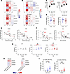 Figure 3
RZV-induced changes in immune cell counts and adaptive immune responses in young and older adults. Heatmaps showing the fold changes in immune cell counts after the (A) first and (B) second dose of RZV. Fold changes at D1, D7, and D60 were compared with D0; fold changes at D61, D67, D120, and D240 were compared with D60 using Wilcoxon’s signed-rank test, with P values corrected for multiple testing using the Benjamini-Hochberg method. The scale displays fold change values; stars on the heatmap represent FDR values. (C) Anti-HZV gE antibody concentrations and (D) comparison of fold changes (D60/D0 and D120/D0) after RZV in young and older individuals. (E) Spearman’s correlation between baseline concentrations of anti-VZV gE antibodies and fold antibody response (D60/D0) after RZV. (F) Spearman’s correlation between pre-second vaccination (D60) anti-VZV gE antibody concentrations and fold antibody response (D120/D60) after second dose. (G) IFN-γ production from PBMCs after 7-day stimulation with 4 μg/mL of the antigen (gE) in RZV. (H) Spearman’s correlation of fold IFN-γ responses (D60/D0 and D120/D0 and D240/D0) and fold antibody production (D60/D0 and D120/D0). The scale indicates the correlation coefficient (r). (I) The frequency of activated (4-1BB+) CD4+ T cells per 106 CD4+ T cells that were positive for CD40L, IL-2, TNF, and IFN-γ after stimulation with the gE antigen in young and older adults. The y axis values in C and G are displayed on a log10 scale; values in D are shown on a log2 scale. Different time points within the same group were compared using Dunn’s multiple-comparison test, and fold changes between young and older adults were compared using the Mann-Whitney U test. *P < 0.05, **P < 0.01, ***P < 0.001, ****P < 0.0001.
RZV led to strong antibody production after the first dose in both age groups (Figure 3C). The second dose further increased anti-VZV gE antibody levels, though the increase in young adults did not reach statistical significance. Young adults showed a greater fold antibody increase only after the first dose (Figure 3D). We also found that prevaccination antibody concentrations were negatively correlated with a fold increase in antibodies after RZV in both age groups (Figure 3E). The concentrations of antibodies before the second vaccination (D60) were negatively associated with fold antibody increase after the second vaccination (Figure 3F).
IFN-γ production after gE antigen stimulation increased significantly after 2 RZV doses (D120) and remained elevated at D240, unlike the placebo, which had no effect (Figure 3G). Importantly, prior to RZV, IFN-γ response was detectable in 55% of the young participants, increasing to 92% after the second dose (D120). In older adults, it increased from 20% at baseline to 77% on D120. No significant relationship was found between IFN-γ and antibody production after RZV exposure (Figure 3H).
RZV induced a robust and significant increase in antigen-specific polyfunctional CD4+ T cells in both age groups, with a lesser degree in the older age group (Supplemental Figure 5). The second dose further increased the frequency of antigen-specific cells. CD40L was the most expressed marker to the antigen among the 4 markers measured. CD40L+IL-2+TNF+IFN-γ+ gE–specific CD4+ T cells had the highest frequency among other marker combinations and were significantly induced after both the first and second RZV dose in young and older adults (Figure 3I). In summary, RZV elicited significant humoral and cellular responses in both young and older individuals.
The impact of IIV4 on circulating immune mediators. We analyzed the impact of IIV4 and a placebo on circulating inflammatory proteins using the Olink 96 Target Inflammation Panel. At 60 days after vaccination, EN-RAGE levels decreased in both the IIV4 placebo and young groups (Figure 4, A and B). In older adults, IIV4 vaccination increased inflammatory mediators such as TNFB, IL-6, CCL25, and TRAIL, while reducing AXIN1, STAMBP, and CD40 (Figure 4C and Supplemental Figure 6A). Since older adults received either the 2021/2022 or the 2022/2023 season of the IIV4 vaccine, we analyzed the effect of these 2 vaccines separately. Interestingly, the 2021/2022 vaccine increased oncostatin M (OSM) and decreased EN-RAGE (Supplemental Figure 6B), while the 2022/2023 vaccine affected other proteins without affecting OSM or EN-RAGE (Supplemental Figure 6, C and D). These results suggest IIV4 has minimal impact on inflammatory proteins in young adults but varies in older adults depending on the vaccine year.
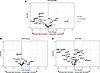 Figure 4
IIV4-induced changes in circulating protein concentrations in young and older adults. Volcano plots displaying the changes in proteins in the (A) placebo group, (B) young adults, and (C) older adults 60 days after vaccination. Blue dots represent the significantly downregulated proteins after vaccination; red dots show the significantly upregulated ones. A moderated 2-tailed t test was used in the R package limma to compare protein concentrations before and after vaccination (nominal P < 0.05).
The association between baseline inflammation and adaptive immunity responses to IIV4. The immune system’s baseline status before vaccination is crucial to predicting vaccine responses (23). Thus, we examined the relationship between baseline inflammation levels as assessed by circulating proteins and the antibody/IFN-γ responses to vaccination in both age groups combined and separately. In the IIV4 cohort, higher baseline inflammation was linked to lower fold-increases in A/Victoria/H1N1 and B/Phuket HAI titers, with proteins like CCL11, MCP-4, and HGF negatively associated with antibody responses (Figure 5, A and B).
 Figure 5
Correlation between circulating protein concentrations at baseline and IIV4-induced immune responses across combined age groups. (A) Heatmap depicting the association between baseline concentrations of circulating immune-related proteins and the fold antibody response (D60/D0) against the A/Victoria/H1N1 and B/Phuket strains after IIV4 vaccination. (B) Example correlation plots showing the relationship between CCL11 and MCP4 with the fold HAI titers to A/Victoria/H1N1 and B/Phuket. (C) Heatmap illustrating the correlation between baseline circulating immune-related proteins and the fold IFN-γ response (D60/D0 and D180/D0) from PBMCs after 7-day stimulation with 1 μg/mL of IIV4. (D) Example correlation plots of baseline CXCL10 levels with D60/D0 fold IFN-γ induction, and IL-10RA with D180/D0 fold IFN-γ production. Spearman’s correlation was used; the scales in A and C, along with the r values on the correlation graphs, represent the Spearman’s correlation coefficients. Stars on heatmaps indicate the nominal P values. NPX, normalized protein expression. *P < 0.05, **P < 0.01, ***P < 0.001.
Age-specific analysis showed baseline inflammation negatively linked to B/Phuket HAI responses in both age groups in general, while A/Victoria/H1N1 showed mixed associations (Supplemental Figure 7, A–C). No significant overlapping proteins were found in young and older adults.
Unlike neutralizing antibody titers, fold changes in IFN-γ were positively correlated with baseline systemic inflammation when both groups were combined (Figure 5, C and D). Higher baseline concentrations of IL-10RA were, however, linked to lower IFN-γ increases in the young group (Supplemental Figure 7D). In older adults, baseline inflammatory proteins like MCP-4, IL-8, CXCL9, and CXCL10 were positively associated with IFN-γ responses (Supplemental Figure 7, E and F).
Our results indicate that baseline inflammation is generally negatively associated with antibody production and positively linked with specific cellular IFN-γ responses to viral strains in IIV4. However, no distinct patterns were observed between age groups.
The impact of RZV on circulating immune mediators. Next, we analyzed the changes in circulating proteins after the placebo and RZV injections. After the first placebo injection, proteins like IL-17C, CXCL5, CXCL1, MCP-4, and MMP-1 increased, but only MMP-1 remained elevated after the second dose (Figure 6, A and B, and Supplemental Figure 8A). RZV had no notable influence on the investigated proteins in young adults (Figure 6, C and D). Interestingly, in older individuals, the first RZV dose reduced proteins such as MCP-1, MCP-4, CXCL5, and CXCL6, while the second dose broadly downregulated inflammation-related proteins like IL-10, TNF, IL-18R1, and TWEAK, among others (Figure 6, E and F, and Supplemental Figure 8B). These results suggest RZV reduces systemic inflammation in older adults.
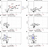 Figure 6
RZV-induced changes in circulating protein concentrations in young and older adults. Volcano plots displaying the changes in proteins in the (A and B) placebo group, (C and D) young adults, and (E and F) older adults after the first or second dose of vaccination compared with baseline (D0). A, C, and E exhibit the protein fold change after the first dose (D60/D0); B, D, and F show the protein fold change after the second dose (D120/D0). Blue dots represent the significantly downregulated proteins after vaccination; red dots show the significantly upregulated ones. A moderated 2-tailed t test was used in the R package limma to compare protein concentrations before and after vaccination (nominal P < 0.05).
The association between baseline inflammation and adaptive immune responses to RZV. Last, we evaluated the relationship between baseline inflammation and immune responses to RZV. Overall, higher levels of circulating immunomodulatory proteins were negatively correlated with antibody responses, even after 2 doses (Figure 7, A and B). Proteins like HGF, IL-8, IL-10RA, CSF-1, and OPG were associated with lower antibody responses in both age groups.
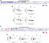 Figure 7
Correlation between circulating protein concentrations at baseline and RZV-induced immune responses across age groups. (A) Heatmap depicting the association between baseline concentrations of circulating immune-related proteins and the fold antibody response (D60/D0 and D120/D0) after RZV. (B) Example correlation plots showing the relationship between baseline levels of HGF and IL-8 with the fold antibody response after the first (D60/D0) and second dose (D120/D0). (C) Heatmap illustrating the correlation between baseline circulating immune-related proteins and the fold IFN-γ response (D60/D0, D120/D0, and D240/D0) from PBMCs after 7-day stimulation with 4 μg/mL of the gE antigen in RZV. (D) Example correlation plots of baseline MMP10 levels with D60/D0 fold IFN-γ, and CCL19 with D120/D0 and D240/D0 fold IFN-γ production. Spearman’s correlation was used; the scale in A and C, along with the r values on the correlation graphs, represent Spearman’s correlation coefficients. Stars on heatmaps indicate the nominal P values. NPX, normalized protein expression. *P < 0.05, **P < 0.01, ***P < 0.001.
Age-specific analysis showed stronger negative correlations in young adults, with proteins like CCL4 and CCL11 linked to lower antibody responses. In older adults, correlations were mixed; higher levels of CXCL9, CXCL10, and IFN-γ were associated with greater antibody induction (Supplemental Figure 9, A–C).
Baseline concentrations of circulating immune mediators were generally negatively correlated with IFN-γ responses at D60 and D240 after RZV, although some positive associations emerged after the second dose (D120/D0) (Figure 7C). Notably, higher baseline concentrations of CD6 and MMP10 were moderately linked to reduced IFN-γ response to the gE antigen after the first dose. In contrast, elevated CCL19 correlated with an increased IFN-γ fold change at D120 and D240 (Figure 7D).
When the age groups were separated, different patterns were observed (Supplemental Figure 9, D and E). In the RZV-young group, higher TWEAK concentrations consistently correlated with weaker IFN-γ responses (Supplemental Figure 9E). Mirroring the overall trend observed in the age-combined RZV group, baseline CD6 was negatively linked to IFN-γ response at D60 in the old group, while CCL19 was positively linked to IFN-γ at D120 and D240 (Supplemental Figure 9F).
Overall, baseline inflammation was negatively associated with RZV-induced adaptive responses when age groups were combined. However, when analyzed separately, different correlation patterns were seen, with older adults displaying a mix of responses, including more positive associations.


-
Discussion
Aging substantially affects vaccine responses, with many vaccines being less effective in older adults. Adjuvants have been developed to address this by enhancing immune activation and memory (24). In this study, we evaluated age-dependent differences in immune system responses to IIV4 and RZV. The results are summarized in Table 2. RZV induced strong antibody and IFN-γ responses in both age groups and reduced systemic inflammation in older adults. In contrast, IIV4 primarily increased neutralizing titers, stronger in young adults, but failed to induce effective cellular responses.
These results align with previous reports of strong cellular and humoral responses induced by RZV in both age groups and weak or no cellular responses from seasonal influenza vaccines (17, 25, 26). Complementing previous findings, we found that both vaccines were well tolerated, with an acceptable safety profile in both age groups (13, 27). Although this is a small cohort, it represents valuable information, as safety data on RZV in young adults has been limited compared with older adults (28).
The efficacy of vaccines is primarily mediated through the induction of specific antibodies and T cell responses. T cell responses play a key role in fighting infections and providing long-term protection. For example, memory T cells are especially important in controlling varicella zoster virus infections, which cause shingles (29). In addition, antigen-specific CD4+ T cells and IFN-γ production from these cells have been linked to protection against influenza, but their numbers decrease significantly in older adults (30). IIV4’s inability to enhance cellular responses likely contributes to its suboptimal effectiveness. An important function of adjuvants is to enhance the T cell memory response (31). For instance, AS01 in RZV contains 2 components: the TLR4 ligand monophosphoryl lipid A (MPLA) and saponin QS-21 derived from the Quillaja saponaria tree (32). MPLA induces the production of proinflammatory cytokines through the MyD88 pathway, and QS-21 activates the inflammasome and IL-18/IL-1β release. The synergistic effect of these 2 components is essential to achieve an optimal T cell response (33). Overall, AS01 triggers strong innate activation in draining lymph nodes via monocytes/macrophages, DCs, and NK cells and early release of IFN-γ in an IL-12/IL-18–dependent manner (33, 34).
Clinical studies with AS01, such as those done with tuberculosis and malaria vaccines, demonstrated strong antigen-specific CD4+ T cell responses and antibody production (35, 36). In this study, we found an RZV-induced robust increase in polyfunctional antigen-specific CD4+ T cells that express different combinations of CD40L, IL-2, TNF, and IFN-γ in both age groups. Notably, T cell polyfunctionality is linked to protection by various vaccines, such as those for COVID-19 and yellow fever (37, 38). Additionally, CD4+ T cell–mediated immunity after RZV was associated with better antibody responses for at least 3 years after vaccination (39). Because AS01 induces a robust and durable T cell response, its use could be further explored in vaccines targeting pathogens where strong cellular immunity is essential, as well as in populations with weakened immunity, including immunocompromised or older individuals. Interestingly, the type and magnitude of immune responses induced by AS01-adjuvanted vaccines vary across different antigens, suggesting that both the adjuvant and antigen contribute to forming the overall immunological profile (32).
An important finding of our study is that both vaccines induce CD40L on antigen-specific CD4+ T cells. This is important because recent work shows that CD40-CD40L signaling does more than provide costimulation: it can program long-lasting functional changes in human monocytes, similar to trained immunity (40). We found that CD40L+ polyfunctional CD4+ T cells are the main responding population after both IIV4 and RZV, and that these responses persist with aging, suggesting that CD40-CD40L interactions may be a common control point linking innate and adaptive immunity after vaccination. RZV, in particular, induces high levels of CD40L+IL-2+TNF+IFN-γ+CD4+ T cells in both age groups. In contrast, weaker CD4+ T cell responses to IIV4 may reflect limited CD40-driven support, helping explain its lower cellular immunogenicity and reduced efficacy in older individuals.
Intriguingly, both IIV4 and RZV modulated the circulating inflammatory proteins, particularly in older individuals. We observed different effects of the IIV4 vaccines administered in fall 2021 and fall 2022 on the proteins in the circulation of older adults, arguing for variation due to differences in the antigenic composition of the vaccines. However, the absence of a placebo group for the 2022/2023 season and the relatively small sample size limit the conclusions. Notably, RZV reduced several inflammatory proteins in older adults, with stronger effects after the second dose. This age-dependent response may be due to the inherently lower baseline levels of inflammatory mediators in young adults.
Consistent with previous studies, we found higher concentrations of inflammatory markers in older adults than in young adults. This is likely partly due to the higher rates of comorbidities and risk factors, such as hypertension and cardiovascular diseases (e.g., atherosclerosis) (41, 42). Elevated concentrations of inflammatory mediators in older adults indicate a state of chronic low-grade inflammation, commonly referred to as “inflammaging” (43). This persistent inflammation may impair the immune system’s ability to mount a robust antigen-specific response upon vaccination, possibly due to immune exhaustion or overactivation (44) due to continuous stimulation leading to diminished function and reduced responsiveness to new antigens. These mediators might also trigger regulatory feedback that weakens immune responses. Lowering systemic inflammation could prevent immune exhaustion and improve both innate and adaptive responses. As an example, the Bacillus Calmette-Guérin vaccine reduced systemic inflammation in a Dutch cohort of healthy participants, which was associated with trained immunity induction — the capacity of innate immune cells to develop a memory-like response (45). Consistently, our study found a strong negative association between inflammatory protein concentrations in the blood and antibody fold-increases in both vaccine groups.
In our study cohort, baseline inflammation was generally negatively correlated with PBMC-derived IFN-γ response after RZV, while it showed a positive association with IFN-γ response after IIV4. Interestingly, in the older volunteers vaccinated with IIV4, higher circulating concentrations of CXCL9, CXCL10, and IFN-γ were linked to higher IFN-γ production. In the old RZV group, higher CXCL9 and CXC10 were also correlated with greater antibody response. Since CXCL9 and CXCL10 are key chemokines involved in activating NK cells and Th1 immunity, it is tempting to speculate that they contribute to the enhanced type II IFN response observed after vaccination (46) and possibly to improved antibody production. Because this study only identifies associations but does not establish causation, future research should aim to understand better how these proteins affect vaccine responses, especially in different age groups. Additionally, future studies should explore strategies to modulate inflammatory proteins prior to or during vaccination by improving or developing adjuvants, with the aim of enhancing vaccine efficacy, especially in older adults.
This study demonstrated the moderate influence of IIV4 and RZV on immune cell subsets. IIV4 significantly increased WBCs in young adults, driven by lymphocyte and monocyte numbers and sustained until D180, whereas older adults showed elevated lymphocytes only at D7. On the other hand, RZV caused transient but stronger changes in cell counts in both age groups. A temporary increase in neutrophil and monocyte numbers and infiltration to the injected muscle has been reported after other vaccinations, suggesting immediate innate immune system activation and response (47, 48). The similar magnitude of changes in immune cell counts between young and older adults suggests that the AS01-adjuvanted RZV can achieve comparable initial innate immune cell activation in both age groups, supporting its effectiveness (34).
It is important to note that the RZV and RZV-associated placebo injections were administered during spring/summer (first and second dose), whereas IIV4 and IIV4-associated placebo injections were administered during fall. Seasonal variations are known to influence immune function and therefore vaccine-induced responses. Although the study design partially accounted for this by including 2 young placebo groups vaccinated in either spring or fall, helping to control for background seasonal effects, we cannot fully determine the impact of seasons on vaccine responses.
A limitation of this study is that a placebo group was only available for young participants, whereas older adults were all vaccinated due to ethical considerations. As a result, direct comparisons between vaccinated and unvaccinated older individuals could not be made, and age-related differences in immune responses must therefore be interpreted with some caution. Particularly for IIV4, where vaccine responses are strongly influenced by prior influenza infections and vaccination history, including an unvaccinated control group of older adults would have helped to better distinguish vaccine effects from those related to preexisting immunity.
In summary, this study highlighted the age-dependent vaccine responses after IIV4 and RZV exposure, focusing on cellular and humoral immunity, inflammatory mediators, and the influence of systemic inflammation prior to vaccination. Although RZV induced potent humoral and cellular immune responses while decreasing systemic inflammation in older adults, IIV4 was able to induce strong antibody production with weaker cellular responses. The incapacity of IIV4 to induce specific cellular immune responses may contribute to its year-dependent suboptimal efficacy. Our findings emphasize the role of adjuvants such as AS01 in enhancing vaccine responses in older adults, with modulating low-grade inflammation being a potential strategy. Further research is needed to explore the underlying transcriptional and epigenetic mechanisms driving different vaccine responses by RZV compared with IIV4. Understanding the immunological pathways contributing to increased vaccine immunogenicity could help refine existing adjuvants and develop new ones.


