
Infectious disease
Abstract
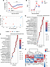
The role of CD8+ T cells in filarial infections remains poorly understood. Given the chronic nature of these infections, individuals living in endemic regions are frequently exposed to other pathogens including viruses. Because CD8 cells are essential for antiviral immunity, understanding how filarial infections shape the cytotoxic compartment is essential to elucidate their impact on bystander immunity. We evaluated the phenotype and function of CD8+ T cells from Loa loa-infected (Fil+) and -uninfected (Fil–) individuals at baseline and following cytomegalovirus (CMV) re-stimulation. Filarial infection was associated with increased activation and proliferation of CD8+ T cells, characterized by elevated expression of Ki-67, CD107a, and production of type 1 and 17 cytokines. Upon CMV re-stimulation, Fil+ individuals showed markedly reduced expansion of antigen-experienced (CD137+) and polyfunctional (CD137+IFN-γ+TNF-α+, CD137+IFN-γ+TNF-α+IL-2+) CD8+ T cells, along with decreased cytotoxic activity. Unbiased clustering analysis revealed markedly reduced frequency of the CD8+CD45RA+CD57+ subset in Fil+ individuals- a population further defined through transcriptomic profiling that showed enrichment for cytolytic gene signatures (GZMB, GNLY, GZMH, CD244, CX3CR1). These results demonstrated that filarial infection is associated with an altered CD8+ T cell profile that is associated with failure to mount effective viral-specific effector responses, including impaired cytokine production and cytotoxic activity.
Authors
Camila Queiroz-Glauss, Justin Lack, Daniel E. Sturdevant, Thomas B. Nutman
Abstract
Pneumocystis jirovecii pneumonia (PCP) remains a major cause of life-threatening respiratory failure in people with advanced HIV, yet the biological factors associated with mortality are incompletely understood. We performed integrated transcriptomic, proteomic, and metabolomic profiling of bronchoalveolar lavage fluid from 42 adults with HIV-associated PCP, relating molecular signatures to mortality. We found that fungal burden, measured by sputum immunofluorescence, Pneumocystis qPCR, and 1,3-β-D-glucan, did not differ between survivors and non-survivors or correlate with baseline gas exchange. Instead, non-survivors exhibited a bronchoalveolar immunometabolic failure signature characterised by marked depletion of immunoglobulins and complement, reduced pattern recognition receptors and fibroblast growth factor family proteins, and broad disruption of extracellular matrix organisation. Metabolomic profiling revealed enrichment of arginine-urea cycle and tricarboxylic acid pathways, with coordinated accumulation of arginine, citrulline, and TCA cycle intermediates consistent with nitrosative and mitochondrial stress. An integrated proteo-metabolomic score summarising this state was significantly higher in non-survivors and did not correlate with fungal burden or cytomegalovirus co-infection. In an immunodeficient precision-cut lung slice model, antigen-screened intravenous immunoglobulin restored macrophage-mediated Pneumocystis clearance, providing proof-of-concept for opsonic augmentation. Our findings indicate that mortality in HIV-PCP is defined by profound immunometabolic failure, highlighting humoral depletion and impaired alveolar repair as potential targets for host-directed adjunctive therapy.
Authors
Peter Rossi-Smith, Ayanda Trevor Mnguni, Dora Pungan, Robert J. Samuels, Vaishnavi R. Kumaran, Leena Syed, Trevor Ferris, Kamil Skirlo, Joseph N. Jarvis, Nelesh P. Govender, Graeme Meintjes, Sean Wasserman, Jay K. Kolls, Rachel P.J. Lai
Abstract
Tuberculosis (TB) caused by Mycobacterium tuberculosis (Mtb) is the leading infectious cause of death globally. Despite wide use of antiretroviral therapy (ART) by people living with HIV, the risk of TB remains increased. To understand immune interactions within lung granulomas, we compared spatial transcriptomics of Mtb and simian immunodeficiency virus (SIV) in co-infected macaques with or without ART as a model for HIV/Mtb co-infection. Spatially differentiated transcriptional profiles were observed in Mtb-only granulomas, with myeloid cells enriched in metabolic/antimicrobial pathways within the inner ring and T cell co-stimulatory/activation pathways enriched in the outer ring. These spatially distinct patterns were lost in SIV/Mtb granulomas with higher enrichment in type I IFN pathways compared to Mtb-only granulomas. SIV/ART/Mtb granulomas had an intermediate transcriptional pattern without restoration to Mtb-only granulomas, despite a lack of viral replication. Cell-cell communication was reduced among SIV/Mtb co-infected groups. These data suggest that HIV disrupts spatially organized immune functions of granulomas, which are not fully restored by ART.
Authors
Jessica M. Medrano, Persis Sunny, Collin R. Diedrich, Pauline Maiello, Christopher Kline, Tara Rutledge, Edwin Klein, Joshua T. Mattila, Jishnu Das, Zandrea Ambrose, Philana Ling Lin
Abstract
Patients who survive sepsis are predisposed to new hospitalizations for respiratory failure, but the underlying mechanisms are unknown. Using a murine model in which prior sepsis predisposes to enhanced lung injury, we previously discovered that classical monocytes persist in the lungs after long-term recovery from sepsis and exhibit enhanced cytokine expression after secondary challenge with intra-nasal lipopolysaccharide. Here, we hypothesized that immune reprogramming of post-sepsis monocytes and altered ontogeny predispose to enhanced lung injury. Monocyte depletion and/or adoptive transfer was performed three weeks and three months after sepsis. Monocytes from post-sepsis mice were necessary and sufficient for enhanced LPS-induced lung injury and promoted neutrophil degranulation. Prior sepsis enhanced JAK-STAT signaling and AP-1 accessibility in monocytes and shifted monocytes toward the neutrophil-like monocyte lineage. Neutrophil-like monocytes demonstrated a pro-inflammatory phenotype with enhanced IL-1β expression and reduced phagocytic capacity. In human sepsis and/or pneumonia survivors, monocytes were predictive of 90-day mortality and exhibit transcriptional and proteomic neutrophil-like signatures. We conclude that sepsis reprograms monocytes into a pro-inflammatory phenotype and skews bone marrow progenitors and monocytes toward the neutrophil-like lineage, predisposing them to induce neutrophil degranulation and lung injury.
Authors
Scott J. Denstaedt, Breanna McBean, Alan P. Boyle, Brett C. Arenberg, Matthias Mack, Bethany B. Moore, Michael W. Newstead, Yamei Deng, Alexey I. Nesvizhskii, Benjamin H. Singer, Jennifer Cano, Hallie C. Prescott, Helen S. Goodridge, Rachel L. Zemans
Abstract
Early antiretroviral therapy (ART) limits viral reservoir establishment in infants but also constrains viral-specific immunity required to clear reactivated cells. In the early ART context, we evaluated whether combining adeno-associated virus serotype 9 (AAV9)–vectored eCD4-IgG1 with pharmacologic latency reversal using the second mitochondria-derived activator of caspases (SMAC) mimetic AZD5582 could promote reservoir reduction and control in simian immunodeficiency virus (SIV)–infected infant macaques. AAV9 delivery achieved sustained eCD4-IgG1 expression and AZD5582 induced on-ART viremia, but the combination did not reduce intact SIV proviral DNA relative to controls. Partial post-intervention control of viremia during Analytical Treatment Interruption (ATI) occurred in 2/6 infants receiving AAV9-eCD4-IgG1 + AZD5582 with restricted reservoir expansion during recrudescence at week 13. Intact reservoir size during ATI correlated inversely with on-ART viremia during AZD5582 treatment. Controllers did not show increased eCD4-IgG1 expression nor increased on-ART viremia during AZD5582 treatment, but harbored less pre-ART intact SIV DNA in PBMCs than non-controllers. Controllers also exhibited higher frequencies and greater polyfunctionality of virus-specific CD8+ T-cells prior to ATI. These findings implicate immune control of reservoir size and post-ART viral dynamics in early-treated infants and suggest that AAV9-eCD4-IgG1 + AZD5582 increases the likelihood of post-ART viral control.
Authors
Jairo A. Fonseca, Alexis C. King, Kaleaha S. Davis, Daniel O'Hagan, Adrian Khoei, Lucas Alves Britto Da Costa, Camryn Cockerham, Mackenzie Cottrell, Stephanie Ehnert, Jennifer S. Wood, Michael D. Alpert, Matthew R. Gardner, Jeffrey D. Lifson, Maud Mavigner, Mauricio A. Martins, Ann Chahroudi
Abstract
Congenital and early-life Zika virus (ZIKV) infection can result in neurologic deficits. Precise mechanisms of injury, especially in the more subtle presentation of postnatal infection, are not fully elucidated. Here, we defined the effects of ZIKV on the developing brain using single cell transcriptomics, histopathology and design-based stereology, diffusion MRI, and neurobehavioral assessments in infant rhesus macaques. ZIKV upregulated interferon-stimulated genes in activated microglia and cell death pathways in neurons and downregulated metabolism and differentiation genes in mature oligodendrocytes. Abnormal micro-organization of the corpus collosum and limbic white matter tracts was seen on diffusion weighted imaging. A curated gene set associated with autism spectrum disorder risk was negatively enriched in inhibitory and excitatory neurons from ZIKV-infected infants, with increased emotional reactivity already evident two weeks following infection. From single cells to organism-level behaviors, these results define the pathways and processes disrupted by early-life ZIKV infection.
Authors
Venkata-Viswanadh Edara, Sienna Freeman, Maureen Sampson, Kathryn M. Moore, Rebecca Richardson, Nils Schoof, Divine Burgess, Michelle J. Lee, Gregory K. Tharp, Roza Vlasova, Shane Taylor, Ariana Hodjatzadeh, Yihana P. Melendez-Alejandro, Samia M. Abdallah, Esther W. Ndungu, Ava K. Mascarenhas, Chao-Hsiung Hsu, Tsang-Wei Tu, Martin Styner, Mehul S. Suthar, Mar M. Sanchez, Steven E. Bosinger, Mark W. Burke, Steven A. Sloan, Jessica Raper, Ann Chahroudi
Abstract
Severe SARS-CoV-2 infection is characterized by lung hyperinflammation, impaired IFN responses, and defective T cell activation, yet the molecular drivers of these immune dysregulations remain incompletely understood. Caspase-11 (CASP11), a key effector of the noncanonical inflammasome, has been shown to mediate an innate hyperinflammatory response and cytokine release in a mild SARS-CoV-2 infection model. However, the role played by CASP11 in severe SARS-CoV-2 disease and how it affects adaptive immunity has not been identified. Here, we found that CASP11 exacerbates severe SARS-CoV-2 pathogenesis by amplifying early innate immune responses while concurrently impairing antiviral CD8+ T cell immunity. Using global KO mice, hematopoietic BM chimeras, and Cx3cr1-expressing mononuclear phagocyte system cell–specific CASP11 deletion models, we show that targeting CASP11 reduces lung inflammation, promotes early NK cell–mediated IFN-γ production, and enhances robust virus-specific effector CD8+ T cell responses. This was associated with enhanced viral clearance and improved survival, even under lethal infection conditions. Importantly, CASP11-KO mice also exhibited faster resolution of postviral inflammation. These findings position CASP11 as a promising immunomodulatory target for acute and delayed manifestations of severe SARS-CoV-2 infection.
Authors
Mostafa M. Eltobgy, Mohamed M. Shamseldin, Owen D. Whitham, Heba M. Amer, Jeffrey R. Atkinson, Asmaa Badr, Jesse M. Hall, Gauruv Gupta, Yara Y. Hassan, Rabab El-Mergawy, Richard Perez, Sarah E. Faber, Maciej Pietrzak, Amy Webb, Xiaoli Zhang, Adam D. Kenney, Destiny Bissell, Jihad I. Omran, Shady Estfanous, Kylene P. Daily, Amir Yousif, Marisa R. Joldrichsen, Andrew McNamara, Mahesh KC, Mark E. Peeples, Emily A. Hemann, Hazem E. Ghoneim, Shahid M. Nimjee, Estelle Cormet-Boyaka, Jianrong Li, Prosper N. Boyaka, Jacob S. Yount, Benjamin M. Segal, Purnima Dubey, Amal O. Amer
Abstract
Mycoplasma pneumoniae pneumonia (MPP) can cause serious extrapulmonary complications, including life-threatening thrombosis. This study aimed to elucidate the roles of neutrophils and neutrophil extracellular traps (NETs) in vascular endothelial cell (EC) activation in pediatric MPP-associated thrombosis. We analyzed the relationship between neutrophils and thrombosis in children with MPP and used mouse models of neutrophilia (Csf3 plasmid injection), neutropenia (Csf3 deficient, Csf3–/–), and defective NETs formation (Pad4 deficient, Pad4–/–). The effects of neutrophils and NETs on EC activation were further examined in vivo, in vitro, and in human samples. Elevated neutrophil count was observed in patients with thrombosis and functioned as a potential diagnostic marker as well as a risk factor for MPP-associated thrombosis. EC activation was enhanced in MPP mice with neutrophilia but attenuated in neutropenic or Pad4–/– mice. NETs activated ECs through TLR2 and JAK/STAT3 signaling, and inhibition of NETs formation (Cl-amidine), TLR2 (C29), and JAK (upadacitinib) each attenuated this response. Strong correlations among neutrophils, NETs, EC activation, and thrombosis were observed in pediatric patients. These findings suggest that neutrophils promoted thrombosis in MPP via NETs-mediated EC activation involving TLR2 and JAK/STAT3 signaling. This study provides mechanistic insights into the inflammatory-thrombotic processes in MPP-associated thrombosis and offers a rationale for further investigation of neutrophils, NETs, TLR2, and JAK/STAT3 signaling in this context.
Authors
Xia Huang, Yifan Zhu, Yun Guo, Tian Lv, Haiyan Gu, Yingying Luo, Dan Li, Hang Sun, Deyu Zhao, Feng Liu
Abstract
Autophagy is a critical host defense mechanism that restricts intracellular pathogens such as Mycobacterium tuberculosis (Mtb). A key step in this process is the ubiquitination of Mtb or Mtb-associated structures. The E3 ligase SMURF1 catalyzes K48-linked ubiquitination, promoting bacterial clearance. However, the function of its homolog, SMURF2, in host defense remains undefined. Here, we demonstrate that Smurf2 deletion in murine macrophages increases SMURF1 levels, enhances LC3B lipidation, augments K48 ubiquitination of Mtb-associated structures, and reduces intracellular Mtb replication. These effects are reversed by Smurf1 deletion, supporting a role for SMURF1 in SMURF2-dependent control of Mtb. Mice with myeloid-specific Smurf2 deletion exhibit modestly prolonged survival following aerosol Mtb infection. In human macrophages, SMURF2 knockdown or its pharmacological inhibition with the HECT E3-ligase inhibitor Heclin reduces Mtb replication. Together, our findings identify SMURF2 as a negative regulator of macrophage control of Mtb and support further investigation of SMURF2 as a potential target for host-directed therapy in tuberculosis.
Authors
Priscila C. Campos, Kathryn C. Rahlwes, Victoria A. Ektnitphong, Beatriz R.S. Dias, Kubra F. Naqvi, Samuel Alvarez-Arguedas, Michael U. Shiloh

Abstract
Despite widespread vaccination, Bordetella pertussis (Bp) cases are resurging globally. Although CD4+ T cells are known to be essential for sustained protection, the antigens they recognize are not fully characterized, hindering vaccine refinement. Using immunopeptidomics, bioinformatics, and functional T cell assays, we identified high-affinity epitopes from reference and clinical Bp strains presented on MHC-II I-Ab. A subset of these epitopes stimulated systemic and mucosal CD4+ T cells of mice immunized with heat-killed Bp, and peripheral blood T cells from humans vaccinated with the whole-cell pertussis vaccine. Mice immunized with a subunit vaccine comprising two recombinant proteins identified in our screen were subsequently challenged with Bp. Bacterial burden was nearly eliminated from the lower respiratory tract and significantly reduced in the upper respiratory tract. Th1/Th17-polarized CD4+ tissue-resident memory T cells (Trms) were induced in nasal and pulmonary tissues. Depleting memory CD4+ T cells before challenge abolished protection, confirming that antigen-specific CD4+ T cells are critical for clearing Bp from the respiratory tract. Our integrated antigen identification and T cell assay approach revealed previously untested Bp antigens that elicit protective CD4+ T cell–mediated immunity, suggesting that incorporating them into new vaccines may help curb the resurgence of pertussis.
Authors
Mohamed M. Shamseldin, Jesse M. Hall, Griffin M. Lawrence, Gabrielle M. Hernandez, Yue Liu, Alexa R. Aubrey, Eva K. Verzani, Rajendar Deora, Amy Lovett-Racke, Jennifer G. Abelin, Purnima Dubey
No posts were found with this tag.


