
Research ArticleCardiologyInflammation
Open Access |  10.1172/jci.insight.197716
Noncoding RNA TY1 reverses fibrosis in systemic sclerosis by attenuating the cGAS/STING pathway
Xaviar M. Jones,1 Salwa Soussi,1 Alessandra Ciullo,1 Kara Tsi,1 Weixin Liu,1 Liang Li,1 Mario Fournier,1 Thassio Mesquita,1 Alberto M. Marchevsky,2 Nunzio Bottini,3 Francesco Boin,3 Ahmed G.E. Ibrahim,1 and Eduardo Marbán1
1Smidt Heart Institute,
2Department of Pathology and Laboratory Medicine, and
3Kao Autoimmunity Institute and Division of Rheumatology, Department of Medicine, Cedars-Sinai Medical Center, Los Angeles, California, USA.
Address correspondence to: Eduardo Marbán, Executive Director, Smidt Heart Institute, Cedars-Sinai Medical Center, 8700 Beverly Blvd., Los Angeles, California, 90048. USA. Phone: 310.423.7558; Email: eduardo.marban@csmc.edu.
Find articles by Jones, X. in: PubMed | Google Scholar
1Smidt Heart Institute,
2Department of Pathology and Laboratory Medicine, and
3Kao Autoimmunity Institute and Division of Rheumatology, Department of Medicine, Cedars-Sinai Medical Center, Los Angeles, California, USA.
Address correspondence to: Eduardo Marbán, Executive Director, Smidt Heart Institute, Cedars-Sinai Medical Center, 8700 Beverly Blvd., Los Angeles, California, 90048. USA. Phone: 310.423.7558; Email: eduardo.marban@csmc.edu.
Find articles by Soussi, S. in: PubMed | Google Scholar
1Smidt Heart Institute,
2Department of Pathology and Laboratory Medicine, and
3Kao Autoimmunity Institute and Division of Rheumatology, Department of Medicine, Cedars-Sinai Medical Center, Los Angeles, California, USA.
Address correspondence to: Eduardo Marbán, Executive Director, Smidt Heart Institute, Cedars-Sinai Medical Center, 8700 Beverly Blvd., Los Angeles, California, 90048. USA. Phone: 310.423.7558; Email: eduardo.marban@csmc.edu.
Find articles by Ciullo, A. in: PubMed | Google Scholar
1Smidt Heart Institute,
2Department of Pathology and Laboratory Medicine, and
3Kao Autoimmunity Institute and Division of Rheumatology, Department of Medicine, Cedars-Sinai Medical Center, Los Angeles, California, USA.
Address correspondence to: Eduardo Marbán, Executive Director, Smidt Heart Institute, Cedars-Sinai Medical Center, 8700 Beverly Blvd., Los Angeles, California, 90048. USA. Phone: 310.423.7558; Email: eduardo.marban@csmc.edu.
Find articles by Tsi, K. in: PubMed | Google Scholar
1Smidt Heart Institute,
2Department of Pathology and Laboratory Medicine, and
3Kao Autoimmunity Institute and Division of Rheumatology, Department of Medicine, Cedars-Sinai Medical Center, Los Angeles, California, USA.
Address correspondence to: Eduardo Marbán, Executive Director, Smidt Heart Institute, Cedars-Sinai Medical Center, 8700 Beverly Blvd., Los Angeles, California, 90048. USA. Phone: 310.423.7558; Email: eduardo.marban@csmc.edu.
Find articles by Liu, W. in: PubMed | Google Scholar
1Smidt Heart Institute,
2Department of Pathology and Laboratory Medicine, and
3Kao Autoimmunity Institute and Division of Rheumatology, Department of Medicine, Cedars-Sinai Medical Center, Los Angeles, California, USA.
Address correspondence to: Eduardo Marbán, Executive Director, Smidt Heart Institute, Cedars-Sinai Medical Center, 8700 Beverly Blvd., Los Angeles, California, 90048. USA. Phone: 310.423.7558; Email: eduardo.marban@csmc.edu.
Find articles by Li, L. in: PubMed | Google Scholar
1Smidt Heart Institute,
2Department of Pathology and Laboratory Medicine, and
3Kao Autoimmunity Institute and Division of Rheumatology, Department of Medicine, Cedars-Sinai Medical Center, Los Angeles, California, USA.
Address correspondence to: Eduardo Marbán, Executive Director, Smidt Heart Institute, Cedars-Sinai Medical Center, 8700 Beverly Blvd., Los Angeles, California, 90048. USA. Phone: 310.423.7558; Email: eduardo.marban@csmc.edu.
Find articles by Fournier, M. in: PubMed | Google Scholar
1Smidt Heart Institute,
2Department of Pathology and Laboratory Medicine, and
3Kao Autoimmunity Institute and Division of Rheumatology, Department of Medicine, Cedars-Sinai Medical Center, Los Angeles, California, USA.
Address correspondence to: Eduardo Marbán, Executive Director, Smidt Heart Institute, Cedars-Sinai Medical Center, 8700 Beverly Blvd., Los Angeles, California, 90048. USA. Phone: 310.423.7558; Email: eduardo.marban@csmc.edu.
Find articles by
Mesquita, T.
in:
PubMed
|
Google Scholar
|

1Smidt Heart Institute,
2Department of Pathology and Laboratory Medicine, and
3Kao Autoimmunity Institute and Division of Rheumatology, Department of Medicine, Cedars-Sinai Medical Center, Los Angeles, California, USA.
Address correspondence to: Eduardo Marbán, Executive Director, Smidt Heart Institute, Cedars-Sinai Medical Center, 8700 Beverly Blvd., Los Angeles, California, 90048. USA. Phone: 310.423.7558; Email: eduardo.marban@csmc.edu.
Find articles by Marchevsky, A. in: PubMed | Google Scholar
1Smidt Heart Institute,
2Department of Pathology and Laboratory Medicine, and
3Kao Autoimmunity Institute and Division of Rheumatology, Department of Medicine, Cedars-Sinai Medical Center, Los Angeles, California, USA.
Address correspondence to: Eduardo Marbán, Executive Director, Smidt Heart Institute, Cedars-Sinai Medical Center, 8700 Beverly Blvd., Los Angeles, California, 90048. USA. Phone: 310.423.7558; Email: eduardo.marban@csmc.edu.
Find articles by
Bottini, N.
in:
PubMed
|
Google Scholar
|
1Smidt Heart Institute,
2Department of Pathology and Laboratory Medicine, and
3Kao Autoimmunity Institute and Division of Rheumatology, Department of Medicine, Cedars-Sinai Medical Center, Los Angeles, California, USA.
Address correspondence to: Eduardo Marbán, Executive Director, Smidt Heart Institute, Cedars-Sinai Medical Center, 8700 Beverly Blvd., Los Angeles, California, 90048. USA. Phone: 310.423.7558; Email: eduardo.marban@csmc.edu.
Find articles by Boin, F. in: PubMed | Google Scholar
1Smidt Heart Institute,
2Department of Pathology and Laboratory Medicine, and
3Kao Autoimmunity Institute and Division of Rheumatology, Department of Medicine, Cedars-Sinai Medical Center, Los Angeles, California, USA.
Address correspondence to: Eduardo Marbán, Executive Director, Smidt Heart Institute, Cedars-Sinai Medical Center, 8700 Beverly Blvd., Los Angeles, California, 90048. USA. Phone: 310.423.7558; Email: eduardo.marban@csmc.edu.
Find articles by Ibrahim, A. in: PubMed | Google Scholar
1Smidt Heart Institute,
2Department of Pathology and Laboratory Medicine, and
3Kao Autoimmunity Institute and Division of Rheumatology, Department of Medicine, Cedars-Sinai Medical Center, Los Angeles, California, USA.
Address correspondence to: Eduardo Marbán, Executive Director, Smidt Heart Institute, Cedars-Sinai Medical Center, 8700 Beverly Blvd., Los Angeles, California, 90048. USA. Phone: 310.423.7558; Email: eduardo.marban@csmc.edu.
Find articles by Marbán, E. in: PubMed | Google Scholar
Published September 8, 2026 - More info
JCI Insight. 2026;11(17):e197716. https://doi.org/10.1172/jci.insight.197716.
© 2026 Jones et al. This work is licensed under the Creative Commons Attribution 4.0 International License. To view a copy of this license, visit http://creativecommons.org/licenses/by/4.0/.
Received: July 14, 2025;

-
Results
TY1 attenuates DNA damage and cGAS/STING in human SSc macrophages. To evaluate DNA damage, we quantified γ-H2AX, a phosphorylated form of the histone protein H2AX, which serves as a sensitive marker for DNA double-stranded breaks in macrophages from healthy human donors (control) or patients with SSc (Supplemental Table 1; supplemental material available online with this article; https://doi.org/10.1172/jci.insight.197716DS1) (14). The experimental protocol to generate human macrophage in culture is depicted in Figure 1A. Compared with control, γ-H2AX was elevated in SSc macrophages exposed to vehicle, while SSc macrophages demonstrated a trend toward increased cGAS/STING signaling compared with healthy donor–derived macrophages (Figure 1, B and C). To probe the role of DNA damage, we used the RNA drug TY1, which enhances the clearance of damaged DNA fragments and attenuates nuclear DNA damage (12). Strikingly, SSc macrophages exposed to TY1 decreased γ-H2AX, reducing it to control levels. To assess whether the effects of TY1 were specific to its sequence, we used a scrambled RNA of the same nucleotide content as TY1 (scramble) but reordered so as to lack any homology to the human or murine genomes. Previous in vitro and in vivo assays have verified the biological inertness of scramble (12). Indeed, transfection with scramble had no effect on the percentage of γ-H2AX+ SSc macrophages (Figure 1, B and C). Similarly, TY1, but not scramble, showed upregulation of RNA expression by qPCR in both healthy and SSc macrophages (Supplemental Figure 1, A and B). TY1’s dramatic attenuation of DNA damage is consistent with its known mechanism of action (12), while the inactivity of scramble affirms the specificity of TY1’s benefits.
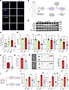 Figure 1
Human SSc macrophages and fibroblasts exhibit attenuation of cGAS-STING and profibrotic cytokines in response to TY1. (A) Schematic describing the experimental protocol for human SSc macrophage generation and exposure to vehicle, scramble or TY1 for 24 hours (n = 6 biological replicates). Confocal microscopy images of γ-H2AX staining in healthy macrophages or SSc macrophages exposed to Vehicle, Scramble or TY1 (n = 5 biological replicates). Magnification ×630. (B and C) Representative images and pooled data for % of γ-H2AX positive cells. (D and E) Representative images and analysis of Western blots showing decreases of cGAS, STING, total TBK-1, and total IRF-3 protein levels in SSc macrophages transfected with TY1 compared with vehicle (n = 6 biological replicates/group). (F and G) Quantification of interferon-β and 2’3 cGAMP in conditioned media from scramble- and TY1-transfected SSc macrophages by ELISA (n = 6 biological replicates/group). (H) Conditioned media from TY1 transfected macrophages show decreased levels of proinflammatory mediators by Proteome Profiler Human Cytokine Array (n = 3 biological replicates/group). (I) Schematic of the experimental protocol for human SSc fibroblast incubation with diluted SSc macrophage-conditioned media (n = 6 biological replicates). (J and K) Downregulation of mRNA levels of pro-fibrotic markers and secreted collagen in SSc fibroblasts exposed to TY1-transfected SSc macrophages. Statistical analysis by repeated-measures ANOVA followed by Tukey post hoc test with 95% CI; *P < 0.05, **P < 0.01, ***P < 0.001, ****P < 0.0001. Figure created with BioRender (biorender.com) under a Cedars Sinai Medical Center license.
We next investigated whether the genoprotective effect of TY1 influences downstream cGAS/STING signaling. Cell lysates from human SSc macrophages in primary culture exhibited reduced protein expression of cGAS, STING, TBK1, and IRF-3 after exposure to TY1, relative to scramble or vehicle (Figure 1, D and E). TY1 also decreased the levels of 2′3′-cGAMP and IFN-β in conditioned media (CM) from SSc macrophages (Figure 1, F and G). Inflammatory cytokines such as CXCL-10 are secreted by SSc macrophages, turning on IFN production and contributing to tissue damage and ultimately fibrosis in SSc. In media conditioned by SSc macrophages, CXCL-10 levels were reduced by TY1 exposure (Figure 1H). To test if TY1 requires Trex1 for its therapeutic effects, we knocked down Trex1 using siRNA in SSc macrophages (Supplemental Figure 1, C–E). When Trex1 was suppressed, TY1 had little effect on fibrosis in SSc fibroblasts, as demonstrated by unchanged gene expression of the profibrotic markers COL1A1, CTGF, POSTN, and TGFβ (Supplemental Figure 1D) as well as unchanged collagen production (Supplemental Figure 1E). Collectively, the data demonstrate that TY1 suppresses DNA damage and cGAS/STING signaling by TY1 in human SSc macrophages and leads to downstream tissue repair.
TY1 prevents fibrotic activation in human fibroblasts. During SSc progression, tissue resident fibroblasts differentiate into myofibroblasts, which secrete factors stimulating canonical and noncanonical profibrotic pathways (15, 16). Interactions between fibroblasts and macrophages modulate this process (17–20), as exemplified by the profibrotic activation of human cardiac fibroblasts upon exposure to human SSc macrophage CM (21). To dissect macrophage-fibroblast cross-talk, we incubated healthy dermal fibroblasts with TGF-β in the presence of TY1 or vehicle (Supplemental Figure 2A). After 24 hours, fibroblasts exposed to TY1 exhibited lower gene expression of the myofibroblast marker α smooth muscle actin (αSMA) and a trend toward lower expression of Collagen 1A1 (COL1A1; Supplemental Figure 2B). The same experimental design, applied to human SSc fibroblasts, produced similar results including reduced expression of Collagen 1A2 (COL1A2; Supplemental Figure 2C). Previous studies using i.v. and oral delivery demonstrate that TY1 is taken up primarily by macrophages, which, in turn, are necessary and sufficient for TY1’s therapeutic effects (12, 13). To look for possible indirect effects mediated by macrophage-secreted factors, we incubated SSc fibroblasts with media conditioned by SSc macrophages transfected with TY1 or scramble (diluted 75% by volume with conventional fibroblast media; Figure 1I). A separate group of SSc fibroblasts, cultured in conventional fibroblast media, but no CM, served as control. Notably, SSc fibroblasts exposed to CM from TY1-transfected SSc macrophages, but not CM from scramble-transfected SSc macrophages, exhibited decreased expression of profibrotic markers CTGF, POSTN, and COL1A1 to transcript levels comparable with those in control. Moreover, SSc fibroblasts exposed to CM from scramble-transfected SSc macrophages expressed elevated col1a1 message levels (Figure 1J). Reductions in levels of profibrotic genes CGTF and POSTN paralleled the TY1-induced changes in collagen expression (Figure 1J). Collagen secretion was also reduced in fibroblasts directly exposed to TY1 (Figure 1K). These findings support the concept that TY1 prevents myofibroblast differentiation through both direct effects and paracrine immunomodulation by SSc macrophages.
TY1 attenuates cachexia and fibrosis in bleomycin-injected mice. If DNA damage and subsequent canonical cGAS/STING activation are pathogenic in SSc, it is logical to predict that TY1 would have disease-modifying bioactivity, as it does in models of myocardial infarction and of heart failure with preserved ejection fraction (12, 22). While no single animal model of SSc reproduces all features of the human disease (23), repeated bleomycin administration causes cachexia, tissue inflammation, and fibrosis of the skin and internal organs, mimicking SSc (24–26). After subdermal bleomycin injections over the course of 3 weeks (at which time the disease phenotype was already evident), mice were randomized to receive vehicle, scramble, or TY1 via oral gavage in casein-chitosan micelles twice weekly for 4 weeks (Figure 2A) (13). Oral administration produced benefits comparable with those of parenteral TY1 (Supplemental Figure 3) and was chosen due to its greater translational relevance. As expected (27), administration of bleomycin led to significant weight loss, but animals fed TY1 showed some weight recovery compared with animals fed vehicle or scramble (Figure 2, B and C). To assess the effects of TY1 on fibrosis, we analyzed lung and skin biopsies from bleomycin-injected mice. Relative to controls, the skin was thickened and fibrotic (histologically and by hydroxyproline content) in bleomycin-injected mice that received vehicle or scramble, but TY1 reversed the skin abnormalities to near-control levels (Figure 2, D–F). Such antifibrotic effects were not confined to skin tissue. Lung samples from TY1 animals also showed less fibrosis (lower Ashcroft score and hydroxyproline content; ref. 28), accompanied by attenuated pulmonary congestion (Figure 2, G–K).
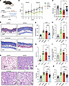 Figure 2
Therapeutic effects of TY1 in mice with skin thickening and SSc-like cardiopulmonary fibrosis after s.c. bleomycin injections. (A) Schematic of the experimental protocol. Vehicle-injected (control) or bleomycin-injected mice were fed with vehicle, scramble or TY1 by oral gavage for 4 weeks (control n = 10; vehicle n = 9; scramble n = 9; TY1 n = 20). (B and C) Changes in body mass over time and pooled endpoint data. (D and E) Representative Masson trichrome images of skin from control mice and bleomycin-injected mice fed vehicle, scramble or TY1, and pooled data for dermal thickness. Scale bar: 2mm. (F) Hydroxyproline content in skin tissue (n = 4–5 samples /group). (G) Representative Masson trichrome images of lung from vehicle- and bleomycin-injected mice fed vehicle, scramble or TY1. Scale bar: 500 µm. (H) Analysis of pulmonary fibrosis by Ashcroft score (n = 4–6 samples/group). (I) Measurement of hydroxyproline content in lung tissue (n = 3–6 samples /group). (J and K) Analysis of lung congestion by lung mass, and lung weight/tibial length. Statistical analysis by repeated-measures ANOVA followed by Tukey post hoc test with 95% CI; *P < 0.05, **P < 0.01, ****P < 0.0001. Figure created with BioRender (biorender.com) under a Cedars Sinai Medical Center license.
Cardiovascular complications are a prominent cause of SSc-related death (29, 30). In particular, cardiac fibrosis and diastolic dysfunction (an early marker of restrictive cardiomyopathy) are diagnosed in 18%–62% of patients with SSc and herald a poor prognosis (31, 32). We (21) and others (33–35) have demonstrated cardiac fibrosis and impaired systolic and diastolic function in bleomycin-induced mice, mimicking the phenotype of patients with SSc with heart involvement. Hearts from TY1-treated mice displayed much less cardiac fibrosis than hearts from mice that had received vehicle or scramble (Figure 3, A and B), without evidence of hypertrophy (Figure 3C). Echocardiography at endpoint revealed diastolic dysfunction in vehicle and scramble groups, but not in TY1 mice (Figure 3D), which exhibited improved e’ and reduced E/e’ ratio (Figure 3, E and F) with preserved ejection fraction (Figure 3G). Furthermore, exercise endurance was impaired in vehicle or scramble groups but restored to control levels in TY1 mice (Figure 3H).
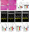 Figure 3
Hearts of mice fed TY1 exhibit antifibrotic effects and attenuation of diastolic dysfunction. (A and B) Representative Masson trichrome cardiac images from vehicle and bleomycin-injected mice treated with vehicle, scramble or TY1, and pooled analysis of percentage of fibrosis. Scale bar: 200 µm. (C) Pooled data for cardiac hypertrophy, measured by heart weight/tibial length. (D) Representative echocardiographic M-mode, tissue Doppler, and pulse wave Doppler images. (E–G) Echocardiographic tissue Doppler early diastolic mitral annular velocity (e’), ratio of transmitral Doppler early filling velocity to tissue Doppler early diastolic mitral annular velocity (E/e’), and ejection fraction (EF, expressed as percent). (H) Maximal distance during treadmill exercise (m) by mice in the various experimental groups. Statistical analysis by Student’s t test or repeated-measures ANOVA followed by Tukey post hoc test with 95% CI; **P < 0.01, ***P < 0.001, ****P < 0.0001.
TY1 prevents skin and cardiac fibrosis in tsk-1 mice. The bleomycin model of SSc exhibits inflammation-induced fibrosis. To determine if TY1 also modifies disease when fibrotic signaling is less directly coupled to inflammation, we used the tight skin (tsk-1) mouse model of SSc, which harbors a mutation in the fibrillin-1 gene, a component of extracellular microfibrils abundant in skin of patients with SSc (36, 37). Homozygous tsk-1 mice die in utero, while heterozygous littermates show elevated circulating autoantibodies and robust dermal and cardiac fibrosis without overt inflammation — a pattern which resembles advanced fibroproliferative stages of SSc. Accordingly, oral gavage of vehicle, scramble, or TY1 was initiated in 5-week-old heterozygous tsk-1 mice (Supplemental Figure 4A). Four weeks later, vehicle-fed tsk-1 mice showed elevated levels of circulating autoantibody Scl-70, as well as prominent skin fibrosis evidenced by increased dermal thickness and hydroxyproline content. TY1 blunted or fully reversed the increases in Scl-70 serum levels (Supplemental Figure 4B), dermal thickness (Supplemental Figure 4, C and D), and skin hydroxyproline content (Supplemental Figure 4E). Cardioprotective effects were also evident: TY1-fed tsk-1 mice had less cardiac fibrosis (Supplemental Figure 4, F and G) and less hypertrophy than vehicle or scramble mice (Supplemental Figure 4H). Thus, TY1 exerts therapeutic benefits not only in human SSc cells but also in 2 complementary mouse models of SSc, consistent with the hypothesis that DNA damage is a central pathogenic feature that transcends the limitations of any single model.
TY1 modulates the immune response and attenuates cGAS/STING in bleomycin-injected mice. To further characterize the benefits of TY1 in SSc, we performed bulk RNA-seq of skin from control (i.e., healthy, no bleomycin), vehicle, and TY1 animals at study endpoint (Figure 4). Heatmaps reveal numerous transcriptional differences between control and vehicle-injected skin, most of which were reversed, in a remarkably coordinated manner, by TY1 (Figure 4A). By gene ontology analysis, pathways related to inflammatory activation were especially attenuated by TY1; these include responses to IFN-β, IL-1, and IL-6 production and regulation of the innate immune response (Figure 4B). Genes of the canonical cGAS/STING pathway and the type I IFN response were markedly downregulated in TY1 mice, again in a strikingly coordinated fashion (Figure 4C). Figure 4D shows qPCR confirmation of the pattern of changes in expression of inflammatory genes IRF7, IFI44, Mx1, IFN-β, and IL-6: upregulation in vehicle samples but attenuation in the TY1 group. In contrast, DNA exonuclease Trex1 was suppressed in vehicle but upregulated by TY1 (Figure 4E), consistent with the RNA drug’s upstream mechanism of action to suppress DNA damage (12). At the protein level, vehicle mice displayed increased levels of circulating proinflammatory mediators C-reactive protein (CRP), CXCL-2, EGF, IL-7, and LIF, while TY1 blunted these responses (Supplemental Figure 5). To determine whether dysregulated inflammation and skin fibrosis result from activated canonical cGAS/STING activity, we analyzed whole-skin lysates. Protein levels of cGAS and STING, and downstream effectors TBK1 and IRF-3, were elevated in skin samples of vehicle mice compared with controls but reduced to normal levels in the skin of TY1 mice (Figure 4, F and G). TY1 prominently reduced the protein levels of cGAS and STING in skin, while TBK1 and IRF-3 showed nonsignificant trends toward normalization (Figure 4, F and G). In contrast, Trex1 protein levels remain unchanged across groups in this bulk tissue assay, not inconsistent with the concept that upregulation of TREX1 in macrophages underlies the effects of TY1. IFN-β levels were markedly elevated in skin lysates from vehicle mice compared with healthy controls but blunted by TY1 (albeit insignificantly; Figure 4H). Taken together, these data further support the hypothesis that activation of cGAS/STING contributes to skin fibrosis in the bleomycin SSc model. The molecular changes in mice faithfully reproduce, and extend, those seen in human SSc macrophages and fibroblasts exposed to TY1 (Figure 1).
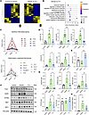 Figure 4
Transcriptomic and protein-level attenuation of cGAS-STING and downstream effectors by TY1 in skin from bleomycin-injected mice. (A) Heatmap of transcriptomic sequencing from lesional skin tissue from control and bleomycin-injected mice treated with vehicle or TY1 (n = 3–4 biological replicates/group). (B) Gene ontology analysis identifies downregulation of DNA damage response, IFN, and inflammatory pathways as major implicated in lesional skin in TY1-treated mice. (C) Downregulation of interferon-stimulated and inflammatory genes. (D) qPCR validation TY1 inflammatory modulation in skin tissue from control and bleomycin-injected mice treated with vehicle or TY1 (n = 3–4 biological replicates/group). (E) Trex1 expression in skin tissue from control and bleomycin-injected mice treated with vehicle, scramble, or TY1 by qPCR (n = 3–4 biological replicates/group). (F and G) Representative images and analysis of Western blots showing decreases of cGAS, STING, TBK-1, and IRF-3 protein levels in skin tissue of animals receiving TY1 compared with vehicle (n = 3 biological replicates/group). (H) Measurement of IFN-β in lesional skin from control and bleomycin-injected mice treated with vehicle or TY1 by ELISA (n = 3 biological replicates/group). Statistical analysis by repeated-measures ANOVA followed by Tukey post hoc test with 95% CI; *P < 0.05, **P < 0.01.


