Issue published September 8, 2026 Previous issue | Next issue

- Volume 11, Issue 17
On the cover:
Genetic background influences developmental airway smooth muscle program and susceptibility to airway hyperresponsiveness in mice
Otoshi et al. report a genetic background-dependent hyperactive TGF-β/PDGFRA/TNC program unleashed by a brief in utero exposure to a vitamin A/retinoic acid–deficient environment, resulting in structural remodeling of the airway smooth muscle and hyperresponsiveness in adult mice. The cover image shows the aberrant ectopic expansion of PDGFRA (red) where airway smooth muscle emerges in the embryonic lung near distal epithelial buds (SOX9, green) and airway epithelial progenitors (SOX2, cyan).
Otoshi et al. report a genetic background-dependent hyperactive TGF-β/PDGFRA/TNC program unleashed by a brief in utero exposure to a vitamin A/retinoic acid–deficient environment, resulting in structural remodeling of the airway smooth muscle and hyperresponsiveness in adult mice. The cover image shows the aberrant ectopic expansion of PDGFRA (red) where airway smooth muscle emerges in the embryonic lung near distal epithelial buds (SOX9, green) and airway epithelial progenitors (SOX2, cyan).
Go to section:
-
Research Letters
×
Abstract
Authors
Ewa A. Ziółkowska, Letitia Williams, Elizabeth Eultgen, Grace R. Kick, Xukai Ding, Alexander Sorensen, Steven Le, Balraj Doray, Patricia I. Dickson, Robert O. Heuckeroth, Jonathan D. Cooper
×Abstract
Authors
Lun Li, Rhonda Lightle, Bader Ali, Georgeio Sader, Robert Shenkar, Sean P. Polster, Douglas A. Marchuk, Jan-Karl Burkhardt, Issam A. Awad, Mark L. Kahn


-
Review
×
Abstract
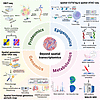
Coordinated changes in gene expression, epigenetic regulation, protein and metabolic activities together drive disease progression and determine clinical outcomes. While spatially resolved transcriptomics has been widely adopted across biomedical fields, it offers an incomplete picture limited to transcriptomic levels. Here, we survey the latest developments in spatial multiomics technologies, with particular emphasis on platforms that extend beyond conventional transcriptomics and profile genomics, epigenomics, proteomics, or metabolomics within intact tissues. These approaches are rapidly becoming commercialized, and here we highlight major technical breakthroughs, enhanced sample compatibility, emerging applications, and computational tools for data analysis. This Review aims to equip researchers with a clear understanding of the current technological landscape and to accelerate the adoption of spatial multiomics methods in biomedical research.
Authors
Xinchen Mao, Zhuo Chen, Emily J. Hwang, Jun Liu, Junrou Huang, Haikuo Li
-
Research Articles
×
Abstract
Estrogen can promote aggressive tumor phenotypes in estrogen receptor–positive (ER+) breast cancer; however, ER– cell lines are not widely considered estrogen responsive. Noncanonical estrogen-stimulated pathways such as the membrane-bound G protein–coupled estrogen receptor (GPR30) can mediate migratory and proliferative phenotypes in breast cancer and are postulated to promote resistance to aromatase therapies. Moreover, dysregulation of UDP-glucose 6-dehydrogenase (UGDH), a ubiquitously expressed enzyme critical to the metabolism of UDP-glucuronic acid into extracellular matrix precursors and hormone regulation, is associated with tumorigenesis. Here, we illustrated the impact of estrogen stimulation on tumor phenotypes in ER+ and ER– cell models in vitro and in vivo. We then demonstrated UGDH’s association with metastatic breast cancer via single-cell sequencing of patient specimens. Genetic knockdown of UGDH blunted estrogen-stimulated tumor phenotypes in vitro, ex vivo, and in vivo using both ER+ and ER– breast cancer lines. Finally, we demonstrated that UGDH knockdown blunted noncanonical estrogen stimulation through GPR30. Ultimately, our study validated prior studies demonstrating estrogen-responsive malignant phenotypes in ER– breast cancer and demonstrated that estrogen-stimulated breast cancer progression can be mediated through noncanonical pathways (e.g., UGDH/GPR30), regardless of ER status.
Authors
Meghan J. Price, Annee D. Nguyen, Corinne H. Strawser, Trupti Trivedi, Cesar C.D. Baeta, Catherine Lavau, Jovita K. Byemerwa, Debarati Mukherjee, Suzanne E. Wardell, Sandeep Artham, Vardhman Kumar, Shyni Varghese, C. Rory. Goodwin
×Abstract
DNA damage and the cGAS/STING innate immunity pathway have been associated with fibrosis in systemic sclerosis (SSc), but a cause-and-effect role has not been established. Here we report the effects of TY1, a noncoding RNA drug of the exomer class that suppresses DNA damage and thereby inhibits cGAS/STING, in human SSc cells and in 2 preclinical models of SSc. Macrophages from patients with SSc exhibited high levels of phosphorylated DNA damage, cGAS, 2’3’-cGAMP, STING, and IFNs, all of which decreased after exposure to TY1. In mice that had been injected s.c. with bleomycin to model SSc, exercise tolerance, cardiac function, lung hydroxyproline, and skin thickness reverted to normal levels after oral administration of TY1. Similar therapeutic benefits were evident in the genetic tsk-1 mouse model of SSc. TY1 attenuated fibrosis and/or fibrotic gene expression in both mouse models of SSc and in human SSc skin fibroblasts. Our findings support the hypothesis that cGAS/STING, activated by DNA damage, is a key driver of fibrosis in SSc.
Authors
Xaviar M. Jones, Salwa Soussi, Alessandra Ciullo, Kara Tsi, Weixin Liu, Liang Li, Mario Fournier, Thassio Mesquita, Alberto M. Marchevsky, Nunzio Bottini, Francesco Boin, Ahmed G.E. Ibrahim, Eduardo Marbán
×Abstract
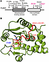
The FLAD1 gene codes for flavin adenine dinucleotide (FAD) synthase. FAD is a cofactor for many redox enzymes involved in vital processes from respiration to signal transduction. In this work, we described a clinical case of 2 siblings carrying compound heterozygous mutations in the FLAD1 gene resulting in the substitutions A418V and R542* at the protein level. The patients demonstrate adrenal insufficiency, which has not previously been associated with FLAD1 protein defects. To verify that adrenal insufficiency is caused by FLAD1 mutations, we created a personalized mouse model carrying the mutations found in the patients. The mutation in the FLAD1 gene, leading to the A418V substitution, appeared viable in the homozygous state, with minimal difference from the WT. The FLAD1 gene mutation leading to the R542* truncation is lethal when homozygous. The mouse model of the compound heterozygous FLAD1A418V/R542* mutations recapitulated the physiological, biochemical, and endocrine manifestations of FLAD1 mutations in patients. The mouse model created demonstrates the causal effect of FLAD1 mutations on the described pathology and potentially paves the way for understanding the disease’s molecular mechanism and developing better therapies.
Authors
Olga A. Averina, Natalia Yu. Kalinchenko, Vitaly A. Ioutsi, Andrey V. Pirogov, Alexander V. Bogachev, Oleg A. Permyakov, Vitaly S. Buev, Ekaterina A. Guseva, Anastasia V. Priymak, Olga A. Bazhanova, Mariia A. Emelianova, Olga O. Grigoryeva, Galina V. Baydakova, Maxim A. Abakumov, Vasily N. Manskikh, Olga A. Dontsova, Petr V. Sergiev, Anatoly N. Tiulpakov
×Abstract
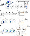
Despite widespread vaccination, Bordetella pertussis (Bp) cases are resurging globally. Although CD4+ T cells are known to be essential for sustained protection, the antigens they recognize are not fully characterized, hindering vaccine refinement. Using immunopeptidomics, bioinformatics, and functional T cell assays, we identified high-affinity epitopes from reference and clinical Bp strains presented on MHC-II I-Ab. A subset of these epitopes stimulated systemic and mucosal CD4+ T cells of mice immunized with heat-killed Bp, and peripheral blood T cells from humans vaccinated with the whole-cell pertussis vaccine. Mice immunized with a subunit vaccine comprising two recombinant proteins identified in our screen were subsequently challenged with Bp. Bacterial burden was nearly eliminated from the lower respiratory tract and significantly reduced in the upper respiratory tract. Th1/Th17-polarized CD4+ tissue-resident memory T cells (Trms) were induced in nasal and pulmonary tissues. Depleting memory CD4+ T cells before challenge abolished protection, confirming that antigen-specific CD4+ T cells are critical for clearing Bp from the respiratory tract. Our integrated antigen identification and T cell assay approach revealed previously untested Bp antigens that elicit protective CD4+ T cell–mediated immunity, suggesting that incorporating them into new vaccines may help curb the resurgence of pertussis.
Authors
Mohamed M. Shamseldin, Jesse M. Hall, Griffin M. Lawrence, Gabrielle M. Hernandez, Yue Liu, Alexa R. Aubrey, Eva K. Verzani, Rajendar Deora, Amy Lovett-Racke, Jennifer G. Abelin, Purnima Dubey
×Abstract
Foxp3 deficiency causes a profound loss of immune tolerance, unleashing autoreactive T and B cells, lymphoproliferation, cytokine-driven inflammation, and autoantibody production. This autoimmune pathology is fueled by increased glutamine usage, but it remains unresolved whether glutamine is necessary to produce energy or for intermediate metabolite biosynthesis responsible for immunomodulation. Here, we demonstrate that glutamine utilization for biosynthetic pathways supported autoimmune inflammation in the settings of Foxp3 deficiency and dextran sodium sulfate–induced colitis. By employing a model of autoimmunity driven by Treg-specific loss of Foxp3, we showed that this effect is independent of pathogenic Foxp3-deficient Treg reprogramming. Mechanistically, glutamine biosynthetic pathways sustained conventional T cell activation and proinflammatory cytokine production by preventing inosine accumulation and signaling, thus implicating adenosine pathway modulation in autoreactive T cell dysregulation. Conversely, autoreactive B cell activation and autoantibody production relied on glutamine-dependent asparagine availability, which we identified as a targetable vulnerability for autoantibody formation. These findings highlighted glutamine-driven biosynthetic processes as critical drivers of autoimmunity and revealed distinct metabolic vulnerabilities in autoreactive T and B cells that could be targeted for therapeutic intervention.
Authors
Mohammad Adeel Zafar, Charlotte N. Hill Machado, Jyotirmaya Behera, Yuelin Zhong, Xiao Li, Shakchhi Joshi, Yassine El Fazaa, Virginia Camacho, Peter Georgiev, Kiran Kurmi, Marcia Haigis, Louis-Marie Charbonnier
×Abstract
Airway structural remodeling and hyperresponsiveness (AHR), hallmarks of asthma, are influenced by genetic variations and adverse exposures. While intrauterine perturbations in lung development have been linked to adult pulmonary disease, the developmental origins of these abnormalities remain poorly understood. Here, we provide evidence of genetic background playing a key role in this process. Using A/J and C57BL/6J mice known for their distinct susceptibility to AHR, we show that A/J embryos selectively develop an aberrant airway smooth muscle (SM) program and AHR in adulthood when exposed transiently to a vitamin A/retinoic acid (RA)–disrupted intrauterine environment in vivo by maternal BMS493 administration. Single-nucleus multiomics identified a mesenchymal cell population overactivating TGF-β targets in response to BMS493 selectively in A/J lungs. These cells, localized to sites of airway SM initiation and p-SMAD2- and -3, exhibited robust BMS493-mediated upregulation of SMAD2/3 targets, including regulators of SM program Pdgfra and Tnc. Functional analyses in vivo and cultured lungs showed aberrant SM formation in areas of overactive TGF-β of BMS493-exposed lungs. These abnormalities were prevented by inhibiting TGF-β signaling in utero in RA-deficient embryos. These findings underscore how distinct genetic backgrounds respond to intrauterine perturbations that program airway structure and function, with potential lasting consequences in postnatal pulmonary function.
Authors
Takehiro Otoshi, Benjamin D. Kotton, Ayyappa K.S. Kameshwar, Yoshinori Seki, Zachary Cardell, Xiangyi Ke, Yuta Matsuno, Pooja Rajaram, Youn-Kyung Kim, Sarah M. Sharpton, Loredana Quadro, Wellington V. Cardoso, Masako Suzuki
×Abstract
Wound healing is a highly dynamic and metabolically demanding process. However, the primary drivers of metabolic alterations involved in this process remain incompletely understood. Here, we employed multiomics profiling of clinical samples to investigate metabolic alterations during wound healing. Our analyses revealed significant activation of the TCA cycle and identified α-ketoglutarate (αKG) as a central regulator orchestrating the reparative phase. Systemic administration of αKG promoted wound closure and re-epithelialization, characterized by enhanced neo-tissue formation with an extended epithelial tongue. Mechanistically, αKG promoted cell proliferation via the cell cycle pathway and enhanced fibroblast-derived TGF-β signaling to induce epithelial-mesenchymal transition–like programs in epithelial cells. To address the spatial metabolic heterogeneity, we developed a transdermal MN platform based on gelatin methacryloyl for localized αKG delivery, further accelerating tissue repair. Collectively, these findings identify αKG as a metabolic driver of wound repair, reveal its dual role in modulating the epithelial-fibroblast microenvironment, and introduce a targeted bioengineering strategy with translational potential for both acute and chronic wound management.
Authors
Yuhan Li, Weimin Lin, Denghao Huang, Yueying Wang, Yimeng Cai, Jie Xiang, Linfeng Liu, Xinxing Shuai, Qi Yin, Shuang Jiang, Malcolm Xing, Yuan Wang, Leixiao Yu, Quan Yuan
×Abstract
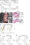
BACKGROUND. Loss of the Y chromosome (LOY) is a frequent event in male tumors and has been linked to cancer progression. However, the degree of mosaic LOY (mLOY) within normal tissues from men with or without cancer remains uncharacterized. METHODS. Here we used a FISH-based assay targeting X- and Y-chromosome centromeres to perform a pan-organ analysis of mLOY in 1,000 male tissue samples from 405 individuals representing 11 organs. Automated image processing generated a quantitative FISH-based mLOY score (YchrFISH) that we validated against a transcriptomic surrogate of Y-chromosome dosage from RNA-seq data. RESULTS. mLOY burden varied by tumor type, with highest degree in colorectal carcinoma. Across tissue groups, YchrFISH scores declined progressively from normal tissues of cancer-free men to histologically normal tissues adjacent to cancer and carcinoma (P < 0.0001). Paired analyses confirmed consistently greater mLOY in malignant compared with tumor-adjacent histologically normal tissue in different organs. Spatially resolved RNA-seq maps of bladders removed for cancer demonstrated a transcriptional gradient of Y-chromosome loss from normal urothelium through intraepithelial neoplasia to invasive carcinoma. CONCLUSION. mLOY gradients exist across histologically normal and malignant tissues, consistent with the concept of field cancerization. Our findings support epithelial mLOY as a biomarker of early malignant transformation and, to our knowledge, a previously unrecognized hallmark of male oncogenesis. FUNDING. NIH grants R35CA294022, P01CA163227, and P50CA97186 (the Pacific Northwest Prostate Cancer SPORE) and the Institute for Prostate Cancer Research.
Authors
Arkadiusz Gertych, Huihui Ye, Xingyu Chen, Eric Vail, V. Krishnan Ramanujan, Lauren Brady, Lawrence D. True, Peter S. Nelson, Peter R. Carroll, Dan Theodorescu
×Abstract
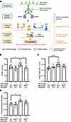
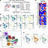
In rheumatoid arthritis (RA), CD4+ T cells specific for citrullinated antigens (cit-antigens) are key drivers of disease, but knowledge about epitopes and phenotypes remains limited. We characterized the frequency and phenotype of cit-specific CD4+ T cells in peripheral blood using HLA class II tetramers combined with computational analysis of phenotypic clusters to simultaneously detect peptides derived from 5 cit-antigens (aggrecan, vimentin, fibrinogen, cartilage intermediate layer protein, and α-enolase) previously implicated in RA pathogenesis. In a cross-sectional cohort, cit-aggrecan–, cit-vimentin–, and cit-fibrinogen–specific T cells were more frequent in participants with RA than healthy volunteers, associated with active disease, and had Th1-like and stem-like lineages in RA. In a longitudinal cohort investigating response to therapy, the frequency of cit-aggrecan–, cit-vimentin–, and cit-fibrinogen–specific CD4+ T cells was significantly higher at baseline and further elevated in responders. Furthermore, the frequency of cit-specific Th1-like cells in responders decreased over time. In contrast, the frequency of Th1-like cells in non-responders increased over time. Collectively, these findings demonstrate that cit-specific CD4+ T cells are expanded in RA and target a broad number of antigens across a breadth of phenotypes. Furthermore, the predominant antigen specificities associate with disease activity and exhibit dynamic changes in phenotype that reflect response to therapy.
Authors
Cliff Rims, Hannah A. DeBerg, Sylvia E. Posso, Virginia S. Muir, Hannes Uchtenhagen, Anne M. Hocking, Heather Bukiri, Jeffrey Carlin, Bernard Ng, Peter S. Linsley, Eddie A. James, Jane H. Buckner
×Abstract
The cholinergic antiinflammatory pathway attenuates lung inflammation via the α7 nicotinic acetylcholine receptor (α7 nAChR) on immune cells. However, the role of α7 nAChR on lung megakaryocytes (Mks) in allergic airway inflammation remains unknown. In this study, allergen-challenged mouse models were used with conditional Mk-specific Chrna7 knockout, pharmacological activation (GTS-21), and Mk reconstitution. IL-33 expression and p38 MAPK signaling were assessed. We found that allergen challenge upregulated α7 nAChR specifically in lung Mks. Mk-specific Chrna7 deletion significantly alleviated allergic airway inflammation, whereas GTS-21 exacerbated inflammation via an Mk-dependent mechanism. Reconstitution with α7 nAChR+ Mks restored airway inflammatory responses. Mechanistically, α7 nAChR activation promoted Mk IL-33 synthesis and secretion through p38 MAPK signaling. Taken together, our results show that α7 nAChR on lung Mks plays a proinflammatory role in allergic airway inflammation, challenging its classical antiinflammatory paradigm and revealing pathogenic mechanisms.
Authors
Hang Wu, Rujia Tao, Shitao Xie, Yao Zhou, Jin-Fu Xu, Zhenwei Xia, Xiao Su
×Abstract
The pericardium plays an important homeostatic role for the neighboring heart, providing both lubrication and structural support. In vivo models have further identified a protective role for the pericardium in modulating cardiac remodeling following myocardial infarction, possibly through the actions of tissue-resident pericardial macrophages. Using patient-derived pericardial samples, we establish that human pericardial immune cells directly inhibit cardiac fibroblast fibrotic activity, and this action is dampened following myocardial infarction. Using single-cell RNA sequencing of patient pericardial fluid cells, we identify two pericardial macrophage subsets that are uniquely altered in response to myocardial infarction, which contributes to a shift in their effector molecule expression profiles. We confirm that fibronectin-expressing human pericardial macrophages are the primary driver of the pericardial antifibrotic actions through the release of cystatin C. Finally, we establish cystatin C as a myeloid cell–derived cardioprotective effector molecule in an in vivo model of myocardial infarction. Collectively, we uncover a molecular mechanism of the local immune environment that regulates cardiac remodeling after myocardial infarction.
Authors
Ali Fatehi Hassanabad, Sarthak Sinha, Arzina Jaffer, Darrell Belke, Nicole L. Rosin, Elodie Labit, Daniel Young, Friederike I. Schoettler, Keerthana Chockalingam, Benjamin Haeyul Lee, Jameson A. Dundas, Emilie de Chantal, Carmina A. Isidoro, Alexander Tam, Hanjoo B. Shim, Anna N. Zarzycki, Afshin Derakhshani, Elisabeth Gorgiogianni, Jeannine D. Turnbull, Antoine Dufour, Shalina S. Ousman, Jeff A. Biernaskie, Paul W.M. Fedak, Justin F. Deniset
×Abstract
Hypercapnia, elevated carbon dioxide (CO2), is common in advanced chronic obstructive pulmonary disease (COPD) and predicts poor clinical outcomes. Traditionally considered a consequence of disease severity, hypercapnia may drive disease progression by promoting airway dysfunction. Here, we show that hypercapnia acts as an active stressor, driving airway smooth muscle (ASM) constriction through a stromal interaction molecule 1–dependent (STIM1-dependent) pathway. Hypercapnia rapidly activates ERK, triggering sarcoplasmic reticulum calcium (Ca2+) release via phosphorylation of the inositol 1,4,5-trisphosphate receptor. ERK also induces nuclear translocation of the transcription factor c-Fos, enhancing STIM1 transcription. These responses were observed under both supraphysiological (~120 mmHg) and clinically relevant (50–60 mmHg) hypercapnia. Increased STIM1 abundance sustains store-operated Ca2+ entry (SOCE), amplifying ASM signaling. In mice, hypercapnia increased ASM and airway contractility in a STIM1-dependent manner. Human genetic analyses revealed noncoding STIM1 variants associated with reduced lung expression that were enriched in patients with COPD. These variants correlated with lower airway resistance under normocapnia; however, this benefit was lost during hypercapnia, indicating a potential gene-environment interaction. Together, our findings position STIM1 as a key mechanistic node linking hypercapnia to Ca2+ dysregulation and airway obstruction, defining a CO2/ERK/STIM1/SOCE axis with translational relevance to chronic lung disease.
Authors
Masahiko Shigemura, Vitalii Kryvenko, Jennifer A. Pacheco, Megan J. Puckelwartz, Milos Aleksic, Natalia D. Magnani, Emma Thompson, Francisco Javier Martin-Romero, Eoin P. Cummins, Werner Seeger, Andreas Bräuninger, Lynn C. Welch, G.R. Scott Budinger, Emilia Lecuona, Laura A. Dada, Ankit Bharat, István Vadász, Murali Prakriya, Jacob I. Sznajder
×Abstract
Schistosomiasis is a common cause of pulmonary hypertension (PH) worldwide. It is known that adaptive immunity and specifically CD4+ T cells are necessary for experimental disease pathogenesis. The lectin complement system is activated in those infected with schistosomiasis. We tested the hypothesis that lectin complement promotes Th2 CD4+ T cell activation, leading to PH in a schistosomiasis exposure model. WT and transgenic mice lacking mannose binding lectin (MBL), and bone marrow chimeras, were experimentally exposed to Schistosoma mansoni eggs. PH severity was assessed by hemodynamics and vascular remodeling, and CD4+ T cell density and phenotype were assessed by flow cytometry. WT recipients of MBL-knockout bone marrow were protected from Schistosoma-induced PH. The protection from PH was associated with fewer Th2 CD4+ T cells. In WT mice exposed to Schistosoma, CD4+ T cell expression of MBL increased. MBL-deficient CD4+ T cells had a suppressed Th2 phenotype when exposed to Schistosoma antigens. Mice with deficiency of C4, which functions downstream of MBL in the lectin complement pathway, were not protected from Schistosoma-induced PH. Mice lacking MBL were not protected from PH caused by hypoxia exposure. MBL in CD4+ T cells promotes Schistosoma-induced PH.
Authors
Claudia Mickael, Dara C. Fonseca Balladares, Rahul Kumar, Michael H. Lee, Kevin Nolan, Linda Sanders, Katie J. Tuscan, Ramraj Prasad, Pilar Londono, Fernanda P. Oliveira, Kennedi B. Pyper, Ari B. Molofsky, Rubin M. Tuder, Kurt R. Stenmark, Brian B. Graham
×Abstract
Gain-of-function (GOF) variants in STAT3 cause a complex disorder characterized by early-onset autoimmunity, lymphoproliferation, recurrent infections, and immune dysregulation. In both primary human and mouse models of STAT3 GOF, CD8+ T cells have been implicated as pathogenic drivers of autoimmunity, though the exact mechanisms remain poorly understood. Here, we found that in patients with STAT3 GOF, CD8+ T cells exist in an activated state. Functional assessment revealed that naive CD8+ T cells have an increased capacity for IFN-γ and TNF-α production, with type I and type II IFN transcriptional signatures. Evaluation of immunoregulatory pathways revealed dysregulation of the purinergic signaling axis in CD8+ T cells: CD39 was increased, whereas downstream purinergic family members, CD73 and the adenosine receptor A2AR, were downregulated, impairing the potential to produce or sense immunosuppressive adenosine. Evaluation of the impact of precision therapy, in the form of JAK inhibition, at a cellular and functional level revealed partial normalization of CD8+ T cell dysregulation in patients, including aberrant cytokine production. Our study suggests that a dysregulated purinergic signaling axis plays a key role in CD8+ T cell dysregulation in STAT3 GOF and may have implications for other rare monogenic immune disorders and common inflammatory disorders.
Authors
Jose S. Campos Duran, Montana S. Knight, Samir U. Sayed, Megan C. Dalalo, Andrea A. Mauracher, Peyton Conrey, Aaron B. Schultz, Ceire A. Hay, Robert B. Lindell, Ilona Neale, Kyle Yeakle, Eric D. Abrams, Erica G. Schmitt, Martin A. Thelin, Christian A. Howard, Sara Bluestein, Christine M. Seroogy, Tamara C. Pozos, Akaluck Thatayatikom, Ingrid S. Lundgren, Amelie Gauthier, Scott W. Canna, Helen C. Su, Michael D. Keller, Ottavia M. Delmonte, Lisa R. Forbes Satter, Steven M. Holland, Jenna R.E. Bergerson, Jennifer W. Leiding, Neil Romberg, Will Bailis, Christopher A. Hunter, Alexandra F. Freeman, Alejandro V. Villarino, Mark S. Anderson, Megan A. Cooper, Tiphanie P. Vogel, Sarah E. Henrickson
×Abstract
Idiopathic pulmonary fibrosis (IPF) is a fatal interstitial lung disease characterized by progressive scarring and respiratory failure. While T cells are elevated in IPF lungs, their contributions to fibrosis beyond inflammation remain poorly understood. Here, we performed multiplex imaging and single-cell RNA and protein profiling on about 90,000 CD3+ T cells from control and fibrotic lungs, revealing 11 distinct subsets of CD4+ and CD8+ T cells, including a rare CD56+ regulatory T cell. In addition to increased T cell numbers in severely fibrotic lungs compared with non-diseased controls, we observed CD4+ and CD8+ T cells localized near epithelial cells and in niches of abnormal epithelium. CXCR4/MIF signaling emerged as a central axis mediating T cell–epithelial interactions, while epidermal growth factor receptor (EGFR) and TGF-β pathways dominated in multiple T cell subsets. Our findings support the concept that T cells in IPF adopt nonclassical activation patterns that are driven by epithelial interactions within the fibrotic microenvironment. These studies provide a foundation for exploring alternative therapeutic strategies in IPF lungs by modulating T cell behavior and communication networks.
Authors
Ana P.M. Serezani, Julia M.R. Bazzano, Bruno D. Pascoalino, Ludmilla da Silva, Abigail J. Dietrich, Chase J. Taylor, Taylor Sherrill, Annika Vannan, Carla L. Calvi, Paula I. Gonzalez-Ericsson, Erin M. Wilfong, Matthew Bacchetta, Ciara M. Shaver, Lorraine B. Ware, Margaret L. Salisbury, Luc Van Kaer, Nicholas E. Banovich, Jonathan A. Kropski, Timothy S. Blackwell
×Abstract
Neonatal sepsis is a predominant cause of neonatal mortality and long-term morbidity that severely affects preterm and low-birth-weight newborns. Antibiotic resistance and long-term developmental issues associated with neonatal sepsis necessitate finding new and improved treatment options. IL-27 has diverse influences on the immune response, is elevated during the neonatal period compared with adulthood, and continues to rise further during infection. Elevated levels of IL-27 early in life predispose the host to impaired control of the pathogen burden and increased mortality. This study explored the therapeutic potential of IL-27p28 antibody administration to improve treatment outcomes during murine neonatal sepsis. Sepsis was induced by subcutaneous inoculation of K1-encapsulated E. coli, and the neonatal pups were rescued with IL-27p28 monoclonal antibody. Pups that received prophylactic antibody prior to the infection demonstrated superior bacterial clearance and significant weight gain compared with controls during infection. The combination of subclinical dose of gentamicin along with IL-27p28 antibody administered 2 hours after infection, significantly improved bacterial clearance and glucose homeostasis, with reduced serum levels of IL-6 and TNF-α, vital organ damage, and improved the survival rate of infected pups compared with gentamicin alone. These findings suggest that IL-27p28 antagonization represents a promising therapeutic tool for treatment of neonatal sepsis.
Authors
Madhavi Annamanedi, Jessica M. Povroznik, Samantha Arevalo-Marcano, Cory M. Robinson
×Abstract
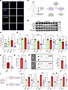
Locally produced nonpituitary growth hormone (npGH) promotes DNA damage accumulation and epithelial-mesenchymal transition (EMT) in aging human colon epithelium. GH receptor (GHR) and npGH are expressed in normal human prostate, and benign prostatic hyperplasia (BPH) prevalence increases with age. We hypothesized that local prostate GH action may promote EMT and contribute to BPH pathogenesis. We show here that the number of patients expressing npGH increases more than 10-fold after age 60, concordant with increased γH2AX, a marker of DNA damage, and EMT activation. GH-treated human primary prostate epithelial cells, normal prostate cells, and primary cell cultures derived from resected BPH specimens exhibited enhanced DNA damage and activated EMT, with induced TWIST2, suppressed E-cadherin, and increased Ki67, cell motility, and proliferation. In mice, prostate tissue adjacent to allografted GH-expressing fibroblasts showed increased γH2AX, TWIST2, and Ki67, along with morphological changes consistent with BPH. While GH and GH-induced IGF-1 both activated EMT, GH triggered DNA damage independently of IGF-1. These results elucidate what we believe to be a novel role for local npGH in aging prostate tissue, whereby npGH increases DNA damage and promotes EMT to enable a microenvironment favoring BPH development. Prostate GHR signaling may be an attractive therapeutic target for BPH.
Authors
Masaki Ryuzaki, Svetlana Zonis, Neil A. Bhowmick, Sandrine Billet, Saravana Kumar Kailasam Mani, Stephen J. Freedland, Hyung L. Kim, Vera Chesnokova, Shlomo Melmed
×Abstract
Early diagnosis of cystinosis is critical to limit disease progression. YKL-40, a protein in the chitinase family, released by inflammatory cells, may be a useful biomarker for cystinosis. In a case-control study of 10 children with cystinosis and 20 without cystinosis, matched by age and baseline eGFR, we measured urine YKL-40, NGAL, and EGF. A lateral flow device (LFD) for YKL-40 was also developed and tested. Urine YKL-40 was over 200-fold higher in children with cystinosis (64.6 ng/mL [IQR: 23.4, 83.8]) compared with controls (0.3 [IQR: 0.3, 0.79]; P = 0.0001) with excellent diagnostic discrimination (AUC = 0.99) that was superior to other biomarkers. LFD measurements for YKL-40 showed similar results (AUC = 0.93). YKL-40 results were verified in 5 cystinosis patients, and YKL-40 staining was markedly higher in kidney biopsies from cystinosis patients than in healthy controls. Urine YKL-40 has excellent diagnostic potential for cystinosis, and point-of-care technologies may facilitate early screening and management of this disease.
Authors
Jason H. Greenberg, Serena D Souza, Heather R. Thiessen Philbrook, Wassim Obeid, Avi Z. Rosenberg, Elena Levtchenko, Koenraad Veys, Susan L. Furth, Chirag R. Parikh
×Abstract
Natural killer (NK) cells undergo stepwise differentiation from multipotent progenitors within secondary lymphoid tissues. Despite the central importance of the tissue microenvironment in their development, little is known about cell-cell interactions that regulate human NK cell trafficking and maturation. Here, we identify the chemokine receptor CXCR4 and its ligand CXCL12 as regulators of stromal–NK cell interactions required for NK cell maturation. We demonstrate that CXCR4 is expressed throughout human NK cell development in peripheral blood and tonsil, and CXCL12 is enriched in stromal niches containing developing NK cells. Pharmacologic blockade or genetic disruption of CXCR4 resulted in diminished adhesion to integrin ligands, and high-resolution imaging demonstrated crosstalk between CXCR4 and integrins, providing a mechanistic basis for chemokine-dependent modulation of adhesion. Further, CXCR4 blockade resulted in altered contact-dependent motility on stromal cells and integrin ligands, with decreased stable stromal engagement and increased cell speed. Consistent with a requirement for these interactions, treatment with the CXCR4 antagonist plerixafor (AMD3100) impaired NK cell generation from CD34+ precursors. Analysis of NK cells from patients with WHIM syndrome with CXCR4 gain-of-function mutations treated with plerixafor revealed similar defects in migration and adhesion, supporting the in vivo relevance of CXCR4-dependent regulation of NK cell adhesion and motility.
Authors
Shira E. Eisman, Francesca E. Grossberg, Batya S. Koenigsberg, David H. McDermott, Frédérique van den Haak, Luis A. Pedroza, Everardo Hegewisch-Solloa, Philip M. Murphy, Emily M. Mace
×Abstract
Polycystic kidney disease (PKD) arises from mutations in cilia-associated genes, such as PKD1 and PKD2, expressed in renal epithelial cells, leading to progressive kidney dysfunction and end-stage kidney disease. Patients with PKD exhibit significant heterogeneity in disease progression, largely due to genetic and environmental modifiers. Like patients, mouse models of PKD also exhibit significant heterogeneity with regards to the gene mutated, age of disease onset, and rate of disease progression. To elucidate the cellular and molecular consequences of these variables, we constructed an integrated single-cell RNA sequencing (scRNA-seq) atlas across mouse models of PKD, mapping changes in cell type composition, gene expression, and intercellular signaling networks across the whole atlas and within individual models. Across models, scRNA-seq data revealed increased Spp1 (osteopontin) expression and signaling from PKD-enriched clusters. Global deletion of Spp1 in Pkd1RC/RC mice resulted in a modest reduction in cyst severity and improved kidney function. From these studies, we created a freely available, searchable website (https://bmblx.bmi.osumc.edu/scPKD/) that can be used to identify cross- and intra-model changes in gene expression, guiding researchers to new therapeutic targets for treating PKD.
Authors
Sarah J. Miller, Hua Zhong, Weidong Wu, Audrey M. Cordova, Morgan E. Yashchenko, Alex Yashchenko, Zhang Li, Daniyal J. Jafree, Chelsea N. Zimmerman, Christa I. DeVette, Vicki Do, Maya E. Hignite, Yohan Park, Fariha Nusrat, Bibi Maryam, Sizhao Lu, Xiaoyan Li, Jenny R. Gipson, Xiaogang Li, David A. Long, Mary C.M. Weiser-Evans, Bradley K. Yoder, Benjamin D. Cowley Jr, Katharina Hopp, Jason R. Stubbs, Qin Ma, Anjun Ma, Kurt A. Zimmerman
×Abstract
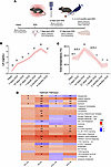
The hypothalamic changes that occur after the loss of ovarian estrogen remain poorly characterized. Here, we performed a comprehensive temporal characterization of the mouse hypothalamus after ovariectomy (OVX), combining physiological measurements with bulk RNA-seq of the posterior hypothalamus (PH) and preoptic area at 14 days and 4 months after OVX. Serum luteinizing hormone levels rose progressively and then declined, and core temperature peaked early and subsequently normalized, recapitulating the endocrine and thermoregulatory dynamics of reproductive aging in humans. Transcriptomic analysis revealed time-dependent activation of inflammatory pathways, glial markers, and KNDy neuron-related gene networks, with the most pronounced changes emerging at 4 months after OVX, particularly in the PH. Immunofluorescence confirmed increased neurokinin B release, declining KNDy neuronal activity, and heightened astrocytic reactivity in the arcuate nucleus after prolonged estrogen withdrawal. To contextualize these findings, we analyzed publicly available human hypothalamic RNA-seq data across chronological age. Age-related transcriptomic patterns, including progressive inflammatory signaling, glial activation, and altered KNDy gene expression, showed significant correlation with the OVX mouse model, particularly at the pathway level. These findings establish a temporal framework for hypothalamic molecular changes after estrogen withdrawal, identify conserved neuroinflammatory signatures across species, and provide a preclinical platform for testing interventions targeting menopause-associated hypothalamic dysfunction.
Authors
Jordana C.B. Bloom, Encarnación Torres, Sidney A. Pereira, Liliana Arvizu-Sanchez, Audrey N. Fontes, Hadine Joffe, David C. Page, Victor M. Navarro
×Abstract
The precise mechanisms underlying the pathogenesis of idiopathic inflammatory myopathy (IIM) remain undefined. However, there has been increasing recognition that tissue-resident memory T cells (TRMs) play an important role in the pathogenesis of systemic autoimmune disease. In IIM, TRM-associated transcriptional signatures have been reported but on a very limited basis. By using multimodal single-cell RNA-sequencing analysis in our established murine model of histidyl-tRNA synthetase–induced myositis, we identified a prominent population of CD4+ TRMs in inflamed skeletal muscle. Muscle CD4+ TRMs exhibited high expression of genes encoding Cd69, Cxcr6, Runx3, and Prdm1, alongside low expression of Klf2, Ccr7, Sell, S1pr1, and Tcf7 — a profile that is generally consistent with previous reports of TRM gene signature and that we validate through comparison with transcriptomic profiles of human muscle tissue. Detailed pathway analysis in our model indicates that muscle CD4+ TRMs contribute to innate immune regulatory pathways enriched for TNF and IFN-γ signaling. Furthermore, analysis of TCR clonotype distribution and CDR3 sequence similarity revealed pronounced clonal expansion of CD4+ TRMs relative to other T cell subsets — a pattern that remained stable from 2 to 6 weeks after immunization. Collectively, these results suggest a potential role for CD4+ TRMs in the pathogenesis of autoimmune myositis.
Authors
Decheng Li, Daniel P. Reay, Iago Pinal-Fernandez, Maria Casal-Dominguez, Andrew L. Mammen, Sarah L. Gaffen, Timothy B. Oriss, Dana P. Ascherman
×Abstract
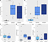
BACKGROUND. Previous studies identified plasma proteins associated with chronic graft-versus-host disease (cGVHD). The goal of this cross-sectional study was to evaluate whether plasma biomarkers were associated with specific organ manifestations to help guide treatment choice. METHODS. Plasma proteins were measured in patients with cGVHD (N = 695) from Chronic GVHD Consortium studies. Correlations of plasma protein levels with individual organ involvement were tested with a P value of 0.05 or less considered significant after Benjamini-Hochberg adjustment and adjustment for 5 baseline clinical variables. RESULTS. Median time from cGVHD diagnosis to blood draw was 0.9 months (IQR 0.1–9.5). Donors were 50% HLA-matched unrelated, 32% matched related, and the remainder were umbilical cord blood, haploidentical, or mismatched unrelated donors. Methotrexate and calcineurin inhibitor prophylaxis for acute GVHD was used in 53% of patients. Overall, 326 (47%) had moderate and 244 (35%) had severe cGVHD with the following organ involvement at time of blood draw: skin (67%), mouth (60%), eye (49%), joint (34%), GI (31%), lung (23%), and liver (17%). After adjustment for batch effects and patient and transplant characteristics, 14 plasma proteins were associated with organ involvement with independent AUCs of 0.7–0.8. All organs except the eye were associated with at least 1 biomarker. However, no combination of plasma proteins improved model fit after adjusting for patient and transplant clinical variables. CONCLUSION. Correlations between plasma proteins and organ involvement were identified but are not actionable. Our future investigations will focus on more granular and immediately proximal determinants of cGVHD biology in both blood and tissue.
Authors
Stephanie J. Lee, Corey Cutler, Ningxin Ma, Timothy W. Randolph, George L. Chen, Joseph Pidala, Betty K. Hamilton, Carrie L. Kitko, Sally Arai, Najla El Jurdi, Lynn Onstad, Motoko Koyama, Catherine J. Lee, Sophie Paczesny, Geoffrey R. Hill