
Research ArticleCell biologyImmunologyNephrology
Open Access |  10.1172/jci.insight.207157
A cross-model single-cell atlas reveals conserved involvement of osteopontin in polycystic kidney disease
Sarah J. Miller,1 Hua Zhong,1 Weidong Wu,2,3 Audrey M. Cordova,1 Morgan E. Yashchenko,1 Alex Yashchenko,1 Zhang Li,4 Daniyal J. Jafree,5,6,7 Chelsea N. Zimmerman,8 Christa I. DeVette,8 Vicki Do,9 Maya E. Hignite,1 Yohan Park,1 Fariha Nusrat,1 Bibi Maryam,1 Sizhao Lu,10 Xiaoyan Li,11,12 Jenny R. Gipson,13 Xiaogang Li,11,12 David A. Long,5,6 Mary C.M. Weiser-Evans,10 Bradley K. Yoder,4 Benjamin D. Cowley Jr,1 Katharina Hopp,10 Jason R. Stubbs,14,15 Qin Ma,2,3 Anjun Ma,2,3 and Kurt A. Zimmerman1
1Department of Internal Medicine, Division of Nephrology, University of Oklahoma Health Sciences Center, Oklahoma City, Oklahoma, USA.
2Department of Biomedical Informatics, College of Medicine, and
3Pelotonia Institute for Immuno-Oncology, The James Comprehensive Cancer Center, The Ohio State University, Columbus, Ohio, USA.
4Department of Cell, Developmental, and Integrative Biology, University of Alabama at Birmingham, Birmingham, Alabama, USA.
5Developmental Biology & Cancer Research & Teaching Department, UCL Great Ormond Street Institute of Child Health, and
6UCL Centre for Kidney and Bladder Health, University College London, London, United Kingdom.
7Cellular Genetics Programme, Wellcome Trust Sanger Institute, Hinxton, United Kingdom.
8Department of Pediatrics, University of Oklahoma Health Science Center, Oklahoma City, Oklahoma, USA.
9Division of Nephrology, Boston Children’s Hospital, Boston, Massachusetts, USA.
10Department of Medicine, Division of Renal Diseases and Hypertension, University of Colorado Anschutz Medical Campus, Aurora, Colorado, USA.
11Department of Internal Medicine, and
12Department of Biochemistry and Molecular Biology, Mayo Clinic, Rochester, Minnesota, USA.
13College of Medicine Core Facilities, University of Oklahoma Health Sciences Center, Oklahoma City, Oklahoma, USA.
14The Jared Grantham Kidney Institute, and
15Division of Nephrology and Hypertension, Department of Internal Medicine, University of Kansas Medical Center, Kansas City, Kansas, USA.
Address correspondence to: Kurt A. Zimmerman, Section of Nephrology, Department of Internal Medicine. University of Oklahoma Health Sciences Center, Biomedical Sciences building, Room 513, Oklahoma City, Oklahoma, 73104, USA. Email: kurt-zimmerman@ouhsc.edu.
Authorship note: SJM and HZ contributed equally to this work.
Find articles by Miller, S. in: PubMed | Google Scholar
1Department of Internal Medicine, Division of Nephrology, University of Oklahoma Health Sciences Center, Oklahoma City, Oklahoma, USA.
2Department of Biomedical Informatics, College of Medicine, and
3Pelotonia Institute for Immuno-Oncology, The James Comprehensive Cancer Center, The Ohio State University, Columbus, Ohio, USA.
4Department of Cell, Developmental, and Integrative Biology, University of Alabama at Birmingham, Birmingham, Alabama, USA.
5Developmental Biology & Cancer Research & Teaching Department, UCL Great Ormond Street Institute of Child Health, and
6UCL Centre for Kidney and Bladder Health, University College London, London, United Kingdom.
7Cellular Genetics Programme, Wellcome Trust Sanger Institute, Hinxton, United Kingdom.
8Department of Pediatrics, University of Oklahoma Health Science Center, Oklahoma City, Oklahoma, USA.
9Division of Nephrology, Boston Children’s Hospital, Boston, Massachusetts, USA.
10Department of Medicine, Division of Renal Diseases and Hypertension, University of Colorado Anschutz Medical Campus, Aurora, Colorado, USA.
11Department of Internal Medicine, and
12Department of Biochemistry and Molecular Biology, Mayo Clinic, Rochester, Minnesota, USA.
13College of Medicine Core Facilities, University of Oklahoma Health Sciences Center, Oklahoma City, Oklahoma, USA.
14The Jared Grantham Kidney Institute, and
15Division of Nephrology and Hypertension, Department of Internal Medicine, University of Kansas Medical Center, Kansas City, Kansas, USA.
Address correspondence to: Kurt A. Zimmerman, Section of Nephrology, Department of Internal Medicine. University of Oklahoma Health Sciences Center, Biomedical Sciences building, Room 513, Oklahoma City, Oklahoma, 73104, USA. Email: kurt-zimmerman@ouhsc.edu.
Authorship note: SJM and HZ contributed equally to this work.
Find articles by Zhong, H. in: PubMed | Google Scholar
1Department of Internal Medicine, Division of Nephrology, University of Oklahoma Health Sciences Center, Oklahoma City, Oklahoma, USA.
2Department of Biomedical Informatics, College of Medicine, and
3Pelotonia Institute for Immuno-Oncology, The James Comprehensive Cancer Center, The Ohio State University, Columbus, Ohio, USA.
4Department of Cell, Developmental, and Integrative Biology, University of Alabama at Birmingham, Birmingham, Alabama, USA.
5Developmental Biology & Cancer Research & Teaching Department, UCL Great Ormond Street Institute of Child Health, and
6UCL Centre for Kidney and Bladder Health, University College London, London, United Kingdom.
7Cellular Genetics Programme, Wellcome Trust Sanger Institute, Hinxton, United Kingdom.
8Department of Pediatrics, University of Oklahoma Health Science Center, Oklahoma City, Oklahoma, USA.
9Division of Nephrology, Boston Children’s Hospital, Boston, Massachusetts, USA.
10Department of Medicine, Division of Renal Diseases and Hypertension, University of Colorado Anschutz Medical Campus, Aurora, Colorado, USA.
11Department of Internal Medicine, and
12Department of Biochemistry and Molecular Biology, Mayo Clinic, Rochester, Minnesota, USA.
13College of Medicine Core Facilities, University of Oklahoma Health Sciences Center, Oklahoma City, Oklahoma, USA.
14The Jared Grantham Kidney Institute, and
15Division of Nephrology and Hypertension, Department of Internal Medicine, University of Kansas Medical Center, Kansas City, Kansas, USA.
Address correspondence to: Kurt A. Zimmerman, Section of Nephrology, Department of Internal Medicine. University of Oklahoma Health Sciences Center, Biomedical Sciences building, Room 513, Oklahoma City, Oklahoma, 73104, USA. Email: kurt-zimmerman@ouhsc.edu.
Authorship note: SJM and HZ contributed equally to this work.
Find articles by Wu, W. in: PubMed | Google Scholar
1Department of Internal Medicine, Division of Nephrology, University of Oklahoma Health Sciences Center, Oklahoma City, Oklahoma, USA.
2Department of Biomedical Informatics, College of Medicine, and
3Pelotonia Institute for Immuno-Oncology, The James Comprehensive Cancer Center, The Ohio State University, Columbus, Ohio, USA.
4Department of Cell, Developmental, and Integrative Biology, University of Alabama at Birmingham, Birmingham, Alabama, USA.
5Developmental Biology & Cancer Research & Teaching Department, UCL Great Ormond Street Institute of Child Health, and
6UCL Centre for Kidney and Bladder Health, University College London, London, United Kingdom.
7Cellular Genetics Programme, Wellcome Trust Sanger Institute, Hinxton, United Kingdom.
8Department of Pediatrics, University of Oklahoma Health Science Center, Oklahoma City, Oklahoma, USA.
9Division of Nephrology, Boston Children’s Hospital, Boston, Massachusetts, USA.
10Department of Medicine, Division of Renal Diseases and Hypertension, University of Colorado Anschutz Medical Campus, Aurora, Colorado, USA.
11Department of Internal Medicine, and
12Department of Biochemistry and Molecular Biology, Mayo Clinic, Rochester, Minnesota, USA.
13College of Medicine Core Facilities, University of Oklahoma Health Sciences Center, Oklahoma City, Oklahoma, USA.
14The Jared Grantham Kidney Institute, and
15Division of Nephrology and Hypertension, Department of Internal Medicine, University of Kansas Medical Center, Kansas City, Kansas, USA.
Address correspondence to: Kurt A. Zimmerman, Section of Nephrology, Department of Internal Medicine. University of Oklahoma Health Sciences Center, Biomedical Sciences building, Room 513, Oklahoma City, Oklahoma, 73104, USA. Email: kurt-zimmerman@ouhsc.edu.
Authorship note: SJM and HZ contributed equally to this work.
Find articles by Cordova, A. in: PubMed | Google Scholar
1Department of Internal Medicine, Division of Nephrology, University of Oklahoma Health Sciences Center, Oklahoma City, Oklahoma, USA.
2Department of Biomedical Informatics, College of Medicine, and
3Pelotonia Institute for Immuno-Oncology, The James Comprehensive Cancer Center, The Ohio State University, Columbus, Ohio, USA.
4Department of Cell, Developmental, and Integrative Biology, University of Alabama at Birmingham, Birmingham, Alabama, USA.
5Developmental Biology & Cancer Research & Teaching Department, UCL Great Ormond Street Institute of Child Health, and
6UCL Centre for Kidney and Bladder Health, University College London, London, United Kingdom.
7Cellular Genetics Programme, Wellcome Trust Sanger Institute, Hinxton, United Kingdom.
8Department of Pediatrics, University of Oklahoma Health Science Center, Oklahoma City, Oklahoma, USA.
9Division of Nephrology, Boston Children’s Hospital, Boston, Massachusetts, USA.
10Department of Medicine, Division of Renal Diseases and Hypertension, University of Colorado Anschutz Medical Campus, Aurora, Colorado, USA.
11Department of Internal Medicine, and
12Department of Biochemistry and Molecular Biology, Mayo Clinic, Rochester, Minnesota, USA.
13College of Medicine Core Facilities, University of Oklahoma Health Sciences Center, Oklahoma City, Oklahoma, USA.
14The Jared Grantham Kidney Institute, and
15Division of Nephrology and Hypertension, Department of Internal Medicine, University of Kansas Medical Center, Kansas City, Kansas, USA.
Address correspondence to: Kurt A. Zimmerman, Section of Nephrology, Department of Internal Medicine. University of Oklahoma Health Sciences Center, Biomedical Sciences building, Room 513, Oklahoma City, Oklahoma, 73104, USA. Email: kurt-zimmerman@ouhsc.edu.
Authorship note: SJM and HZ contributed equally to this work.
Find articles by Yashchenko, M. in: PubMed | Google Scholar
1Department of Internal Medicine, Division of Nephrology, University of Oklahoma Health Sciences Center, Oklahoma City, Oklahoma, USA.
2Department of Biomedical Informatics, College of Medicine, and
3Pelotonia Institute for Immuno-Oncology, The James Comprehensive Cancer Center, The Ohio State University, Columbus, Ohio, USA.
4Department of Cell, Developmental, and Integrative Biology, University of Alabama at Birmingham, Birmingham, Alabama, USA.
5Developmental Biology & Cancer Research & Teaching Department, UCL Great Ormond Street Institute of Child Health, and
6UCL Centre for Kidney and Bladder Health, University College London, London, United Kingdom.
7Cellular Genetics Programme, Wellcome Trust Sanger Institute, Hinxton, United Kingdom.
8Department of Pediatrics, University of Oklahoma Health Science Center, Oklahoma City, Oklahoma, USA.
9Division of Nephrology, Boston Children’s Hospital, Boston, Massachusetts, USA.
10Department of Medicine, Division of Renal Diseases and Hypertension, University of Colorado Anschutz Medical Campus, Aurora, Colorado, USA.
11Department of Internal Medicine, and
12Department of Biochemistry and Molecular Biology, Mayo Clinic, Rochester, Minnesota, USA.
13College of Medicine Core Facilities, University of Oklahoma Health Sciences Center, Oklahoma City, Oklahoma, USA.
14The Jared Grantham Kidney Institute, and
15Division of Nephrology and Hypertension, Department of Internal Medicine, University of Kansas Medical Center, Kansas City, Kansas, USA.
Address correspondence to: Kurt A. Zimmerman, Section of Nephrology, Department of Internal Medicine. University of Oklahoma Health Sciences Center, Biomedical Sciences building, Room 513, Oklahoma City, Oklahoma, 73104, USA. Email: kurt-zimmerman@ouhsc.edu.
Authorship note: SJM and HZ contributed equally to this work.
Find articles by
Yashchenko, A.
in:
PubMed
|
Google Scholar
|

1Department of Internal Medicine, Division of Nephrology, University of Oklahoma Health Sciences Center, Oklahoma City, Oklahoma, USA.
2Department of Biomedical Informatics, College of Medicine, and
3Pelotonia Institute for Immuno-Oncology, The James Comprehensive Cancer Center, The Ohio State University, Columbus, Ohio, USA.
4Department of Cell, Developmental, and Integrative Biology, University of Alabama at Birmingham, Birmingham, Alabama, USA.
5Developmental Biology & Cancer Research & Teaching Department, UCL Great Ormond Street Institute of Child Health, and
6UCL Centre for Kidney and Bladder Health, University College London, London, United Kingdom.
7Cellular Genetics Programme, Wellcome Trust Sanger Institute, Hinxton, United Kingdom.
8Department of Pediatrics, University of Oklahoma Health Science Center, Oklahoma City, Oklahoma, USA.
9Division of Nephrology, Boston Children’s Hospital, Boston, Massachusetts, USA.
10Department of Medicine, Division of Renal Diseases and Hypertension, University of Colorado Anschutz Medical Campus, Aurora, Colorado, USA.
11Department of Internal Medicine, and
12Department of Biochemistry and Molecular Biology, Mayo Clinic, Rochester, Minnesota, USA.
13College of Medicine Core Facilities, University of Oklahoma Health Sciences Center, Oklahoma City, Oklahoma, USA.
14The Jared Grantham Kidney Institute, and
15Division of Nephrology and Hypertension, Department of Internal Medicine, University of Kansas Medical Center, Kansas City, Kansas, USA.
Address correspondence to: Kurt A. Zimmerman, Section of Nephrology, Department of Internal Medicine. University of Oklahoma Health Sciences Center, Biomedical Sciences building, Room 513, Oklahoma City, Oklahoma, 73104, USA. Email: kurt-zimmerman@ouhsc.edu.
Authorship note: SJM and HZ contributed equally to this work.
Find articles by
Li, Z.
in:
PubMed
|
Google Scholar
|
1Department of Internal Medicine, Division of Nephrology, University of Oklahoma Health Sciences Center, Oklahoma City, Oklahoma, USA.
2Department of Biomedical Informatics, College of Medicine, and
3Pelotonia Institute for Immuno-Oncology, The James Comprehensive Cancer Center, The Ohio State University, Columbus, Ohio, USA.
4Department of Cell, Developmental, and Integrative Biology, University of Alabama at Birmingham, Birmingham, Alabama, USA.
5Developmental Biology & Cancer Research & Teaching Department, UCL Great Ormond Street Institute of Child Health, and
6UCL Centre for Kidney and Bladder Health, University College London, London, United Kingdom.
7Cellular Genetics Programme, Wellcome Trust Sanger Institute, Hinxton, United Kingdom.
8Department of Pediatrics, University of Oklahoma Health Science Center, Oklahoma City, Oklahoma, USA.
9Division of Nephrology, Boston Children’s Hospital, Boston, Massachusetts, USA.
10Department of Medicine, Division of Renal Diseases and Hypertension, University of Colorado Anschutz Medical Campus, Aurora, Colorado, USA.
11Department of Internal Medicine, and
12Department of Biochemistry and Molecular Biology, Mayo Clinic, Rochester, Minnesota, USA.
13College of Medicine Core Facilities, University of Oklahoma Health Sciences Center, Oklahoma City, Oklahoma, USA.
14The Jared Grantham Kidney Institute, and
15Division of Nephrology and Hypertension, Department of Internal Medicine, University of Kansas Medical Center, Kansas City, Kansas, USA.
Address correspondence to: Kurt A. Zimmerman, Section of Nephrology, Department of Internal Medicine. University of Oklahoma Health Sciences Center, Biomedical Sciences building, Room 513, Oklahoma City, Oklahoma, 73104, USA. Email: kurt-zimmerman@ouhsc.edu.
Authorship note: SJM and HZ contributed equally to this work.
Find articles by
Jafree, D.
in:
PubMed
|
Google Scholar
|
1Department of Internal Medicine, Division of Nephrology, University of Oklahoma Health Sciences Center, Oklahoma City, Oklahoma, USA.
2Department of Biomedical Informatics, College of Medicine, and
3Pelotonia Institute for Immuno-Oncology, The James Comprehensive Cancer Center, The Ohio State University, Columbus, Ohio, USA.
4Department of Cell, Developmental, and Integrative Biology, University of Alabama at Birmingham, Birmingham, Alabama, USA.
5Developmental Biology & Cancer Research & Teaching Department, UCL Great Ormond Street Institute of Child Health, and
6UCL Centre for Kidney and Bladder Health, University College London, London, United Kingdom.
7Cellular Genetics Programme, Wellcome Trust Sanger Institute, Hinxton, United Kingdom.
8Department of Pediatrics, University of Oklahoma Health Science Center, Oklahoma City, Oklahoma, USA.
9Division of Nephrology, Boston Children’s Hospital, Boston, Massachusetts, USA.
10Department of Medicine, Division of Renal Diseases and Hypertension, University of Colorado Anschutz Medical Campus, Aurora, Colorado, USA.
11Department of Internal Medicine, and
12Department of Biochemistry and Molecular Biology, Mayo Clinic, Rochester, Minnesota, USA.
13College of Medicine Core Facilities, University of Oklahoma Health Sciences Center, Oklahoma City, Oklahoma, USA.
14The Jared Grantham Kidney Institute, and
15Division of Nephrology and Hypertension, Department of Internal Medicine, University of Kansas Medical Center, Kansas City, Kansas, USA.
Address correspondence to: Kurt A. Zimmerman, Section of Nephrology, Department of Internal Medicine. University of Oklahoma Health Sciences Center, Biomedical Sciences building, Room 513, Oklahoma City, Oklahoma, 73104, USA. Email: kurt-zimmerman@ouhsc.edu.
Authorship note: SJM and HZ contributed equally to this work.
Find articles by Zimmerman, C. in: PubMed | Google Scholar
1Department of Internal Medicine, Division of Nephrology, University of Oklahoma Health Sciences Center, Oklahoma City, Oklahoma, USA.
2Department of Biomedical Informatics, College of Medicine, and
3Pelotonia Institute for Immuno-Oncology, The James Comprehensive Cancer Center, The Ohio State University, Columbus, Ohio, USA.
4Department of Cell, Developmental, and Integrative Biology, University of Alabama at Birmingham, Birmingham, Alabama, USA.
5Developmental Biology & Cancer Research & Teaching Department, UCL Great Ormond Street Institute of Child Health, and
6UCL Centre for Kidney and Bladder Health, University College London, London, United Kingdom.
7Cellular Genetics Programme, Wellcome Trust Sanger Institute, Hinxton, United Kingdom.
8Department of Pediatrics, University of Oklahoma Health Science Center, Oklahoma City, Oklahoma, USA.
9Division of Nephrology, Boston Children’s Hospital, Boston, Massachusetts, USA.
10Department of Medicine, Division of Renal Diseases and Hypertension, University of Colorado Anschutz Medical Campus, Aurora, Colorado, USA.
11Department of Internal Medicine, and
12Department of Biochemistry and Molecular Biology, Mayo Clinic, Rochester, Minnesota, USA.
13College of Medicine Core Facilities, University of Oklahoma Health Sciences Center, Oklahoma City, Oklahoma, USA.
14The Jared Grantham Kidney Institute, and
15Division of Nephrology and Hypertension, Department of Internal Medicine, University of Kansas Medical Center, Kansas City, Kansas, USA.
Address correspondence to: Kurt A. Zimmerman, Section of Nephrology, Department of Internal Medicine. University of Oklahoma Health Sciences Center, Biomedical Sciences building, Room 513, Oklahoma City, Oklahoma, 73104, USA. Email: kurt-zimmerman@ouhsc.edu.
Authorship note: SJM and HZ contributed equally to this work.
Find articles by DeVette, C. in: PubMed | Google Scholar
1Department of Internal Medicine, Division of Nephrology, University of Oklahoma Health Sciences Center, Oklahoma City, Oklahoma, USA.
2Department of Biomedical Informatics, College of Medicine, and
3Pelotonia Institute for Immuno-Oncology, The James Comprehensive Cancer Center, The Ohio State University, Columbus, Ohio, USA.
4Department of Cell, Developmental, and Integrative Biology, University of Alabama at Birmingham, Birmingham, Alabama, USA.
5Developmental Biology & Cancer Research & Teaching Department, UCL Great Ormond Street Institute of Child Health, and
6UCL Centre for Kidney and Bladder Health, University College London, London, United Kingdom.
7Cellular Genetics Programme, Wellcome Trust Sanger Institute, Hinxton, United Kingdom.
8Department of Pediatrics, University of Oklahoma Health Science Center, Oklahoma City, Oklahoma, USA.
9Division of Nephrology, Boston Children’s Hospital, Boston, Massachusetts, USA.
10Department of Medicine, Division of Renal Diseases and Hypertension, University of Colorado Anschutz Medical Campus, Aurora, Colorado, USA.
11Department of Internal Medicine, and
12Department of Biochemistry and Molecular Biology, Mayo Clinic, Rochester, Minnesota, USA.
13College of Medicine Core Facilities, University of Oklahoma Health Sciences Center, Oklahoma City, Oklahoma, USA.
14The Jared Grantham Kidney Institute, and
15Division of Nephrology and Hypertension, Department of Internal Medicine, University of Kansas Medical Center, Kansas City, Kansas, USA.
Address correspondence to: Kurt A. Zimmerman, Section of Nephrology, Department of Internal Medicine. University of Oklahoma Health Sciences Center, Biomedical Sciences building, Room 513, Oklahoma City, Oklahoma, 73104, USA. Email: kurt-zimmerman@ouhsc.edu.
Authorship note: SJM and HZ contributed equally to this work.
Find articles by Do, V. in: PubMed | Google Scholar
1Department of Internal Medicine, Division of Nephrology, University of Oklahoma Health Sciences Center, Oklahoma City, Oklahoma, USA.
2Department of Biomedical Informatics, College of Medicine, and
3Pelotonia Institute for Immuno-Oncology, The James Comprehensive Cancer Center, The Ohio State University, Columbus, Ohio, USA.
4Department of Cell, Developmental, and Integrative Biology, University of Alabama at Birmingham, Birmingham, Alabama, USA.
5Developmental Biology & Cancer Research & Teaching Department, UCL Great Ormond Street Institute of Child Health, and
6UCL Centre for Kidney and Bladder Health, University College London, London, United Kingdom.
7Cellular Genetics Programme, Wellcome Trust Sanger Institute, Hinxton, United Kingdom.
8Department of Pediatrics, University of Oklahoma Health Science Center, Oklahoma City, Oklahoma, USA.
9Division of Nephrology, Boston Children’s Hospital, Boston, Massachusetts, USA.
10Department of Medicine, Division of Renal Diseases and Hypertension, University of Colorado Anschutz Medical Campus, Aurora, Colorado, USA.
11Department of Internal Medicine, and
12Department of Biochemistry and Molecular Biology, Mayo Clinic, Rochester, Minnesota, USA.
13College of Medicine Core Facilities, University of Oklahoma Health Sciences Center, Oklahoma City, Oklahoma, USA.
14The Jared Grantham Kidney Institute, and
15Division of Nephrology and Hypertension, Department of Internal Medicine, University of Kansas Medical Center, Kansas City, Kansas, USA.
Address correspondence to: Kurt A. Zimmerman, Section of Nephrology, Department of Internal Medicine. University of Oklahoma Health Sciences Center, Biomedical Sciences building, Room 513, Oklahoma City, Oklahoma, 73104, USA. Email: kurt-zimmerman@ouhsc.edu.
Authorship note: SJM and HZ contributed equally to this work.
Find articles by Hignite, M. in: PubMed | Google Scholar
1Department of Internal Medicine, Division of Nephrology, University of Oklahoma Health Sciences Center, Oklahoma City, Oklahoma, USA.
2Department of Biomedical Informatics, College of Medicine, and
3Pelotonia Institute for Immuno-Oncology, The James Comprehensive Cancer Center, The Ohio State University, Columbus, Ohio, USA.
4Department of Cell, Developmental, and Integrative Biology, University of Alabama at Birmingham, Birmingham, Alabama, USA.
5Developmental Biology & Cancer Research & Teaching Department, UCL Great Ormond Street Institute of Child Health, and
6UCL Centre for Kidney and Bladder Health, University College London, London, United Kingdom.
7Cellular Genetics Programme, Wellcome Trust Sanger Institute, Hinxton, United Kingdom.
8Department of Pediatrics, University of Oklahoma Health Science Center, Oklahoma City, Oklahoma, USA.
9Division of Nephrology, Boston Children’s Hospital, Boston, Massachusetts, USA.
10Department of Medicine, Division of Renal Diseases and Hypertension, University of Colorado Anschutz Medical Campus, Aurora, Colorado, USA.
11Department of Internal Medicine, and
12Department of Biochemistry and Molecular Biology, Mayo Clinic, Rochester, Minnesota, USA.
13College of Medicine Core Facilities, University of Oklahoma Health Sciences Center, Oklahoma City, Oklahoma, USA.
14The Jared Grantham Kidney Institute, and
15Division of Nephrology and Hypertension, Department of Internal Medicine, University of Kansas Medical Center, Kansas City, Kansas, USA.
Address correspondence to: Kurt A. Zimmerman, Section of Nephrology, Department of Internal Medicine. University of Oklahoma Health Sciences Center, Biomedical Sciences building, Room 513, Oklahoma City, Oklahoma, 73104, USA. Email: kurt-zimmerman@ouhsc.edu.
Authorship note: SJM and HZ contributed equally to this work.
Find articles by Park, Y. in: PubMed | Google Scholar
1Department of Internal Medicine, Division of Nephrology, University of Oklahoma Health Sciences Center, Oklahoma City, Oklahoma, USA.
2Department of Biomedical Informatics, College of Medicine, and
3Pelotonia Institute for Immuno-Oncology, The James Comprehensive Cancer Center, The Ohio State University, Columbus, Ohio, USA.
4Department of Cell, Developmental, and Integrative Biology, University of Alabama at Birmingham, Birmingham, Alabama, USA.
5Developmental Biology & Cancer Research & Teaching Department, UCL Great Ormond Street Institute of Child Health, and
6UCL Centre for Kidney and Bladder Health, University College London, London, United Kingdom.
7Cellular Genetics Programme, Wellcome Trust Sanger Institute, Hinxton, United Kingdom.
8Department of Pediatrics, University of Oklahoma Health Science Center, Oklahoma City, Oklahoma, USA.
9Division of Nephrology, Boston Children’s Hospital, Boston, Massachusetts, USA.
10Department of Medicine, Division of Renal Diseases and Hypertension, University of Colorado Anschutz Medical Campus, Aurora, Colorado, USA.
11Department of Internal Medicine, and
12Department of Biochemistry and Molecular Biology, Mayo Clinic, Rochester, Minnesota, USA.
13College of Medicine Core Facilities, University of Oklahoma Health Sciences Center, Oklahoma City, Oklahoma, USA.
14The Jared Grantham Kidney Institute, and
15Division of Nephrology and Hypertension, Department of Internal Medicine, University of Kansas Medical Center, Kansas City, Kansas, USA.
Address correspondence to: Kurt A. Zimmerman, Section of Nephrology, Department of Internal Medicine. University of Oklahoma Health Sciences Center, Biomedical Sciences building, Room 513, Oklahoma City, Oklahoma, 73104, USA. Email: kurt-zimmerman@ouhsc.edu.
Authorship note: SJM and HZ contributed equally to this work.
Find articles by Nusrat, F. in: PubMed | Google Scholar
1Department of Internal Medicine, Division of Nephrology, University of Oklahoma Health Sciences Center, Oklahoma City, Oklahoma, USA.
2Department of Biomedical Informatics, College of Medicine, and
3Pelotonia Institute for Immuno-Oncology, The James Comprehensive Cancer Center, The Ohio State University, Columbus, Ohio, USA.
4Department of Cell, Developmental, and Integrative Biology, University of Alabama at Birmingham, Birmingham, Alabama, USA.
5Developmental Biology & Cancer Research & Teaching Department, UCL Great Ormond Street Institute of Child Health, and
6UCL Centre for Kidney and Bladder Health, University College London, London, United Kingdom.
7Cellular Genetics Programme, Wellcome Trust Sanger Institute, Hinxton, United Kingdom.
8Department of Pediatrics, University of Oklahoma Health Science Center, Oklahoma City, Oklahoma, USA.
9Division of Nephrology, Boston Children’s Hospital, Boston, Massachusetts, USA.
10Department of Medicine, Division of Renal Diseases and Hypertension, University of Colorado Anschutz Medical Campus, Aurora, Colorado, USA.
11Department of Internal Medicine, and
12Department of Biochemistry and Molecular Biology, Mayo Clinic, Rochester, Minnesota, USA.
13College of Medicine Core Facilities, University of Oklahoma Health Sciences Center, Oklahoma City, Oklahoma, USA.
14The Jared Grantham Kidney Institute, and
15Division of Nephrology and Hypertension, Department of Internal Medicine, University of Kansas Medical Center, Kansas City, Kansas, USA.
Address correspondence to: Kurt A. Zimmerman, Section of Nephrology, Department of Internal Medicine. University of Oklahoma Health Sciences Center, Biomedical Sciences building, Room 513, Oklahoma City, Oklahoma, 73104, USA. Email: kurt-zimmerman@ouhsc.edu.
Authorship note: SJM and HZ contributed equally to this work.
Find articles by Maryam, B. in: PubMed | Google Scholar
1Department of Internal Medicine, Division of Nephrology, University of Oklahoma Health Sciences Center, Oklahoma City, Oklahoma, USA.
2Department of Biomedical Informatics, College of Medicine, and
3Pelotonia Institute for Immuno-Oncology, The James Comprehensive Cancer Center, The Ohio State University, Columbus, Ohio, USA.
4Department of Cell, Developmental, and Integrative Biology, University of Alabama at Birmingham, Birmingham, Alabama, USA.
5Developmental Biology & Cancer Research & Teaching Department, UCL Great Ormond Street Institute of Child Health, and
6UCL Centre for Kidney and Bladder Health, University College London, London, United Kingdom.
7Cellular Genetics Programme, Wellcome Trust Sanger Institute, Hinxton, United Kingdom.
8Department of Pediatrics, University of Oklahoma Health Science Center, Oklahoma City, Oklahoma, USA.
9Division of Nephrology, Boston Children’s Hospital, Boston, Massachusetts, USA.
10Department of Medicine, Division of Renal Diseases and Hypertension, University of Colorado Anschutz Medical Campus, Aurora, Colorado, USA.
11Department of Internal Medicine, and
12Department of Biochemistry and Molecular Biology, Mayo Clinic, Rochester, Minnesota, USA.
13College of Medicine Core Facilities, University of Oklahoma Health Sciences Center, Oklahoma City, Oklahoma, USA.
14The Jared Grantham Kidney Institute, and
15Division of Nephrology and Hypertension, Department of Internal Medicine, University of Kansas Medical Center, Kansas City, Kansas, USA.
Address correspondence to: Kurt A. Zimmerman, Section of Nephrology, Department of Internal Medicine. University of Oklahoma Health Sciences Center, Biomedical Sciences building, Room 513, Oklahoma City, Oklahoma, 73104, USA. Email: kurt-zimmerman@ouhsc.edu.
Authorship note: SJM and HZ contributed equally to this work.
Find articles by
Lu, S.
in:
PubMed
|
Google Scholar
|
1Department of Internal Medicine, Division of Nephrology, University of Oklahoma Health Sciences Center, Oklahoma City, Oklahoma, USA.
2Department of Biomedical Informatics, College of Medicine, and
3Pelotonia Institute for Immuno-Oncology, The James Comprehensive Cancer Center, The Ohio State University, Columbus, Ohio, USA.
4Department of Cell, Developmental, and Integrative Biology, University of Alabama at Birmingham, Birmingham, Alabama, USA.
5Developmental Biology & Cancer Research & Teaching Department, UCL Great Ormond Street Institute of Child Health, and
6UCL Centre for Kidney and Bladder Health, University College London, London, United Kingdom.
7Cellular Genetics Programme, Wellcome Trust Sanger Institute, Hinxton, United Kingdom.
8Department of Pediatrics, University of Oklahoma Health Science Center, Oklahoma City, Oklahoma, USA.
9Division of Nephrology, Boston Children’s Hospital, Boston, Massachusetts, USA.
10Department of Medicine, Division of Renal Diseases and Hypertension, University of Colorado Anschutz Medical Campus, Aurora, Colorado, USA.
11Department of Internal Medicine, and
12Department of Biochemistry and Molecular Biology, Mayo Clinic, Rochester, Minnesota, USA.
13College of Medicine Core Facilities, University of Oklahoma Health Sciences Center, Oklahoma City, Oklahoma, USA.
14The Jared Grantham Kidney Institute, and
15Division of Nephrology and Hypertension, Department of Internal Medicine, University of Kansas Medical Center, Kansas City, Kansas, USA.
Address correspondence to: Kurt A. Zimmerman, Section of Nephrology, Department of Internal Medicine. University of Oklahoma Health Sciences Center, Biomedical Sciences building, Room 513, Oklahoma City, Oklahoma, 73104, USA. Email: kurt-zimmerman@ouhsc.edu.
Authorship note: SJM and HZ contributed equally to this work.
Find articles by Li, X. in: PubMed | Google Scholar
1Department of Internal Medicine, Division of Nephrology, University of Oklahoma Health Sciences Center, Oklahoma City, Oklahoma, USA.
2Department of Biomedical Informatics, College of Medicine, and
3Pelotonia Institute for Immuno-Oncology, The James Comprehensive Cancer Center, The Ohio State University, Columbus, Ohio, USA.
4Department of Cell, Developmental, and Integrative Biology, University of Alabama at Birmingham, Birmingham, Alabama, USA.
5Developmental Biology & Cancer Research & Teaching Department, UCL Great Ormond Street Institute of Child Health, and
6UCL Centre for Kidney and Bladder Health, University College London, London, United Kingdom.
7Cellular Genetics Programme, Wellcome Trust Sanger Institute, Hinxton, United Kingdom.
8Department of Pediatrics, University of Oklahoma Health Science Center, Oklahoma City, Oklahoma, USA.
9Division of Nephrology, Boston Children’s Hospital, Boston, Massachusetts, USA.
10Department of Medicine, Division of Renal Diseases and Hypertension, University of Colorado Anschutz Medical Campus, Aurora, Colorado, USA.
11Department of Internal Medicine, and
12Department of Biochemistry and Molecular Biology, Mayo Clinic, Rochester, Minnesota, USA.
13College of Medicine Core Facilities, University of Oklahoma Health Sciences Center, Oklahoma City, Oklahoma, USA.
14The Jared Grantham Kidney Institute, and
15Division of Nephrology and Hypertension, Department of Internal Medicine, University of Kansas Medical Center, Kansas City, Kansas, USA.
Address correspondence to: Kurt A. Zimmerman, Section of Nephrology, Department of Internal Medicine. University of Oklahoma Health Sciences Center, Biomedical Sciences building, Room 513, Oklahoma City, Oklahoma, 73104, USA. Email: kurt-zimmerman@ouhsc.edu.
Authorship note: SJM and HZ contributed equally to this work.
Find articles by Gipson, J. in: PubMed | Google Scholar
1Department of Internal Medicine, Division of Nephrology, University of Oklahoma Health Sciences Center, Oklahoma City, Oklahoma, USA.
2Department of Biomedical Informatics, College of Medicine, and
3Pelotonia Institute for Immuno-Oncology, The James Comprehensive Cancer Center, The Ohio State University, Columbus, Ohio, USA.
4Department of Cell, Developmental, and Integrative Biology, University of Alabama at Birmingham, Birmingham, Alabama, USA.
5Developmental Biology & Cancer Research & Teaching Department, UCL Great Ormond Street Institute of Child Health, and
6UCL Centre for Kidney and Bladder Health, University College London, London, United Kingdom.
7Cellular Genetics Programme, Wellcome Trust Sanger Institute, Hinxton, United Kingdom.
8Department of Pediatrics, University of Oklahoma Health Science Center, Oklahoma City, Oklahoma, USA.
9Division of Nephrology, Boston Children’s Hospital, Boston, Massachusetts, USA.
10Department of Medicine, Division of Renal Diseases and Hypertension, University of Colorado Anschutz Medical Campus, Aurora, Colorado, USA.
11Department of Internal Medicine, and
12Department of Biochemistry and Molecular Biology, Mayo Clinic, Rochester, Minnesota, USA.
13College of Medicine Core Facilities, University of Oklahoma Health Sciences Center, Oklahoma City, Oklahoma, USA.
14The Jared Grantham Kidney Institute, and
15Division of Nephrology and Hypertension, Department of Internal Medicine, University of Kansas Medical Center, Kansas City, Kansas, USA.
Address correspondence to: Kurt A. Zimmerman, Section of Nephrology, Department of Internal Medicine. University of Oklahoma Health Sciences Center, Biomedical Sciences building, Room 513, Oklahoma City, Oklahoma, 73104, USA. Email: kurt-zimmerman@ouhsc.edu.
Authorship note: SJM and HZ contributed equally to this work.
Find articles by Li, X. in: PubMed | Google Scholar
1Department of Internal Medicine, Division of Nephrology, University of Oklahoma Health Sciences Center, Oklahoma City, Oklahoma, USA.
2Department of Biomedical Informatics, College of Medicine, and
3Pelotonia Institute for Immuno-Oncology, The James Comprehensive Cancer Center, The Ohio State University, Columbus, Ohio, USA.
4Department of Cell, Developmental, and Integrative Biology, University of Alabama at Birmingham, Birmingham, Alabama, USA.
5Developmental Biology & Cancer Research & Teaching Department, UCL Great Ormond Street Institute of Child Health, and
6UCL Centre for Kidney and Bladder Health, University College London, London, United Kingdom.
7Cellular Genetics Programme, Wellcome Trust Sanger Institute, Hinxton, United Kingdom.
8Department of Pediatrics, University of Oklahoma Health Science Center, Oklahoma City, Oklahoma, USA.
9Division of Nephrology, Boston Children’s Hospital, Boston, Massachusetts, USA.
10Department of Medicine, Division of Renal Diseases and Hypertension, University of Colorado Anschutz Medical Campus, Aurora, Colorado, USA.
11Department of Internal Medicine, and
12Department of Biochemistry and Molecular Biology, Mayo Clinic, Rochester, Minnesota, USA.
13College of Medicine Core Facilities, University of Oklahoma Health Sciences Center, Oklahoma City, Oklahoma, USA.
14The Jared Grantham Kidney Institute, and
15Division of Nephrology and Hypertension, Department of Internal Medicine, University of Kansas Medical Center, Kansas City, Kansas, USA.
Address correspondence to: Kurt A. Zimmerman, Section of Nephrology, Department of Internal Medicine. University of Oklahoma Health Sciences Center, Biomedical Sciences building, Room 513, Oklahoma City, Oklahoma, 73104, USA. Email: kurt-zimmerman@ouhsc.edu.
Authorship note: SJM and HZ contributed equally to this work.
Find articles by
Long, D.
in:
PubMed
|
Google Scholar
|
1Department of Internal Medicine, Division of Nephrology, University of Oklahoma Health Sciences Center, Oklahoma City, Oklahoma, USA.
2Department of Biomedical Informatics, College of Medicine, and
3Pelotonia Institute for Immuno-Oncology, The James Comprehensive Cancer Center, The Ohio State University, Columbus, Ohio, USA.
4Department of Cell, Developmental, and Integrative Biology, University of Alabama at Birmingham, Birmingham, Alabama, USA.
5Developmental Biology & Cancer Research & Teaching Department, UCL Great Ormond Street Institute of Child Health, and
6UCL Centre for Kidney and Bladder Health, University College London, London, United Kingdom.
7Cellular Genetics Programme, Wellcome Trust Sanger Institute, Hinxton, United Kingdom.
8Department of Pediatrics, University of Oklahoma Health Science Center, Oklahoma City, Oklahoma, USA.
9Division of Nephrology, Boston Children’s Hospital, Boston, Massachusetts, USA.
10Department of Medicine, Division of Renal Diseases and Hypertension, University of Colorado Anschutz Medical Campus, Aurora, Colorado, USA.
11Department of Internal Medicine, and
12Department of Biochemistry and Molecular Biology, Mayo Clinic, Rochester, Minnesota, USA.
13College of Medicine Core Facilities, University of Oklahoma Health Sciences Center, Oklahoma City, Oklahoma, USA.
14The Jared Grantham Kidney Institute, and
15Division of Nephrology and Hypertension, Department of Internal Medicine, University of Kansas Medical Center, Kansas City, Kansas, USA.
Address correspondence to: Kurt A. Zimmerman, Section of Nephrology, Department of Internal Medicine. University of Oklahoma Health Sciences Center, Biomedical Sciences building, Room 513, Oklahoma City, Oklahoma, 73104, USA. Email: kurt-zimmerman@ouhsc.edu.
Authorship note: SJM and HZ contributed equally to this work.
Find articles by
Weiser-Evans, M.
in:
PubMed
|
Google Scholar
|
1Department of Internal Medicine, Division of Nephrology, University of Oklahoma Health Sciences Center, Oklahoma City, Oklahoma, USA.
2Department of Biomedical Informatics, College of Medicine, and
3Pelotonia Institute for Immuno-Oncology, The James Comprehensive Cancer Center, The Ohio State University, Columbus, Ohio, USA.
4Department of Cell, Developmental, and Integrative Biology, University of Alabama at Birmingham, Birmingham, Alabama, USA.
5Developmental Biology & Cancer Research & Teaching Department, UCL Great Ormond Street Institute of Child Health, and
6UCL Centre for Kidney and Bladder Health, University College London, London, United Kingdom.
7Cellular Genetics Programme, Wellcome Trust Sanger Institute, Hinxton, United Kingdom.
8Department of Pediatrics, University of Oklahoma Health Science Center, Oklahoma City, Oklahoma, USA.
9Division of Nephrology, Boston Children’s Hospital, Boston, Massachusetts, USA.
10Department of Medicine, Division of Renal Diseases and Hypertension, University of Colorado Anschutz Medical Campus, Aurora, Colorado, USA.
11Department of Internal Medicine, and
12Department of Biochemistry and Molecular Biology, Mayo Clinic, Rochester, Minnesota, USA.
13College of Medicine Core Facilities, University of Oklahoma Health Sciences Center, Oklahoma City, Oklahoma, USA.
14The Jared Grantham Kidney Institute, and
15Division of Nephrology and Hypertension, Department of Internal Medicine, University of Kansas Medical Center, Kansas City, Kansas, USA.
Address correspondence to: Kurt A. Zimmerman, Section of Nephrology, Department of Internal Medicine. University of Oklahoma Health Sciences Center, Biomedical Sciences building, Room 513, Oklahoma City, Oklahoma, 73104, USA. Email: kurt-zimmerman@ouhsc.edu.
Authorship note: SJM and HZ contributed equally to this work.
Find articles by
Yoder, B.
in:
PubMed
|
Google Scholar
|
1Department of Internal Medicine, Division of Nephrology, University of Oklahoma Health Sciences Center, Oklahoma City, Oklahoma, USA.
2Department of Biomedical Informatics, College of Medicine, and
3Pelotonia Institute for Immuno-Oncology, The James Comprehensive Cancer Center, The Ohio State University, Columbus, Ohio, USA.
4Department of Cell, Developmental, and Integrative Biology, University of Alabama at Birmingham, Birmingham, Alabama, USA.
5Developmental Biology & Cancer Research & Teaching Department, UCL Great Ormond Street Institute of Child Health, and
6UCL Centre for Kidney and Bladder Health, University College London, London, United Kingdom.
7Cellular Genetics Programme, Wellcome Trust Sanger Institute, Hinxton, United Kingdom.
8Department of Pediatrics, University of Oklahoma Health Science Center, Oklahoma City, Oklahoma, USA.
9Division of Nephrology, Boston Children’s Hospital, Boston, Massachusetts, USA.
10Department of Medicine, Division of Renal Diseases and Hypertension, University of Colorado Anschutz Medical Campus, Aurora, Colorado, USA.
11Department of Internal Medicine, and
12Department of Biochemistry and Molecular Biology, Mayo Clinic, Rochester, Minnesota, USA.
13College of Medicine Core Facilities, University of Oklahoma Health Sciences Center, Oklahoma City, Oklahoma, USA.
14The Jared Grantham Kidney Institute, and
15Division of Nephrology and Hypertension, Department of Internal Medicine, University of Kansas Medical Center, Kansas City, Kansas, USA.
Address correspondence to: Kurt A. Zimmerman, Section of Nephrology, Department of Internal Medicine. University of Oklahoma Health Sciences Center, Biomedical Sciences building, Room 513, Oklahoma City, Oklahoma, 73104, USA. Email: kurt-zimmerman@ouhsc.edu.
Authorship note: SJM and HZ contributed equally to this work.
Find articles by Cowley, B. in: PubMed | Google Scholar
1Department of Internal Medicine, Division of Nephrology, University of Oklahoma Health Sciences Center, Oklahoma City, Oklahoma, USA.
2Department of Biomedical Informatics, College of Medicine, and
3Pelotonia Institute for Immuno-Oncology, The James Comprehensive Cancer Center, The Ohio State University, Columbus, Ohio, USA.
4Department of Cell, Developmental, and Integrative Biology, University of Alabama at Birmingham, Birmingham, Alabama, USA.
5Developmental Biology & Cancer Research & Teaching Department, UCL Great Ormond Street Institute of Child Health, and
6UCL Centre for Kidney and Bladder Health, University College London, London, United Kingdom.
7Cellular Genetics Programme, Wellcome Trust Sanger Institute, Hinxton, United Kingdom.
8Department of Pediatrics, University of Oklahoma Health Science Center, Oklahoma City, Oklahoma, USA.
9Division of Nephrology, Boston Children’s Hospital, Boston, Massachusetts, USA.
10Department of Medicine, Division of Renal Diseases and Hypertension, University of Colorado Anschutz Medical Campus, Aurora, Colorado, USA.
11Department of Internal Medicine, and
12Department of Biochemistry and Molecular Biology, Mayo Clinic, Rochester, Minnesota, USA.
13College of Medicine Core Facilities, University of Oklahoma Health Sciences Center, Oklahoma City, Oklahoma, USA.
14The Jared Grantham Kidney Institute, and
15Division of Nephrology and Hypertension, Department of Internal Medicine, University of Kansas Medical Center, Kansas City, Kansas, USA.
Address correspondence to: Kurt A. Zimmerman, Section of Nephrology, Department of Internal Medicine. University of Oklahoma Health Sciences Center, Biomedical Sciences building, Room 513, Oklahoma City, Oklahoma, 73104, USA. Email: kurt-zimmerman@ouhsc.edu.
Authorship note: SJM and HZ contributed equally to this work.
Find articles by
Hopp, K.
in:
PubMed
|
Google Scholar
|
1Department of Internal Medicine, Division of Nephrology, University of Oklahoma Health Sciences Center, Oklahoma City, Oklahoma, USA.
2Department of Biomedical Informatics, College of Medicine, and
3Pelotonia Institute for Immuno-Oncology, The James Comprehensive Cancer Center, The Ohio State University, Columbus, Ohio, USA.
4Department of Cell, Developmental, and Integrative Biology, University of Alabama at Birmingham, Birmingham, Alabama, USA.
5Developmental Biology & Cancer Research & Teaching Department, UCL Great Ormond Street Institute of Child Health, and
6UCL Centre for Kidney and Bladder Health, University College London, London, United Kingdom.
7Cellular Genetics Programme, Wellcome Trust Sanger Institute, Hinxton, United Kingdom.
8Department of Pediatrics, University of Oklahoma Health Science Center, Oklahoma City, Oklahoma, USA.
9Division of Nephrology, Boston Children’s Hospital, Boston, Massachusetts, USA.
10Department of Medicine, Division of Renal Diseases and Hypertension, University of Colorado Anschutz Medical Campus, Aurora, Colorado, USA.
11Department of Internal Medicine, and
12Department of Biochemistry and Molecular Biology, Mayo Clinic, Rochester, Minnesota, USA.
13College of Medicine Core Facilities, University of Oklahoma Health Sciences Center, Oklahoma City, Oklahoma, USA.
14The Jared Grantham Kidney Institute, and
15Division of Nephrology and Hypertension, Department of Internal Medicine, University of Kansas Medical Center, Kansas City, Kansas, USA.
Address correspondence to: Kurt A. Zimmerman, Section of Nephrology, Department of Internal Medicine. University of Oklahoma Health Sciences Center, Biomedical Sciences building, Room 513, Oklahoma City, Oklahoma, 73104, USA. Email: kurt-zimmerman@ouhsc.edu.
Authorship note: SJM and HZ contributed equally to this work.
Find articles by Stubbs, J. in: PubMed | Google Scholar
1Department of Internal Medicine, Division of Nephrology, University of Oklahoma Health Sciences Center, Oklahoma City, Oklahoma, USA.
2Department of Biomedical Informatics, College of Medicine, and
3Pelotonia Institute for Immuno-Oncology, The James Comprehensive Cancer Center, The Ohio State University, Columbus, Ohio, USA.
4Department of Cell, Developmental, and Integrative Biology, University of Alabama at Birmingham, Birmingham, Alabama, USA.
5Developmental Biology & Cancer Research & Teaching Department, UCL Great Ormond Street Institute of Child Health, and
6UCL Centre for Kidney and Bladder Health, University College London, London, United Kingdom.
7Cellular Genetics Programme, Wellcome Trust Sanger Institute, Hinxton, United Kingdom.
8Department of Pediatrics, University of Oklahoma Health Science Center, Oklahoma City, Oklahoma, USA.
9Division of Nephrology, Boston Children’s Hospital, Boston, Massachusetts, USA.
10Department of Medicine, Division of Renal Diseases and Hypertension, University of Colorado Anschutz Medical Campus, Aurora, Colorado, USA.
11Department of Internal Medicine, and
12Department of Biochemistry and Molecular Biology, Mayo Clinic, Rochester, Minnesota, USA.
13College of Medicine Core Facilities, University of Oklahoma Health Sciences Center, Oklahoma City, Oklahoma, USA.
14The Jared Grantham Kidney Institute, and
15Division of Nephrology and Hypertension, Department of Internal Medicine, University of Kansas Medical Center, Kansas City, Kansas, USA.
Address correspondence to: Kurt A. Zimmerman, Section of Nephrology, Department of Internal Medicine. University of Oklahoma Health Sciences Center, Biomedical Sciences building, Room 513, Oklahoma City, Oklahoma, 73104, USA. Email: kurt-zimmerman@ouhsc.edu.
Authorship note: SJM and HZ contributed equally to this work.
Find articles by
Ma, Q.
in:
PubMed
|
Google Scholar
|
1Department of Internal Medicine, Division of Nephrology, University of Oklahoma Health Sciences Center, Oklahoma City, Oklahoma, USA.
2Department of Biomedical Informatics, College of Medicine, and
3Pelotonia Institute for Immuno-Oncology, The James Comprehensive Cancer Center, The Ohio State University, Columbus, Ohio, USA.
4Department of Cell, Developmental, and Integrative Biology, University of Alabama at Birmingham, Birmingham, Alabama, USA.
5Developmental Biology & Cancer Research & Teaching Department, UCL Great Ormond Street Institute of Child Health, and
6UCL Centre for Kidney and Bladder Health, University College London, London, United Kingdom.
7Cellular Genetics Programme, Wellcome Trust Sanger Institute, Hinxton, United Kingdom.
8Department of Pediatrics, University of Oklahoma Health Science Center, Oklahoma City, Oklahoma, USA.
9Division of Nephrology, Boston Children’s Hospital, Boston, Massachusetts, USA.
10Department of Medicine, Division of Renal Diseases and Hypertension, University of Colorado Anschutz Medical Campus, Aurora, Colorado, USA.
11Department of Internal Medicine, and
12Department of Biochemistry and Molecular Biology, Mayo Clinic, Rochester, Minnesota, USA.
13College of Medicine Core Facilities, University of Oklahoma Health Sciences Center, Oklahoma City, Oklahoma, USA.
14The Jared Grantham Kidney Institute, and
15Division of Nephrology and Hypertension, Department of Internal Medicine, University of Kansas Medical Center, Kansas City, Kansas, USA.
Address correspondence to: Kurt A. Zimmerman, Section of Nephrology, Department of Internal Medicine. University of Oklahoma Health Sciences Center, Biomedical Sciences building, Room 513, Oklahoma City, Oklahoma, 73104, USA. Email: kurt-zimmerman@ouhsc.edu.
Authorship note: SJM and HZ contributed equally to this work.
Find articles by
Ma, A.
in:
PubMed
|
Google Scholar
|
1Department of Internal Medicine, Division of Nephrology, University of Oklahoma Health Sciences Center, Oklahoma City, Oklahoma, USA.
2Department of Biomedical Informatics, College of Medicine, and
3Pelotonia Institute for Immuno-Oncology, The James Comprehensive Cancer Center, The Ohio State University, Columbus, Ohio, USA.
4Department of Cell, Developmental, and Integrative Biology, University of Alabama at Birmingham, Birmingham, Alabama, USA.
5Developmental Biology & Cancer Research & Teaching Department, UCL Great Ormond Street Institute of Child Health, and
6UCL Centre for Kidney and Bladder Health, University College London, London, United Kingdom.
7Cellular Genetics Programme, Wellcome Trust Sanger Institute, Hinxton, United Kingdom.
8Department of Pediatrics, University of Oklahoma Health Science Center, Oklahoma City, Oklahoma, USA.
9Division of Nephrology, Boston Children’s Hospital, Boston, Massachusetts, USA.
10Department of Medicine, Division of Renal Diseases and Hypertension, University of Colorado Anschutz Medical Campus, Aurora, Colorado, USA.
11Department of Internal Medicine, and
12Department of Biochemistry and Molecular Biology, Mayo Clinic, Rochester, Minnesota, USA.
13College of Medicine Core Facilities, University of Oklahoma Health Sciences Center, Oklahoma City, Oklahoma, USA.
14The Jared Grantham Kidney Institute, and
15Division of Nephrology and Hypertension, Department of Internal Medicine, University of Kansas Medical Center, Kansas City, Kansas, USA.
Address correspondence to: Kurt A. Zimmerman, Section of Nephrology, Department of Internal Medicine. University of Oklahoma Health Sciences Center, Biomedical Sciences building, Room 513, Oklahoma City, Oklahoma, 73104, USA. Email: kurt-zimmerman@ouhsc.edu.
Authorship note: SJM and HZ contributed equally to this work.
Find articles by Zimmerman, K. in: PubMed | Google Scholar
Published July 28, 2026 - More info
JCI Insight. 2026;11(17):e207157. https://doi.org/10.1172/jci.insight.207157.
© 2026 Miller et al. This work is licensed under the Creative Commons Attribution 4.0 International License. To view a copy of this license, visit http://creativecommons.org/licenses/by/4.0/.
Received: March 25, 2026; Accepted: July 20, 2026


-
Results
Generation of a cross-model, scRNA-seq atlas of mouse PKD. We analyzed scRNA-seq or single-nucleus RNA-seq (snRNA-seq) data collected from 6 different mouse models of PKD, including published data — Pax8 tetOcre Pkd1fl/fl mice harvested 66, 100, or 130 days after doxycycline induction (6); adult induced Ift88 mice (induced at 8 weeks of age) harvested approximately 7 months after induction (7); adult induced Pkd2 mice harvested approximately 4 months after induction (8); Pkhd1cre Pkd1fl/fl mice harvested at 7, 14, and 21 days of age (9); and adult induced Ift88 mice harvested 8 weeks after ischemia reperfusion injury (7) — and unpublished data (Pkd1RC/RC mice harvested at 1 year of age; Figure 1A; harvest times depicted with a red arrow). A detailed description of grouping, genetic mutation, cell types impacted by the genetic mutation, time points of harvest, technology used, biological sex, total number of cells analyzed per sample, and PKD severity, when available, can be found in Supplemental Table 1; supplemental material available online with this article; https://doi.org/10.1172/jci.insight.207157DS1 After removal of doublets, ambient RNA, and low-quality cells (see Supplemental Methods), we clustered and annotated 265,644 cells from 41 samples (19 control; 22 PKD), identifying all major cell types including tubular epithelial cells, immune cells, microvasculature, fibroblasts, and podocytes (Figure 1, B and C). Identification and annotation of major cell types was accomplished using cell type–specific marker genes previously identified in the literature (10, 11).
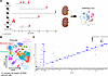 Figure 1
Single-cell atlas of PKD in mice. (A) Schematic of experimental design used to generate the single-cell atlas of PKD in mice. Red arrows indicate time points of harvest. (B) Uniform Manifold Approximation and Projection (UMAP) of all cells collected from mouse models of PKD. (C) Dot plot showing the top 5 markers of each cluster from the whole atlas. Pax8cre Pkd1fl/fl model (8 control samples, 9 PKD samples; all male), adult induced (AI) Ift88 model (2 controls, 2 PKD; all female), Pkd1RC/RC model (2 controls, 3 PKD; 3 males [1 control, 2 PKD], 2 females [1 control, 1PKD]), AI Pkd2 (2 controls, 2 PKD; all female), Pkhd1cre Pkd1fl/fl (3 controls, 3 PKD; mix of males and females), AI injured Ift88 (4 controls [2 sham operated Ift88, 2 injured cre negative control], 2 PKD; all female).
To better understand how mutations in PKD-related genes impacted tubular epithelium across models, we subsetted epithelial cells from the whole atlas and analyzed them in isolation. Following subsetting, epithelial cells were reclustered to determine what specific subpopulations were present. Analysis of the data revealed 12 transcriptionally distinct clusters of cells spanning the entire length of the nephron, including proximal tubule (PT; clusters 0, 1, 2, and 5), loop of Henle (LOH; clusters 3, 7, and 9), distal tubule (clusters 6, 8, and 11), and collecting duct (clusters 4 and 10; Figure 2, A and B). The proportion of S1–S3 PT, collecting duct principal cells (PCs), lower limb of the LOH, and thin descending limb of the LOH were increased in combined PKD samples compared with controls, with changes in the lower limb LOH being the most substantial (Figure 2, C and D). Across all nephron segments examined, tubular epithelial cells from PKD samples had increased expression of genes associated with tubular injury (Lcn2 and Havcr1), complement (C3), inflammation (Cxcl1), and fibrosis (Col1a1, Spp1, and Sparc), in agreement with previous data (Figure 2E and Supplemental Table 2) (12, 13). Pathway and transcription factor inference revealed that several clusters expressed genes associated with proinflammatory pathways (JAK-STAT, NF-κB, PI3K, and TNF) and transcription factors (Figure 2, F and G).
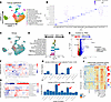 Figure 2
Analyses of scRNA-seq data from the nephron. (A) UMAP of tubular epithelial cells subclustered from Figure 1. (B) Dot plot showing the top 5 markers of each cluster of cells. (C) UMAP of tubular epithelial cells based on experimental group. (D) Quantification of cluster abundance in control and PKD samples from whole atlas shown as log2(fold change). (E) DEGs in all tubular epithelial cells when comparing control and PKD samples. (F and G) Pathway and transcription factor inference from DecoupleR. (H and I) Quantification of the number of DEGs in each cluster (control vs. PKD) in pseudobulked scRNA-seq data determined using DESeq2. (J) DEGs between control and PKD tubular cell clusters. Wilcoxon rank-sum test (E and H–J).
To better understand how PKD impacted gene expression in each nephron segment, we performed pseudobulk analysis of our scRNA-seq data, followed by identification of differentially expressed genes (DEGs) in each cluster using DESeq2 (14). This analysis revealed that PCs and cells from the lower limb LOH had the most DEGs when comparing control and PKD samples (Figure 2, H and I), independent of the P value threshold that was used (adjusted P value < 0.05 or P value < 0.05). Further investigation of DEGs (using adjusted P value < 0.05) in the lower limb LOH revealed that PKD mice had increased expression of genes associated with kidney injury (Lcn2, Havcr1, and Vcam1), complement (Cfi and C4b), inflammation (Csf1 and Ccl2), fibrosis (Pdgfb and Admats1), and fluid/ion secretion (Cftr and Kcnip4) compared with controls (Figure 2J, red arrows). We also found that expression of Gprc5a, a recently identified marker of cyst-lining epithelial cells (6), was increased in PCs from PKD mice (Figure 2J, blue arrow).
When analyzing the data, we noted inconsistencies in gene expression across individual PKD replicates, even within the same disease model. To better understand this variability, we grouped DEGs (adjusted P value < 0.05) based on variables that may be driving this effect, including sex, experimental model, time point of harvest, rate of disease progression, and genetic mutation (orthologous vs. non-orthologous) and replotted the data using heatmaps. For rate of disease progression, mice were considered to have rapidly progressing PKD if they developed cysts of any size in 8 weeks or less (Pkhd1cre Pkd1fl/fl model [3 samples]; Injured Ift88 model [2 samples]). In both the lower limb LOH and PCs, we found that DEGs most strongly segregated based on the rate of disease progression rather than sex, experimental model, time point of harvest, or genetic mutation (Supplemental Figures 1 and 2), suggesting that rate of PKD progression, and not the type of genetic mutation, is the most important variable driving the observed DEGs.
Individual-model-level comparison of cluster abundance and DEGs in relation to one another and patients with ADPKD. In addition to identifying commonalities in gene expression across PKD mouse models, we also sought to understand model-specific differences in nephron segment abundance and gene expression and how those differences related to patients with ADPKD. As such, we subsetted our atlas at the individual model level and quantified cell proportion and DEGs within each nephron segment (Figure 3A). For this quantification, we excluded samples from the S1 and S2 PT, as we found that the sequencing technology (single cell vs. single nucleus) heavily impacted cluster abundance, independently of phenotype (Supplemental Figure 3). When we analyzed the data, we found that the proportion of PCs, S3 PT, distal convoluted tubule (DCT), lower limb LOH, and nephron connecting tubule was significantly different when comparing control to PKD samples, but only in a few select models (Figure 3B). Of note, we found that the proportion of lower limb LOH cells was increased in the Ift88 and Pkd2 models (vs. respective non-cystic controls) and that the proportion of DCT was decreased in Pkd1RC/RC and Pkd2 mice relative to their non-cystic controls (Figure 3B). PCs were increased in the Pkhd1cre Pkd1fl/fl model but decreased in the injured Ift88 model in relation to non-cystic controls (Figure 3B). The observed differences in cluster abundance occurred despite the fact that the gene of interest is impacted in all nephron segments across PKD models, although some models, such as the Pkhd1cre Pkd1fl/fl model, have greater Cre recombinase activity in the distal nephron (15), which may be reflected by the fact that PCs were the only cluster in this model in which the proportion was different when comparing control and PKD samples.
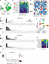 Figure 3
Model-level analysis of cluster abundance and DEGs. (A) UMAP of tubular epithelial cells based on individual models. (B) Quantification of cluster abundance in individual models. (C) Jaccard indices of intersected genes that were increased or decreased in the lower limb LOH and principal cells (PCs) of individual PKD models. (D and E) Upset plot showing shared genes that are increased or decreased across models in (D) lower limb LOH and (E) PCs. (F) UMAP of control and ADPKD single-cell data. (G) Quantification of cluster proportion. (H) Dot plot showing the percentage of intersected DEGs when comparing lower limb LOH and PCs between individual mouse models and ADPKD patients. *P < 0.05 by the Wilcoxon rank-sum test (B and G).
We next calculated DEGs (adjusted P < 0.05) in each nephron segment from each cystic model in relation to non-cystic controls from the same model using the Wilcoxon rank-sum test and plotted the data using Jaccard indices. We focused our analyses on the lower limb LOH and PCs due to the fact that these clusters had the highest number of DEGs at the whole-atlas level. Expectedly, we found that DEG overlap across models was relatively limited, ranging from no overlap to 25.6% of DEGs (Figure 3C). However, quite unexpectedly, we found that the models with the most DEG overlap did not share a common genetic mutation. For example, we found that the Ift88 and Pkhd1cre Pkd1fl/fl models were most similar in terms of increased DEGs in PCs, while Ift88 and Pkd1RC/RC models were most similar in terms of increased DEGs in the lower limb LOH (Figure 3C). In contrast, the Ift88 model was most similar to injured Ift88 and Pkhd1cre Pkd1fl/fl models in terms of decreased DEGs in PCs (Figure 3C). We also visualized intersected genes in these 2 nephron segments using upset plots and found that no genes were increased or decreased in all models, although some genes, such as Spp1, were increased in multiple models in both nephron segments (Figure 3, D and E). Thus, while there are certain genes that are enriched in multiple models, each model is unique in terms of DEGs that are altered across the 2 nephron segments analyzed.
Next, we set out to quantify which mouse model was most similar to patients with ADPKD in terms of cluster proportion and gene expression. To do this, we analyzed published snRNA-seq data from patients with ADPKD (16), identifying all nephron segments as well as immune cells, vasculature, and fibroblasts (Figure 3F and Supplemental Figure 4A). Unlike the mouse models analyzed, quantification of human data revealed that patients with ADPKD had a significant decrease in the proportion of cells from the LOH (Figure 3G). The proportion of PCs was increased in patients with ADPKD, similar to what was observed in the Pkhd1cre Pkd1fl/fl model. Patients with ADPKD also had a decreased proportion of DCT cells, similar to what was observed in the Pkd1RC/RC and Pkd2 mouse models (Figure 3G).
Next, we calculated DEGs in each nephron segment of ADPKD kidneys in relation to non-cystic controls using the Wilcoxon rank-sum test and quantified the percentage of overlapping genes in the same nephron segment of each mouse model. In both the lower limb LOH and PCs, the Pkhd1cre Pkd1fl/fl model had the highest frequency of DEG overlap with ADPKD patients while the Pax8 Pkd1fl/fl had the highest number of intersected genes, likely owing to the fact that this model has the highest number of DEGs when comparing control to PKD samples (Figure 3, D and H). We also found that the Ift88 model had the second highest frequency of DEG overlap with ADPKD patients in both nephron segments, while the orthologous Pkd1RC/RC model ranked near the bottom in terms of DEG overlap with ADPKD patients (Figure 3H). The Pkhd1cre Pkd1fl/fl model had the highest frequency of DEG overlap with ADPKD patients in several other nephron segments, including DCTs, intercalated cells (ICs), and the thin limb LOH (Supplemental Figure 4B). PT DEG overlap with ADPKD patients was similar across most models (Supplemental Figure 4B). It should be noted that the patient samples used in this analysis represent a stage of advanced disease, as is typical in ADPKD patient samples (16). Thus, based on the above data, we conclude that the Pkhd1cre Pkd1fl/fl model is most similar to patients with ADPKD in terms of cluster abundance and DEG overlap at a stage of advanced human disease.
Identification of PKD-specific clusters in the lower limb LOH and PCs. In our initial clustering, we grouped all cells from each nephron segment into one homogeneous population, without making assumptions about PKD-specific subsets that may be embedded within these nephron segments. To determine whether there were PKD-enriched subsets embedded within each nephron segment, we subsetted, reclustered, and quantified the abundance of each nephron segment at high resolution (Supplemental Figure 5A). We focused our analysis on the lower limb LOH and PCs due to the fact that these nephron segments were the most impacted in PKD samples, both at the whole-atlas and individual-model level. This analysis revealed that the lower limb LOH had 2 subsetted clusters that were greater than 2-fold enriched in PKD samples relative to non-cystic controls (Figure 4, A and B). These segments, which we annotated as lower limb PKD cluster 1 and lower limb PKD cluster 2, had highly disparate enrichment among individual PKD models (Figure 4, C and D). For example, lower limb PKD cluster 1 was almost exclusively derived from the Pax8 Pkd1fl/fl model, while lower limb PKD cluster 2 was derived from the Ift88, Pkd1RC/RC, and Pkhd1cre Pkd1fl/fl models (Figure 4D). DEGs in each of the PKD-enriched clusters from the lower limb were unique (Figure 4, E and F). Pathway and transcription factor inference supported this claim, showing that lower limb PKD cluster 1 had an enrichment of genes associated with the TGF-β and WNT pathways, whereas lower limb PKD cluster 2 had an enrichment of genes associated with EGFR, p53, and VEGF pathways (Figure 4G). Both PKD clusters were enriched for TNF-α pathway genes.
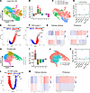 Figure 4
PKD-enriched clusters across mouse models. (A) UMAP showing high-resolution clusters and groups in the lower limb LOH. (B) Quantification of subsetted, high-resolution lower limb LOH cluster. Two segments were greater than 2-fold increased in PKD samples. (C) Re-annotated high-resolution clusters from lower limb LOH. (D) Quantification of PKD-enriched cluster abundance in lower limb LOH from individual models in relation to model specific, non-cystic controls. (E and F) Volcano plot showing DEGs in (E) PKD cluster 1 and (F) PKD cluster 2 in relation to all other cells in the lower limb LOH. (G) Pathway and transcription factor inference of genes enriched in PKD cluster 1 and PKD cluster 2. (H) UMAP showing high-resolution clusters and groups in principal cells (PCs). (I) Quantification of subsetted, high-resolution PC cluster. One segment was greater than 2-fold increased in PKD samples versus non-cystic controls. (J) Re-annotated high-resolution clusters from PCs. (K) Quantification of PKD-enriched cluster abundance in PCs from individual models in relation to model specific, non-cystic controls. (L) Volcano plot showing DEGs in PC PKD cluster in relation to all other PCs. (M) Pathway and transcription factor inference of genes enriched in the PC PKD cluster.
Analysis of subsetted PCs revealed that a single cluster, cluster 0, was enriched in combined PKD samples compared with controls (Figure 4, H–J). Enrichment of this cluster in PKD samples was largely driven by the Pax8 Pkd1fl/fl model, although several other models also had increased numbers in PKD samples compared with controls (Figure 4K). The PC PKD cluster had enrichment of mitochondrial genes as well as Spp1 and Lcn2 (Figure 4L). Pathways enriched in the PC PKD cluster include EGFR, p53, TGF-β, TNF-α, and JAK-STAT (Figure 4M). In contrast to previous data (6), we did not find an obvious PKD-enriched cluster in PTs, although we did find 2 PKD-enriched clusters in the thin descending limb LOH (Supplemental Figure 5, B and C).
Atlas- and model-level analyses of cell-cell communication show consistent enrichment of SPP1 signaling in PKD clusters. Our data indicate that the lower limb LOH and PCs are the nephron segments most altered in PKD. Furthermore, by subsetting and reclustering at high resolution, we were able to identify 3 PKD-specific clusters (lower limb PKD cluster 1, lower limb PKD cluster 2, and the PC PKD cluster) in the combined segments. To understand whole-atlas- and model-level signaling between PKD-specific clusters and other cell types, we performed CellChat on the fully annotated single-cell atlas (Supplemental Figure 6). In the whole atlas, we found that the number and strength of interactions were increased between lower limb PKD cluster 1 and fibroblasts in PKD samples (Supplemental Figure 7A). The strength, albeit not the number, of interactions was also increased in PKD samples from lower limb PKD cluster 2 and PC PKD cluster in the whole atlas (Supplemental Figure 7A). When we analyzed signaling interactions at the model level, we found that the number and strength of interactions were highest in lower limb PKD cluster 1 in the majority of models, with the exception of the injured Ift88 and Pkhd1cre Pkd1fl/fl models (Supplemental Figure 7, B–G). Of interest, we noted that the number and strength of signaling interactions were most frequently increased between lower limb PKD cluster 1, immune cells, and fibroblasts (Supplemental Figure 7, B–G).
We next analyzed outgoing signaling from PKD-enriched clusters at the atlas and model level. Analysis of the data revealed that SPP1 was the strongest outgoing signaling pattern in the whole atlas and in each individual model (red arrows) (Figure 5A). With regards to cell types, we found that lower limb PKD cluster 2 had the highest outgoing signaling strength across models (Figure 5A). Other outgoing signaling pathways of interest that were shared in multiple models included complement (green arrows), CSF (dark blue arrows), SEMA3 (purple arrows), WNT (light blue arrows), and MIF (yellow arrows). These pathways along with SPP1 signaling have been associated with PKD in the past, confirming the robustness of our analysis (17–22).
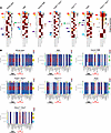 Figure 5
CellChat analysis of cell-cell communication in PKD-enriched clusters at the whole-atlas and model level reveals elevated outgoing SPP1 signaling. (A) Top outgoing signaling pathways from PKD-enriched clusters at the whole-atlas or model level. Arrows indicate pathways that are conserved across multiple models. (B) SPP1 signaling from PKD-enriched clusters to all other cell types at the whole-atlas or model level.
When we analyzed incoming pathways to the PKD-specific clusters, we once again found that SPP1 was the top pathway (Supplemental Figure 8). Incoming WNT and MIF signaling was also observed in multiple models. Other pathways of interest shared across multiple models included galectin (orange arrows), TGF-β (black arrows), IGF (pink arrows), FGF (light green arrows), MK (gray arrows), and TWEAK (salmon arrows), several of which have also been associated with PKD (Supplemental Figure 8) (21, 23–26). These data suggest that PKD-enriched clusters receive signaling from multiple growth factor signaling pathways, which may alter cyst growth.
We next analyzed SPP1 signaling from the PKD clusters, as this pathway was the most consistently enriched outgoing pathway across PKD models. The data indicate that SPP1 from PKD-enriched clusters, particularly lower limb PKD cluster 2, most frequently interacted with fibroblasts and monocytes across models (Figure 5B). A more detailed analyses of SPP1 signaling from lower limb PKD cluster 2 to fibroblasts and monocytes revealed distinct ligand-receptor pairs in each model (Supplemental Figures 9–12). However, we did note consistent Spp1–Itgα4+Itgβ1 and Spp1–Cd44 signaling between lower limb PKD cluster 2 and monocytes across models (Supplemental Figures 9–12), in line with data indicating that SPP1 serves as a monocyte chemoattractant (27, 28). The Spp1–Cd44 signaling axis in particular has also been implicated in kidney injury models as a key pathway involved in neutrophil recruitment and impaired tubular regeneration (29). Across models we also observed consistent signaling between neutrophils and lower limb PKD cluster 2, suggesting the pathway may function in a similar manner in PKD (Supplemental Figures 9–12).
Loss of Spp1 improves PKD in the orthologous Pkd1RC/RC model. Analysis of scRNA-seq data indicated that SPP1 gene expression and signaling is enriched in PKD enriched clusters and signals to monocytes and fibroblasts. In order to investigate the functional importance of SPP1 signaling in mouse models of PKD, we crossed the orthologous Pkd1RC/RC model to commercially available Spp1-knockout mice and analyzed the PKD phenotype at 1 year of age. We chose this model due to its orthologous nature and the fact that the rate of PKD progression mimics that of patients with ADPKD (30). We first confirmed that Spp1 expression was strongly increased in PKD-enriched clusters from Pkd1RC/RC scRNA-seq data (Figure 6A, red arrows) and was also present in cyst-lining cells of Pkd1RC/RC mice at the protein level (Figure 6B). Analyses of 1-year-old Pkd1RC/RC mice showed that loss of Spp1 modestly reduced 2KW/BW, cystic index, and cyst number compared with age-matched, littermate controls (Figure 6, C–F). Despite the fact that no significant differences were found in fibrosis between groups as indicated by Picrosirius red staining, kidney function was improved in Pkd1RC/RC Spp1-knockout mice compared with Pkd1RC/RC Spp1 control mice (Figure 6, G–I). Loss of Spp1 did not impact Ly6chi monocyte numbers but reduced the number of Ly6clo monocytes in Pkd1RC/RC mice, while other immune cell numbers remained unchanged (Supplemental Figure 13). Whether the loss of Spp1 and corresponding improvement in PKD severity is due to reduced SPP1-mediated Ly6clo monocyte recruitment is unknown.
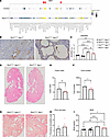 Figure 6
Loss of Spp1 improves PKD severity in the Pkd1RC/RC model. (A) Dot plot showing expression of Spp1 in all cell types of Pkd1RC/RC mice. (B) Immunohistochemistry staining of SPP1 (osteopontin) in Pkd1RC/WT and Pkd1RC/RC sections. Original magnification, ×20. (C) Quantification of 2 kidney weight to body weight (2KW/BW) ratio in Pkd1RC/WT Spp1cont, Pkd1RC/WT Spp1–/–, Pkd1RC/RC Spp1cont, and Pkd1RC/RC Spp1–/– mice at 1 year of age. (D–F) H&E-stained images of Pkd1RC/RC Spp1cont, and Pkd1RC/RC Spp1–/– mice along with quantification of (E) cystic index and (F) cyst number. (G and H) Picrosirius red–stained sections (G) and quantification (H). (I) Quantification of kidney function as measured by blood urea nitrogen (BUN). *P < 0.05; **P < 0.01; ***P < 0.001; ****P < 0.0001 by 1-way ANOVA (B and I) or Student’s t test (all others). Pkd1RC/WT Spp1cont = 6 mice, Pkd1RC/WT Spp1–/– = 8 mice, Pkd1RC/RC Spp1cont = 10 mice, Pkd1RC/RC Spp1–/– = 12 mice; 36 total Pkd1RC/RC mice.
Validation of enriched SPP1 expression and signaling in ADPKD tissue. To validate the therapeutic potential of targeting Spp1 in patients with ADPKD, we first analyzed expression of SPP1 in scRNA-seq data collected from control and ADPKD tissue. The data indicate that SPP1 expression was increased across nephron segments, including LOHs and PCs (Figure 7A). We also found that SPP1 expression was increased, albeit not specific to cyst-lining epithelia, in ADPKD kidney tissue compared with control tissue (Figure 7B). Next, we performed CellChat analysis on scRNA-seq data from control and ADPKD kidney tissue. Analysis of the data indicated that SPP1 signaling is the second strongest outgoing signaling pathway in ADPKD kidney tissue (Figure 7C). Similar to what was observed in animal models, SPP1 produced by tubular cells predominantly signaled to immune cells and fibroblasts (Figure 7D). Collectively, these data indicate that SPP1 expression and signaling is enriched in ADPKD kidney tissue.
 Figure 7
SPP1 expression and signaling are enriched in patients with ADPKD. (A) Dot plot showing expression of SPP1 in nephron segments from control and ADPKD patients. (B) RNA scope images showing SPP1 expression in control and ADPKD tissue. (C) Heatmap showing top outgoing signaling pathways in each cell type from control and ADPKD scRNA-seq data. (D) SPP1 signaling in control and ADPKD clusters.


