
Research ArticleImmunologyPulmonology
Open Access |  10.1172/jci.insight.202919
Role of CD4+ T cell mannose binding lectin in Schistosoma-induced pulmonary hypertension
Claudia Mickael,1 Dara C. Fonseca Balladares,2,3 Rahul Kumar,4 Michael H. Lee,2,3 Kevin Nolan,2,3 Linda Sanders,1 Katie J. Tuscan,1 Ramraj Prasad,5 Pilar Londono,1 Fernanda P. Oliveira,6 Kennedi B. Pyper,7 Ari B. Molofsky,7 Rubin M. Tuder,1,5 Kurt R. Stenmark,5 and Brian B. Graham2,3
1Division of Pulmonary Sciences and Critical Care Medicine, Department of Medicine, University of Colorado Anschutz Medical Campus, Aurora, Colorado, USA.
2Department of Medicine, UCSF, San Francisco, California, USA.
3Lung Biology Center, Zuckerberg San Francisco General Hospital, San Francisco, California, USA.
4Department of Translational Immunology, Genentech Inc., South San Francisco, California, USA.
5Cardiovascular Pulmonary Research Lab, University of Colorado School of Medicine, Aurora, Colorado, USA.
6Division of Respiratory Diseases, Federal University of São Paulo, São Paulo, Brazil.
7Department of Laboratory Medicine, UCSF, San Francisco, California, USA.
Address correspondence to: Claudia Mickael, Division of Pulmonary Sciences and Critical Care Medicine/Cardiovascular Research Laboratories, Department of Medicine, University of Colorado Denver/Anschutz Medical Campus, 12700 E. 19th Ave., Aurora, Colorado, 80045, USA. Phone: 303.724.5871; Email: claudia.mickael@cuanschutz.edu. Or to: Brian B. Graham, Division of Pulmonary and Critical Care, Zuckerberg San Francisco General Hospital, UCSF, San Francisco, California, 94110, USA. Phone: 628.206.4422; Email: brian.graham@ucsf.edu.
Find articles by Mickael, C. in: PubMed | Google Scholar
1Division of Pulmonary Sciences and Critical Care Medicine, Department of Medicine, University of Colorado Anschutz Medical Campus, Aurora, Colorado, USA.
2Department of Medicine, UCSF, San Francisco, California, USA.
3Lung Biology Center, Zuckerberg San Francisco General Hospital, San Francisco, California, USA.
4Department of Translational Immunology, Genentech Inc., South San Francisco, California, USA.
5Cardiovascular Pulmonary Research Lab, University of Colorado School of Medicine, Aurora, Colorado, USA.
6Division of Respiratory Diseases, Federal University of São Paulo, São Paulo, Brazil.
7Department of Laboratory Medicine, UCSF, San Francisco, California, USA.
Address correspondence to: Claudia Mickael, Division of Pulmonary Sciences and Critical Care Medicine/Cardiovascular Research Laboratories, Department of Medicine, University of Colorado Denver/Anschutz Medical Campus, 12700 E. 19th Ave., Aurora, Colorado, 80045, USA. Phone: 303.724.5871; Email: claudia.mickael@cuanschutz.edu. Or to: Brian B. Graham, Division of Pulmonary and Critical Care, Zuckerberg San Francisco General Hospital, UCSF, San Francisco, California, 94110, USA. Phone: 628.206.4422; Email: brian.graham@ucsf.edu.
Find articles by Fonseca Balladares, D. in: PubMed | Google Scholar
1Division of Pulmonary Sciences and Critical Care Medicine, Department of Medicine, University of Colorado Anschutz Medical Campus, Aurora, Colorado, USA.
2Department of Medicine, UCSF, San Francisco, California, USA.
3Lung Biology Center, Zuckerberg San Francisco General Hospital, San Francisco, California, USA.
4Department of Translational Immunology, Genentech Inc., South San Francisco, California, USA.
5Cardiovascular Pulmonary Research Lab, University of Colorado School of Medicine, Aurora, Colorado, USA.
6Division of Respiratory Diseases, Federal University of São Paulo, São Paulo, Brazil.
7Department of Laboratory Medicine, UCSF, San Francisco, California, USA.
Address correspondence to: Claudia Mickael, Division of Pulmonary Sciences and Critical Care Medicine/Cardiovascular Research Laboratories, Department of Medicine, University of Colorado Denver/Anschutz Medical Campus, 12700 E. 19th Ave., Aurora, Colorado, 80045, USA. Phone: 303.724.5871; Email: claudia.mickael@cuanschutz.edu. Or to: Brian B. Graham, Division of Pulmonary and Critical Care, Zuckerberg San Francisco General Hospital, UCSF, San Francisco, California, 94110, USA. Phone: 628.206.4422; Email: brian.graham@ucsf.edu.
Find articles by
Kumar, R.
in:
PubMed
|
Google Scholar
|

1Division of Pulmonary Sciences and Critical Care Medicine, Department of Medicine, University of Colorado Anschutz Medical Campus, Aurora, Colorado, USA.
2Department of Medicine, UCSF, San Francisco, California, USA.
3Lung Biology Center, Zuckerberg San Francisco General Hospital, San Francisco, California, USA.
4Department of Translational Immunology, Genentech Inc., South San Francisco, California, USA.
5Cardiovascular Pulmonary Research Lab, University of Colorado School of Medicine, Aurora, Colorado, USA.
6Division of Respiratory Diseases, Federal University of São Paulo, São Paulo, Brazil.
7Department of Laboratory Medicine, UCSF, San Francisco, California, USA.
Address correspondence to: Claudia Mickael, Division of Pulmonary Sciences and Critical Care Medicine/Cardiovascular Research Laboratories, Department of Medicine, University of Colorado Denver/Anschutz Medical Campus, 12700 E. 19th Ave., Aurora, Colorado, 80045, USA. Phone: 303.724.5871; Email: claudia.mickael@cuanschutz.edu. Or to: Brian B. Graham, Division of Pulmonary and Critical Care, Zuckerberg San Francisco General Hospital, UCSF, San Francisco, California, 94110, USA. Phone: 628.206.4422; Email: brian.graham@ucsf.edu.
Find articles by Lee, M. in: PubMed | Google Scholar
1Division of Pulmonary Sciences and Critical Care Medicine, Department of Medicine, University of Colorado Anschutz Medical Campus, Aurora, Colorado, USA.
2Department of Medicine, UCSF, San Francisco, California, USA.
3Lung Biology Center, Zuckerberg San Francisco General Hospital, San Francisco, California, USA.
4Department of Translational Immunology, Genentech Inc., South San Francisco, California, USA.
5Cardiovascular Pulmonary Research Lab, University of Colorado School of Medicine, Aurora, Colorado, USA.
6Division of Respiratory Diseases, Federal University of São Paulo, São Paulo, Brazil.
7Department of Laboratory Medicine, UCSF, San Francisco, California, USA.
Address correspondence to: Claudia Mickael, Division of Pulmonary Sciences and Critical Care Medicine/Cardiovascular Research Laboratories, Department of Medicine, University of Colorado Denver/Anschutz Medical Campus, 12700 E. 19th Ave., Aurora, Colorado, 80045, USA. Phone: 303.724.5871; Email: claudia.mickael@cuanschutz.edu. Or to: Brian B. Graham, Division of Pulmonary and Critical Care, Zuckerberg San Francisco General Hospital, UCSF, San Francisco, California, 94110, USA. Phone: 628.206.4422; Email: brian.graham@ucsf.edu.
Find articles by Nolan, K. in: PubMed | Google Scholar
1Division of Pulmonary Sciences and Critical Care Medicine, Department of Medicine, University of Colorado Anschutz Medical Campus, Aurora, Colorado, USA.
2Department of Medicine, UCSF, San Francisco, California, USA.
3Lung Biology Center, Zuckerberg San Francisco General Hospital, San Francisco, California, USA.
4Department of Translational Immunology, Genentech Inc., South San Francisco, California, USA.
5Cardiovascular Pulmonary Research Lab, University of Colorado School of Medicine, Aurora, Colorado, USA.
6Division of Respiratory Diseases, Federal University of São Paulo, São Paulo, Brazil.
7Department of Laboratory Medicine, UCSF, San Francisco, California, USA.
Address correspondence to: Claudia Mickael, Division of Pulmonary Sciences and Critical Care Medicine/Cardiovascular Research Laboratories, Department of Medicine, University of Colorado Denver/Anschutz Medical Campus, 12700 E. 19th Ave., Aurora, Colorado, 80045, USA. Phone: 303.724.5871; Email: claudia.mickael@cuanschutz.edu. Or to: Brian B. Graham, Division of Pulmonary and Critical Care, Zuckerberg San Francisco General Hospital, UCSF, San Francisco, California, 94110, USA. Phone: 628.206.4422; Email: brian.graham@ucsf.edu.
Find articles by Sanders, L. in: PubMed | Google Scholar
1Division of Pulmonary Sciences and Critical Care Medicine, Department of Medicine, University of Colorado Anschutz Medical Campus, Aurora, Colorado, USA.
2Department of Medicine, UCSF, San Francisco, California, USA.
3Lung Biology Center, Zuckerberg San Francisco General Hospital, San Francisco, California, USA.
4Department of Translational Immunology, Genentech Inc., South San Francisco, California, USA.
5Cardiovascular Pulmonary Research Lab, University of Colorado School of Medicine, Aurora, Colorado, USA.
6Division of Respiratory Diseases, Federal University of São Paulo, São Paulo, Brazil.
7Department of Laboratory Medicine, UCSF, San Francisco, California, USA.
Address correspondence to: Claudia Mickael, Division of Pulmonary Sciences and Critical Care Medicine/Cardiovascular Research Laboratories, Department of Medicine, University of Colorado Denver/Anschutz Medical Campus, 12700 E. 19th Ave., Aurora, Colorado, 80045, USA. Phone: 303.724.5871; Email: claudia.mickael@cuanschutz.edu. Or to: Brian B. Graham, Division of Pulmonary and Critical Care, Zuckerberg San Francisco General Hospital, UCSF, San Francisco, California, 94110, USA. Phone: 628.206.4422; Email: brian.graham@ucsf.edu.
Find articles by Tuscan, K. in: PubMed | Google Scholar
1Division of Pulmonary Sciences and Critical Care Medicine, Department of Medicine, University of Colorado Anschutz Medical Campus, Aurora, Colorado, USA.
2Department of Medicine, UCSF, San Francisco, California, USA.
3Lung Biology Center, Zuckerberg San Francisco General Hospital, San Francisco, California, USA.
4Department of Translational Immunology, Genentech Inc., South San Francisco, California, USA.
5Cardiovascular Pulmonary Research Lab, University of Colorado School of Medicine, Aurora, Colorado, USA.
6Division of Respiratory Diseases, Federal University of São Paulo, São Paulo, Brazil.
7Department of Laboratory Medicine, UCSF, San Francisco, California, USA.
Address correspondence to: Claudia Mickael, Division of Pulmonary Sciences and Critical Care Medicine/Cardiovascular Research Laboratories, Department of Medicine, University of Colorado Denver/Anschutz Medical Campus, 12700 E. 19th Ave., Aurora, Colorado, 80045, USA. Phone: 303.724.5871; Email: claudia.mickael@cuanschutz.edu. Or to: Brian B. Graham, Division of Pulmonary and Critical Care, Zuckerberg San Francisco General Hospital, UCSF, San Francisco, California, 94110, USA. Phone: 628.206.4422; Email: brian.graham@ucsf.edu.
Find articles by Prasad, R. in: PubMed | Google Scholar
1Division of Pulmonary Sciences and Critical Care Medicine, Department of Medicine, University of Colorado Anschutz Medical Campus, Aurora, Colorado, USA.
2Department of Medicine, UCSF, San Francisco, California, USA.
3Lung Biology Center, Zuckerberg San Francisco General Hospital, San Francisco, California, USA.
4Department of Translational Immunology, Genentech Inc., South San Francisco, California, USA.
5Cardiovascular Pulmonary Research Lab, University of Colorado School of Medicine, Aurora, Colorado, USA.
6Division of Respiratory Diseases, Federal University of São Paulo, São Paulo, Brazil.
7Department of Laboratory Medicine, UCSF, San Francisco, California, USA.
Address correspondence to: Claudia Mickael, Division of Pulmonary Sciences and Critical Care Medicine/Cardiovascular Research Laboratories, Department of Medicine, University of Colorado Denver/Anschutz Medical Campus, 12700 E. 19th Ave., Aurora, Colorado, 80045, USA. Phone: 303.724.5871; Email: claudia.mickael@cuanschutz.edu. Or to: Brian B. Graham, Division of Pulmonary and Critical Care, Zuckerberg San Francisco General Hospital, UCSF, San Francisco, California, 94110, USA. Phone: 628.206.4422; Email: brian.graham@ucsf.edu.
Find articles by Londono, P. in: PubMed | Google Scholar
1Division of Pulmonary Sciences and Critical Care Medicine, Department of Medicine, University of Colorado Anschutz Medical Campus, Aurora, Colorado, USA.
2Department of Medicine, UCSF, San Francisco, California, USA.
3Lung Biology Center, Zuckerberg San Francisco General Hospital, San Francisco, California, USA.
4Department of Translational Immunology, Genentech Inc., South San Francisco, California, USA.
5Cardiovascular Pulmonary Research Lab, University of Colorado School of Medicine, Aurora, Colorado, USA.
6Division of Respiratory Diseases, Federal University of São Paulo, São Paulo, Brazil.
7Department of Laboratory Medicine, UCSF, San Francisco, California, USA.
Address correspondence to: Claudia Mickael, Division of Pulmonary Sciences and Critical Care Medicine/Cardiovascular Research Laboratories, Department of Medicine, University of Colorado Denver/Anschutz Medical Campus, 12700 E. 19th Ave., Aurora, Colorado, 80045, USA. Phone: 303.724.5871; Email: claudia.mickael@cuanschutz.edu. Or to: Brian B. Graham, Division of Pulmonary and Critical Care, Zuckerberg San Francisco General Hospital, UCSF, San Francisco, California, 94110, USA. Phone: 628.206.4422; Email: brian.graham@ucsf.edu.
Find articles by Oliveira, F. in: PubMed | Google Scholar
1Division of Pulmonary Sciences and Critical Care Medicine, Department of Medicine, University of Colorado Anschutz Medical Campus, Aurora, Colorado, USA.
2Department of Medicine, UCSF, San Francisco, California, USA.
3Lung Biology Center, Zuckerberg San Francisco General Hospital, San Francisco, California, USA.
4Department of Translational Immunology, Genentech Inc., South San Francisco, California, USA.
5Cardiovascular Pulmonary Research Lab, University of Colorado School of Medicine, Aurora, Colorado, USA.
6Division of Respiratory Diseases, Federal University of São Paulo, São Paulo, Brazil.
7Department of Laboratory Medicine, UCSF, San Francisco, California, USA.
Address correspondence to: Claudia Mickael, Division of Pulmonary Sciences and Critical Care Medicine/Cardiovascular Research Laboratories, Department of Medicine, University of Colorado Denver/Anschutz Medical Campus, 12700 E. 19th Ave., Aurora, Colorado, 80045, USA. Phone: 303.724.5871; Email: claudia.mickael@cuanschutz.edu. Or to: Brian B. Graham, Division of Pulmonary and Critical Care, Zuckerberg San Francisco General Hospital, UCSF, San Francisco, California, 94110, USA. Phone: 628.206.4422; Email: brian.graham@ucsf.edu.
Find articles by Pyper, K. in: PubMed | Google Scholar
1Division of Pulmonary Sciences and Critical Care Medicine, Department of Medicine, University of Colorado Anschutz Medical Campus, Aurora, Colorado, USA.
2Department of Medicine, UCSF, San Francisco, California, USA.
3Lung Biology Center, Zuckerberg San Francisco General Hospital, San Francisco, California, USA.
4Department of Translational Immunology, Genentech Inc., South San Francisco, California, USA.
5Cardiovascular Pulmonary Research Lab, University of Colorado School of Medicine, Aurora, Colorado, USA.
6Division of Respiratory Diseases, Federal University of São Paulo, São Paulo, Brazil.
7Department of Laboratory Medicine, UCSF, San Francisco, California, USA.
Address correspondence to: Claudia Mickael, Division of Pulmonary Sciences and Critical Care Medicine/Cardiovascular Research Laboratories, Department of Medicine, University of Colorado Denver/Anschutz Medical Campus, 12700 E. 19th Ave., Aurora, Colorado, 80045, USA. Phone: 303.724.5871; Email: claudia.mickael@cuanschutz.edu. Or to: Brian B. Graham, Division of Pulmonary and Critical Care, Zuckerberg San Francisco General Hospital, UCSF, San Francisco, California, 94110, USA. Phone: 628.206.4422; Email: brian.graham@ucsf.edu.
Find articles by Molofsky, A. in: PubMed | Google Scholar
1Division of Pulmonary Sciences and Critical Care Medicine, Department of Medicine, University of Colorado Anschutz Medical Campus, Aurora, Colorado, USA.
2Department of Medicine, UCSF, San Francisco, California, USA.
3Lung Biology Center, Zuckerberg San Francisco General Hospital, San Francisco, California, USA.
4Department of Translational Immunology, Genentech Inc., South San Francisco, California, USA.
5Cardiovascular Pulmonary Research Lab, University of Colorado School of Medicine, Aurora, Colorado, USA.
6Division of Respiratory Diseases, Federal University of São Paulo, São Paulo, Brazil.
7Department of Laboratory Medicine, UCSF, San Francisco, California, USA.
Address correspondence to: Claudia Mickael, Division of Pulmonary Sciences and Critical Care Medicine/Cardiovascular Research Laboratories, Department of Medicine, University of Colorado Denver/Anschutz Medical Campus, 12700 E. 19th Ave., Aurora, Colorado, 80045, USA. Phone: 303.724.5871; Email: claudia.mickael@cuanschutz.edu. Or to: Brian B. Graham, Division of Pulmonary and Critical Care, Zuckerberg San Francisco General Hospital, UCSF, San Francisco, California, 94110, USA. Phone: 628.206.4422; Email: brian.graham@ucsf.edu.
Find articles by Tuder, R. in: PubMed | Google Scholar
1Division of Pulmonary Sciences and Critical Care Medicine, Department of Medicine, University of Colorado Anschutz Medical Campus, Aurora, Colorado, USA.
2Department of Medicine, UCSF, San Francisco, California, USA.
3Lung Biology Center, Zuckerberg San Francisco General Hospital, San Francisco, California, USA.
4Department of Translational Immunology, Genentech Inc., South San Francisco, California, USA.
5Cardiovascular Pulmonary Research Lab, University of Colorado School of Medicine, Aurora, Colorado, USA.
6Division of Respiratory Diseases, Federal University of São Paulo, São Paulo, Brazil.
7Department of Laboratory Medicine, UCSF, San Francisco, California, USA.
Address correspondence to: Claudia Mickael, Division of Pulmonary Sciences and Critical Care Medicine/Cardiovascular Research Laboratories, Department of Medicine, University of Colorado Denver/Anschutz Medical Campus, 12700 E. 19th Ave., Aurora, Colorado, 80045, USA. Phone: 303.724.5871; Email: claudia.mickael@cuanschutz.edu. Or to: Brian B. Graham, Division of Pulmonary and Critical Care, Zuckerberg San Francisco General Hospital, UCSF, San Francisco, California, 94110, USA. Phone: 628.206.4422; Email: brian.graham@ucsf.edu.
Find articles by
Stenmark, K.
in:
PubMed
|
Google Scholar
|
1Division of Pulmonary Sciences and Critical Care Medicine, Department of Medicine, University of Colorado Anschutz Medical Campus, Aurora, Colorado, USA.
2Department of Medicine, UCSF, San Francisco, California, USA.
3Lung Biology Center, Zuckerberg San Francisco General Hospital, San Francisco, California, USA.
4Department of Translational Immunology, Genentech Inc., South San Francisco, California, USA.
5Cardiovascular Pulmonary Research Lab, University of Colorado School of Medicine, Aurora, Colorado, USA.
6Division of Respiratory Diseases, Federal University of São Paulo, São Paulo, Brazil.
7Department of Laboratory Medicine, UCSF, San Francisco, California, USA.
Address correspondence to: Claudia Mickael, Division of Pulmonary Sciences and Critical Care Medicine/Cardiovascular Research Laboratories, Department of Medicine, University of Colorado Denver/Anschutz Medical Campus, 12700 E. 19th Ave., Aurora, Colorado, 80045, USA. Phone: 303.724.5871; Email: claudia.mickael@cuanschutz.edu. Or to: Brian B. Graham, Division of Pulmonary and Critical Care, Zuckerberg San Francisco General Hospital, UCSF, San Francisco, California, 94110, USA. Phone: 628.206.4422; Email: brian.graham@ucsf.edu.
Find articles by
Graham, B.
in:
PubMed
|
Google Scholar
|
Published July 28, 2026 - More info
JCI Insight. 2026;11(17):e202919. https://doi.org/10.1172/jci.insight.202919.
© 2026 Mickael et al. This work is licensed under the Creative Commons Attribution 4.0 International License. To view a copy of this license, visit http://creativecommons.org/licenses/by/4.0/.
Received: December 1, 2025; Accepted: July 21, 2026

-
Results
MBL deficiency protects mice from Schistosoma-induced PH. To investigate the functional role of the complement factor MBL in the development of Schistosoma-induced PH, we examined mice that lack both isoforms, Mbl1 and Mbl2. These double-knockout mice are hereafter referred to as MblKO mice. WT isogenic mice were used as experimental controls. The mouse model of Schistosoma-induced PH used involves sensitizing to Schistosoma egg antigen through i.p. injection, followed by challenge with i.v. egg injection. This leads to embolization in the precapillary pulmonary vasculature, while i.p. sensitization is necessary before i.v. challenge for the PH phenotype to develop (10), indicating a requirement for adaptive immunity.
Complement factors including the MBLs are typically described as being expressed by hepatocytes (11, 12), but other organs can also express complement including the BM and BM-derived cells (13). Here, we focused on the immune system and BM-derived complement. We investigated this potential source of MBL by performing BM transplantation (BMT) using MblKO donors and lethally irradiated WT recipients. The recipients, referred to as MblKO-chimeras, were compared with control mice that received WT BM and were treated in a similar manner, denoted as MblWT-chimeras. The MblWT-chimera mice did not exhibit any evidence of a baseline PH phenotype. When the MblWT-chimera mice were subjected to Schistosoma egg i.p. sensitization followed by Schistosoma egg i.v. challenge, they developed significant PH, as evident by an increase in right ventricle (RV) systolic pressure (RVSP) measured by right heart catheterization, an increase in the RV mass relative to the left ventricle plus septum (Fulton index), and an increase in the thickness of the medial compartment of the pulmonary vasculature, as determined by quantitative analysis of immunostained lung specimens (Figure 1). At baseline, the MblKO-chimera mice also exhibited no discernible pulmonary vascular phenotype. In contrast to the WT-chimeras, following Schistosoma egg sensitization and challenge, the MblKO-chimera mice displayed a relative resistance to PH, with no significant increase in RVSP, right ventricular hypertrophy, or medial wall remodeling (Figure 1). These data indicate that the combination of Mbl1 and Mbl2, as expressed by BM-derived cells, is critical for the development of schistosomiasis-induced PH.
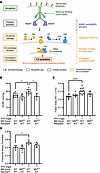 Figure 1
PH phenotype of WT-chimera and MblKO-chimera mice following Schistosoma exposure. (A) Schematic of the lectin pathway. Mannose on foreign carbohydrates and glycoproteins binds mannose binding lectin (MBL), which forms a complex with mannan-binding lectin-associated serine proteases 1 and 2 (MASP1 and MASP2). This complex enzymatically degrades C4 releasing C4b fragment and C2 releasing C2a fragment. C4b and C2a together form the C4b2a complex, which is functionally a C3 convertase, resulting in C3 activation. C4a and C2b together form the C4a2b complex, which is functionally a C3 convertase, resulting in C3 activation. Active C3 is the point at which the classical and alternative complement pathways converge. This figure was generated by ChatGPT. (B) RVSP, (C) Fulton index, and (D) fractional media thickness in WT recipients of WT or MblKO bone marrow. N = 5–12/group; data are shown as the mean ± SD. ANOVA with post hoc Tukey’s test shown, *P < 0.05, **P < 0.01, ***P < 0.001. BM, bone marrow; LV, left ventricle; MblKO, deficiency of both Mbl1 and Mbl2; RV, right ventricle; RVSP, right ventricle systolic pressure; S, septum.
BM MBL deficiency suppresses complement deposition in the lungs of Schistosoma-exposed mice. Activation of the lectin complement pathway results in C4 and C2 cleavage, the assembly of the C3 and C5 convertases, and fragmentation of C3 and C5. We thus analyzed the lung tissue of the Schistosoma-induced PH mice to look for evidence of C3 complement fragment deposition in the lung tissue, using Western blot on whole lung lysates. This revealed in the lung tissue of MblWT-chimeras an increase in the deposition of the terminal C3 degradation product C3d following Schistosoma challenge (Supplemental Figure 1; supplemental material available online with this article; https://doi.org/10.1172/jci.insight.202919DS1). In contrast, MblKO-chimera mice challenged with Schistosoma had relatively less C3d in the lung tissue. These data suggest that deleting both Mbl1 and Mbl2 in the BM compartment suppresses complement activation in the lung tissue.
BM MBL deficiency reduces CD4+ T cell activation. Given our suspicion that complement may contribute mechanistically to CD4+ T cell activation, we analyzed the phenotype of CD4+ T cells in the lung tissue of the Schistosoma-challenged mice. To exclude intravascular lymphocytes, we administered a fluorescently labeled anti-CD45 antibody i.v. to mark intravascular cells and utilized flow cytometry gating techniques to exclude the marked cells (Supplemental Figure 2A).
In the MblWT-chimera mice, we observed that Schistosoma sensitization and challenge resulted in a 2.9-fold increase in total CD4+ T cells and a 39-fold increase in Th2 CD4+ T cells in the lungs as compared with that in unchallenged MblWT-chimera mice (Figure 2, A and B). In contrast, there was no significant increase in either total or Th2 CD4+ T cells in the MblKO-chimera mice following Schistosoma exposure. We also investigated other CD4+ T cell phenotypes and found that both Th17 and Treg (Foxp3+) CD4+ T cells were significantly increased in the MblWT-chimera mice following Schistosoma exposure, but this was not seen in the MblKO-chimera mice (Figure 2, C and D). However, both the MblWT-chimera and MblKO-chimera mice displayed a modest increase in Th1 CD4+ T cells (Figure 2E), suggesting that lectin complement was upstream of type 2 but not type 1 immunity in Schistosoma-exposed mice, although it could also contribute to Th17 and Treg phenotypes as well.
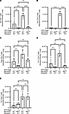 Figure 2
Absolute number of CD4+ T cells in WT and MblKO-chimera mice following Schistosoma exposure. Absolute number of (A) CD4+ T cells, (B) IL-4+ Th2 CD4+ T cells, (C) IFN-γ+ Th1 CD4+ T cells, (D) IL-17A+ Th17 CD4+ T cells, and (E) FOXP3+ Treg CD4+ T cells. N = 3–5/group; data are shown as the mean ± SD. ANOVA with post hoc Tukey’s test shown, *P < 0.05, **P < 0.01, ***P < 0.001, ****P < 0.0001. BM, bone marrow; MblKO, deficiency of both Mbl1 and Mbl2.
As we had previously observed a requirement for Th2 cells specifically in Schistosoma-induced PH (2), we focused on the role of MBL in contributing to type 2 immunity. We conducted IL-4 and IL-13 ELISA assays to evaluate whole lung type 2 inflammation in the mice, finding that the Schistosoma-exposed MblKO-chimera exhibited lower levels of IL-4 compared with Schistosoma-exposed MblWT-chimera mice (Supplemental Figure 3A). The effect on IL-13 suppression was less pronounced (Supplemental Figure 3B). As an additional measure of type 2 inflammation, we also estimated the volume of peri-egg granulomas using stereology and found that the granuloma volumes were not suppressed in the MblKO-chimera relative to MblWT-chimera mice (Supplemental Figure 3C). These data suggest that the effect of deleting the lectin pathway may relate particularly to the expression of IL-4 by Th2 CD4+ T cells.
MBL expression promotes a Th2 CD4+ T cell phenotype by a cell-intrinsic pathway. We then sought to identify which cells within the BM compartment could be necessary to express MBL. We specifically considered the potential guiding role of MBL in the phenotyping of CD4+ T cells through an autocrine or cell-intrinsic processes. To test this hypothesis, we first quantified MBL expression by CD4+ T cells in Schistosoma-exposed mice as compared with control WT mice using flow cytometry (gating strategy in Supplemental Figure 2B). Consistent with this hypothesis, Schistosoma-exposed mice had a significant increase in all CD4+ T cells, and specifically memory and central memory CD4+ T cells, which also expressed higher levels of MBL as compared with control, unexposed mice (as well as naive CD4+ T cells, to a more modest extent; Figure 3).
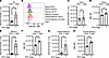 Figure 3
MBL expression by CD4+ T cells increases with Schistosoma exposure. (A) Absolute number of all CD4+ T cells. (B) Representative expression of MBL in different CD4+ T cell populations by flow cytometry, unchallenged or challenged with i.p./i.v. eggs. Absolute number and median fluorescent intensity (MFI) of (C and D) naive CD4+ T cells, (E and F) memory CD4+ T cells, and (G and H) central memory CD4+ T cells. N = 5/group; data are shown as the mean ± SD. t test, **P < 0.01, ****P < 0.0001.
We also queried if Th2 cells are located around the vessels of Schistosoma-induced PH mice, which would facilitate pathologic signaling by adjacency. To do so, we used Il5Cre;Ai14 (IL-5 reporter) mice costained with CD3 to identify Th2 cells. We observed that Schistosoma-sensitized and challenged mice had more Th2 cells throughout the entire lung, but in particular many Th2 cells were located immediately adjacent to vessels (Supplemental Figure 4).
We then assessed whether the source of MBL in the CD4+ T cells is extrinsic via the extracellular environment versus intrinsic to the CD4 cells. To do so, we first performed a BM chimera experiment, in which a 1:1 ratio of MblKO BM (with the cells marked by CD45.2) was mixed with WT BM (from a CD45.1 donor) and transplanted into a lethally irradiated CD45.1 recipient (Supplemental Figure 5A, and gating strategy in Supplemental Figure 2C). In this manner, we would be able to identify T cells that are MBL expressing (CD45.1) or MBL deficient (CD45.2). We posited that if MBL expression extrinsic to the T cells is necessary, the CD45.1 and CD45.2 T cells would have the same phenotype; whereas if MBL expression intrinsic to the T cells was necessary, then the CD45.1 and CD45.2 T cells would have different phenotypes. Although the sample sizes are very small, we observed trends in fewer CD45.2-marked CD4+ T cells, and specifically Th2 CD4+ T cells (on average a 9-fold reduction, P = 0.08 by paired t test), than CD45.1-marked cells (Supplemental Figure 5, B and C). These in vivo data indicated that intrinsic MBL expression by the T cells themselves could contribute to their activation.
Based on these suggestive in vivo data, we sought to confirm the requirement for MBL to polarize CD4+ T cells to a Th2 phenotype using an in vitro approach. We isolated CD4+ T cells and splenocytes from the spleens of Schistosoma-sensitized WT and MblKO mice. The CD4+ T cells were labeled with CellTrace Violet (CTV) ex vivo and then cultured with the splenocytes (serving as antigen-presenting cells) and S. mansoni soluble egg antigen (SEA) for 96 hours (see summary of experimental design in Supplemental Figure 6A). We read out the phenotype of the CD4+ T cells by flow cytometry, quantifying CTV dilution as an indicator of cellular proliferation (Supplemental Figure 6B). We observed that WT CD4+ T cells mixed with WT splenocytes underwent significant proliferation (as evidenced by CTV dilution) only after the addition of SEA (Figure 4, A and C). In contrast, MblKO CD4+ T cells mixed with MblKO splenocytes and SEA underwent significantly less proliferation (Figure 4, B and C). To confirm that MBL is functionally important in CD4+ T cells but not splenocytes, we combined WT CD4+ T cells with MBL-deficient splenocytes and observed that the T cell proliferation was comparable to that in cells cocultured with WT splenocytes (Figure 4C).
 Figure 4
MBL expression by CD4+ T cells is required for a Th2 phenotype. (A) Histogram of CellTrace Violet expression by WT CD4+ T cells, cocultured with WT splenocytes, under conditions of with SEA or no SEA added, demonstrating proliferation with SEA. (B) Histogram of CellTrace Violet expression by MblKO CD4+ T cells, cocultured with MblKO splenocytes, under conditions of with SEA or no SEA added, demonstrating no T cell proliferation in either condition. (C) Quantification of the fraction of CD4+ T cells that are divided (have dilution of the CellTrace Violet signal), for WT and MblKO CD4+ T cells and splenocytes, all with SEA added, in the combinations shown. N = 5–8/group; data are shown as the mean ± SD. ANOVA with post hoc Tukey’s test shown, ***P < 0.001. SEA, soluble egg antigen; MblKO, deficiency of both Mbl1 and Mbl2.
We considered alternative mechanisms by which T cell activation could be modulated, such as the complement pathway impacting DC function, which would thus indirectly affect the T cell phenotype (14). There are 2 types of conventional DCs, types 1 and 2, referred to as cDC1 and cDC2, respectively. Prototypically, cDC1s activate CD8+ T cells, and cDC2s activate CD4+ T cells; we recently observed that cDC2s contribute to Schistosoma-induced PH (15). By flow cytometry (gating strategy in Supplemental Figure 7), we found that cDC1s but not cDC2s were decreased in the MblKO-chimera mice (Supplemental Figure 8, A and B). We did not observe a change in the number of interstitial macrophages in the MblKO-chimera mice (Supplemental Figure 8C).
C4-deficient BM does not protect from Schistosoma-induced PH. Activation of complement factor 4 (C4) is a critical mediator downstream of MBL in the lectin complement pathway: the fragment C4a together with C2b (also activated by the MBL-MASP1-MASP2 complex) forms the C3 convertase that leads to C3 activation converging with the classical and alternative complement pathways. To determine if the canonical extracellular lectin pathway is required for CD4+ T cell activation in Schistosoma-induced PH, we assessed the phenotype of lethally irradiated WT mice that received C4-knockout (C4KO) BM, subject to the same i.p. sensitization and i.v. challenge protocol. Interestingly, these mice were not protected from Schistosoma-induced PH (Figure 5), suggesting that the extracellular downstream lectin pathway is not required for the function of MBL in activating Th2 cells.
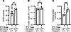 Figure 5
Bone marrow C4 deficiency, a downstream target of MBL, is not protective in Schistosoma-induced PH. (A) RVSP, (B) Fulton index, and (C) fractional pulmonary vascular media thickness of C4KO (C4-deficient) chimera mice, unchallenged or challenged with i.p./i.v. eggs. N = 4–6/group; data are shown as the mean ± SD. Unpaired t test, **P < 0.01, ***P < 0.001.
MBL BM deficiency does not protect from hypoxia-induced PH. As described above, there was reasonable evidence to support the hypothesis that the lectin complement cascade contributes to the pathology present in pulmonary complications of schistosomiasis. We then studied the potential role of the lectin complement pathway in an alternative form of experimental PH, hypoxia-induced disease, which does not have a clear biologic rationale for lectin complement pathway function. We found that the MblKO-chimera mice developed significant PH following 21 days of hypoxia exposure, as evidenced by a significant increase in both RVSP and RV hypertrophy in the hypoxia-challenged mice, as compared with control MblKO-chimera mice maintained in normoxia (Figure 6). These data indicate that MBL in the BM does not contribute to hypoxia-induced PH, which is driven by Th17 immunity (16).
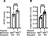 Figure 6
Bone marrow MBL deficiency is not protective in chronic hypoxia-induced PH. (A) RVSP and (B) Fulton index of WT-chimera and MblKO-chimera mice. N = 8–9/group; data are shown as the mean ± SD. Unpaired t test, ****P < 0.0001. BM, bone marrow; MblKO, deficiency of both Mbl1 and Mbl2.

-
Discussion
We found evidence that the lectin complement system is activated in Schistosoma-exposed mice and promotes the development of experimental Schistosoma-induced PH (Figure 7). MBL proteins expressed by BM-derived cells were required, as deleting both Mbl1 and Mbl2 in the BM was found to be protective. We further identified a critical function of MBL in the ability of CD4+ T cells to activate to a Th2 phenotype in the context of Schistosoma exposure. The requirement for lectin complement pathway activation in the pathogenesis of PH due to Schistosoma was dispensable in a noninfectious form of PH following hypoxia exposure, suggesting differential roles of the lectin pathway in different forms of PH. The role of MBL may also be relevant in other Th2-driven pathologies, such as type 2 asthma, or ovalbumin-induced inflammation.
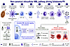 Figure 7
Schematic of the immune cascade promoting Schistosoma-induced PH. Schistosoma antigens are presented by classical DCs to CD4+ T cells, which use MBL to develop a Th2 phenotype. Through the expression of IL-4 and IL-13, the Th2 cells result in the recruitment of classical monocytes, which become interstitial macrophages that express TSP1, causing the activation of TGF-β. In the setting of chronic infection, TGF-β likely promotes a degree of immune tolerance that is beneficial for the host. However, in the context of the pulmonary vasculature, TGF-β causes deleterious proremodeling phenotypes that result in vascular cell proliferation, vasoconstriction, and pulmonary hypertension. Figure generated by ChatGPT.
The relevance to human disease is supported by prior observations of more CD4+ T cells and evidence of persistent type 2 inflammation in the autopsy lung tissue of individuals who died of Schistosoma-induced PH (1, 17). It is possible that targeting type 2 inflammation could benefit those with Schistosoma-induced PH, or the initial inflammation could trigger a downstream, self-perpetuating pathology that is now resistant to antiinflammatory treatments. In individuals without concern for active infection, blocking the MBL pathway such as by anti-MBL antibodies could be of benefit (18, 19). However, in individuals who live in an endemic setting and are at risk for reinfection, targeting the lectin complement system would be contraindicated owing to the possibility of compromising the host response to the parasite.
Complement is a critical part of the innate immune system; as such it promotes cell lysis through the formation of the membrane attack complex (MAC) on bacterial membranes, facilitating phagocytosis, promoting inflammation, and helping guide the adaptive immune response. Here, we targeted the lectin pathway, which is specifically activated in response to foreign material by the binding of pattern-recognition plasma molecules such as MBL to carbohydrates or acetylated residues present on organisms such as parasites (20). The bound MBL then activates MBL-associated serine proteases 1 and 2 (MASP-1 and MASP-2), leading to C4 and C2 cleavage. This leads to the assembly of the C3 and C5 convertases, with subsequent cleavage of C3 and C5, which enter the common complement pathway that ends with MAC assembly. Humans have 1 MBL isoform, whereas mice have 2 isoforms; the 2 mouse forms appear to be similar in function (21), with 1 of the 2 (MBL1) lost in the evolution of modern humans (22).
Molecular members of the classical, alternative, and lectin complement pathway have signaling function in expressing cells, independent of activation of MAC. Intracellular C3 and C5, termed the “complosome,” have been described to contribute to the phenotype of Th1 and Th17 CD4+ T cells (23–27). C3 and C5 impact T cell phenotypes by regulating metabolism, including through mitochondrial function (23, 25, 28–31). However, a complosome role in Th2 biology has not been previously described to our knowledge. The observation that C4 deficiency in the BM compartment does not protect from Schistosoma-induced PH in the same way that MBL deficiency does suggests that the function of MBL does not require downstream C4 activation in this model. Complosome C3 and C5 in Th1 and Th17 cells also do not require the downstream activation of the complement pathway. This is also similar to the observation that DC function requires DC expression of C3AR1 and C5AR1 (32), but C4-deficient DCs do not have suppressed ability to activate T cells (33). Our data that C4 in BM-derived cells appears dispensable for Schistosoma-induced PH does not preclude a role for liver-expressed C4 (the majority of C4 is made by the liver) (34, 35), which may contribute to the host response to the parasite.
We observed MBL deficiency also reduced Tregs. In other PH models, Tregs appear to be protective, as their deletion contributes to PH, and reconstituting Treg-deficient animals is protective (36, 37). In the context of Schistosoma-induced PH, Th2 cells mechanistically contribute to pulmonary vascular pathology without being affected by levels or activation of Tregs (2), as compared with the protective role of Tregs in other forms of PH.
There are several potential mechanisms by which MBL expression in CD4+ T cells could be increased. MBL is an acute phase protein, with increased expression in settings of inflammation, such as ischemia-reperfusion and oxidative stress (38), as well as infection. Hypoxic pathway mechanisms are another candidate driver (39). In CD4+ T cells, the HIF2α isoform has been linked to a Th2 phenotype (40).
Complement links innate and adaptive immunity. We have found Th2 CD4+ T cells are required for Schistosoma-induced PH (1, 2). C3aR, C5aR, and CD46 (a regulator of complement activation) are all expressed by T cells (41), and all act as costimulatory signals for T cells (32, 42). C3aR is expressed in the lysosomes of naive T cells and transported to the cell membrane of activated T cells (43). We observed the function of CD4+ T cells is controlled by intrinsic MBL expression, likely acting at differentiation stages prior to their full activation, as MBL expression was lower in activated cells. There is evidence in other settings of an autocrine function for immune cell-derived complement, as T cells from C3-deficient patients cannot generate IFN-γ even in C3-sufficient serum (43). C3ar1–/– and C5ar1–/– mice have defective T cell activation (32, 44) and shift their phenotype to Tregs (45). Tregs express C3aR and C5aR, and signaling through these receptors decreases Foxp3 expression and inhibits Treg function (45). We found that MBL-deficient chimeras had fewer cDC1s, although this does not explain the protection observed, as experimentally depleting cDC1s using Batf3–/– mice resulted in more severe S. mansoni–induced PH (15). We previously observed that cDC2s in particular contribute to Schistosoma-induced PH (15); here, we found that the MBL depletion in CD4+ T cells did not impact cDC2 density, consistent with the role of cDC2s being proximate to T cell activation.
There are broader implications related to the relevance of MBL in schistosomiasis infection. Genetic polymorphisms in the MBL promoter that lead to decreased expression and lower plasma MBL are associated with increased schistosomiasis infection (6). MBL genetic polymorphisms and higher plasma levels are also associated with more severe liver disease (46, 47), which likely contributes to the development of clinical PH by promoting egg embolization via portocaval shunts (48).
A limitation in our work is that we did not precisely knockout MBL in CD4+ T cells in vivo. We are not aware of these mice being developed and would be challenged by needing to target both MBL isoforms in a cell-specific manner. We were able to isolate the requirement to the BM compartment and then focused specifically on the role of MBL in CD4+ T cells using our in vitro system.
In the BM transplant experiments, we used mixtures of male and female donor cells transplanted into only female recipients. Sex-mismatch status between donor and recipient can independently stimulate the immune system and is a risk factor for graft-versus-host disease (49, 50); thus, some of the immune pathways stimulated and modulated by MBL could be independent of Schistosoma-driven immunity. However, the greatest signal reported is for female donors into male recipients, potentially related to increased immune memory from prior pregnancies in the donor and/or Y chromosome–derived antigens in the recipient (49, 50). None of the donor or recipient mice we used had been previously pregnant: all were dedicated for experimental purposes.
Another limitation is the uncertainty of why whole-lung IL-4 and IL-13 concentration did not correlate as strongly with CD4+ Th2 density and the PH phenotype. This may be due to other cells expressing IL-4 and IL-13, which could include ILC2s, macrophages, mast cells, eosinophils, and basophils. This observation may also relate to location of cytokine expression, either concentrated in the pulmonary vasculature or that in other lung compartments, possibly in relation to peri-egg granulomas. Creating lung lysates will result in average cytokine concentrations across the entire tissue, leading to an apparent discrepancy as compared to assessing perivascular cells specifically.
In conclusion, we found that lectin complement pathway MBL proteins contribute to the pathogenesis of experimental schistosomiasis-induced PH by mechanistically contributing to the activation of CD4+ T cells to a Th2 phenotype. Therapeutically targeting this pathway to prevent or treat clinical Schistosoma-induced PH specifically should be considered with caution, as this pathway’s contribution is likely at the earliest stages of disease development, and host immunity should ideally be preserved in the event of ongoing infection or reinfection with the parasite. However, this may be a promising therapeutic target in other Th2-driven diseases such as asthma.


