
Research ArticleCell biologyMuscle biologyPulmonology
Open Access |  10.1172/jci.insight.202475
Hypercapnia enhances airway smooth muscle contractility via STIM1-dependent Ca2+ signaling
Masahiko Shigemura,1,2 Vitalii Kryvenko,3 Jennifer A. Pacheco,4 Megan J. Puckelwartz,4,5 Milos Aleksic,6 Natalia D. Magnani,7 Emma Thompson,8 Francisco Javier Martin-Romero,9 Eoin P. Cummins,10 Werner Seeger,3 Andreas Bräuninger,11 Lynn C. Welch,12 G.R. Scott Budinger,12 Emilia Lecuona,1 Laura A. Dada,12 Ankit Bharat,1 István Vadász,3 Murali Prakriya,5 and Jacob I. Sznajder12
1Division of Thoracic Surgery, Department of Surgery, Feinberg School of Medicine, Northwestern University, Chicago, Illinois, USA.
2Laboratory of Homeostatic Physiology, Department of Physiology and Cell Biology, Graduate School of Medicine, Kobe University, Kobe, Japan.
3Department of Internal Medicine, Justus Liebig University, Universities of Giessen and Marburg Lung Center, German Center for Lung Research, Cardio-Pulmonary Institute, and Institute for Lung Health, Giessen, Germany.
4Center for Genetic Medicine,
5Department of Pharmacology, and
6Department of Neuroscience, Feinberg School of Medicine, Northwestern University, Chicago, Illinois, USA.
7Faculty of Pharmacy and Biochemistry, University of Buenos Aires, Buenos Aires, Argentina.
8Department of Human Genetics, University of Chicago, Chicago, Illinois, USA.
9Department of Biochemistry and Molecular Biology, Universidad de Extremadura, Badajoz, Spain.
10University College Dublin School of Medicine, UCD Conway Institute of Biomolecular and Biomedical Research, Dublin, Ireland.
11Institute of Pathology, Justus Liebig University, Giessen, Germany.
12Division of Pulmonary and Critical Care Medicine, Department of Medicine, Feinberg School of Medicine, Northwestern University, Chicago, Illinois, USA.
Address correspondence to: Masahiko Shigemura, Division of Thoracic Surgery, Feinberg School of Medicine, Northwestern University, Chicago, Illinois 60611, USA. Phone: 312.503.5033; Email: masahiko.shigemura@northwestern.edu; also to: Masahiko Shigemura, Laboratory of Homeostatic Physiology, Department of Physiology and Cell Biology, Graduate School of Medicine, Kobe University, Kobe, Japan. Email: mkshige@med.kobe-u.ac.jp.
Find articles by
Shigemura, M.
in:
PubMed
|
Google Scholar
|

1Division of Thoracic Surgery, Department of Surgery, Feinberg School of Medicine, Northwestern University, Chicago, Illinois, USA.
2Laboratory of Homeostatic Physiology, Department of Physiology and Cell Biology, Graduate School of Medicine, Kobe University, Kobe, Japan.
3Department of Internal Medicine, Justus Liebig University, Universities of Giessen and Marburg Lung Center, German Center for Lung Research, Cardio-Pulmonary Institute, and Institute for Lung Health, Giessen, Germany.
4Center for Genetic Medicine,
5Department of Pharmacology, and
6Department of Neuroscience, Feinberg School of Medicine, Northwestern University, Chicago, Illinois, USA.
7Faculty of Pharmacy and Biochemistry, University of Buenos Aires, Buenos Aires, Argentina.
8Department of Human Genetics, University of Chicago, Chicago, Illinois, USA.
9Department of Biochemistry and Molecular Biology, Universidad de Extremadura, Badajoz, Spain.
10University College Dublin School of Medicine, UCD Conway Institute of Biomolecular and Biomedical Research, Dublin, Ireland.
11Institute of Pathology, Justus Liebig University, Giessen, Germany.
12Division of Pulmonary and Critical Care Medicine, Department of Medicine, Feinberg School of Medicine, Northwestern University, Chicago, Illinois, USA.
Address correspondence to: Masahiko Shigemura, Division of Thoracic Surgery, Feinberg School of Medicine, Northwestern University, Chicago, Illinois 60611, USA. Phone: 312.503.5033; Email: masahiko.shigemura@northwestern.edu; also to: Masahiko Shigemura, Laboratory of Homeostatic Physiology, Department of Physiology and Cell Biology, Graduate School of Medicine, Kobe University, Kobe, Japan. Email: mkshige@med.kobe-u.ac.jp.
Find articles by Kryvenko, V. in: PubMed | Google Scholar
1Division of Thoracic Surgery, Department of Surgery, Feinberg School of Medicine, Northwestern University, Chicago, Illinois, USA.
2Laboratory of Homeostatic Physiology, Department of Physiology and Cell Biology, Graduate School of Medicine, Kobe University, Kobe, Japan.
3Department of Internal Medicine, Justus Liebig University, Universities of Giessen and Marburg Lung Center, German Center for Lung Research, Cardio-Pulmonary Institute, and Institute for Lung Health, Giessen, Germany.
4Center for Genetic Medicine,
5Department of Pharmacology, and
6Department of Neuroscience, Feinberg School of Medicine, Northwestern University, Chicago, Illinois, USA.
7Faculty of Pharmacy and Biochemistry, University of Buenos Aires, Buenos Aires, Argentina.
8Department of Human Genetics, University of Chicago, Chicago, Illinois, USA.
9Department of Biochemistry and Molecular Biology, Universidad de Extremadura, Badajoz, Spain.
10University College Dublin School of Medicine, UCD Conway Institute of Biomolecular and Biomedical Research, Dublin, Ireland.
11Institute of Pathology, Justus Liebig University, Giessen, Germany.
12Division of Pulmonary and Critical Care Medicine, Department of Medicine, Feinberg School of Medicine, Northwestern University, Chicago, Illinois, USA.
Address correspondence to: Masahiko Shigemura, Division of Thoracic Surgery, Feinberg School of Medicine, Northwestern University, Chicago, Illinois 60611, USA. Phone: 312.503.5033; Email: masahiko.shigemura@northwestern.edu; also to: Masahiko Shigemura, Laboratory of Homeostatic Physiology, Department of Physiology and Cell Biology, Graduate School of Medicine, Kobe University, Kobe, Japan. Email: mkshige@med.kobe-u.ac.jp.
Find articles by Pacheco, J. in: PubMed | Google Scholar
1Division of Thoracic Surgery, Department of Surgery, Feinberg School of Medicine, Northwestern University, Chicago, Illinois, USA.
2Laboratory of Homeostatic Physiology, Department of Physiology and Cell Biology, Graduate School of Medicine, Kobe University, Kobe, Japan.
3Department of Internal Medicine, Justus Liebig University, Universities of Giessen and Marburg Lung Center, German Center for Lung Research, Cardio-Pulmonary Institute, and Institute for Lung Health, Giessen, Germany.
4Center for Genetic Medicine,
5Department of Pharmacology, and
6Department of Neuroscience, Feinberg School of Medicine, Northwestern University, Chicago, Illinois, USA.
7Faculty of Pharmacy and Biochemistry, University of Buenos Aires, Buenos Aires, Argentina.
8Department of Human Genetics, University of Chicago, Chicago, Illinois, USA.
9Department of Biochemistry and Molecular Biology, Universidad de Extremadura, Badajoz, Spain.
10University College Dublin School of Medicine, UCD Conway Institute of Biomolecular and Biomedical Research, Dublin, Ireland.
11Institute of Pathology, Justus Liebig University, Giessen, Germany.
12Division of Pulmonary and Critical Care Medicine, Department of Medicine, Feinberg School of Medicine, Northwestern University, Chicago, Illinois, USA.
Address correspondence to: Masahiko Shigemura, Division of Thoracic Surgery, Feinberg School of Medicine, Northwestern University, Chicago, Illinois 60611, USA. Phone: 312.503.5033; Email: masahiko.shigemura@northwestern.edu; also to: Masahiko Shigemura, Laboratory of Homeostatic Physiology, Department of Physiology and Cell Biology, Graduate School of Medicine, Kobe University, Kobe, Japan. Email: mkshige@med.kobe-u.ac.jp.
Find articles by
Puckelwartz, M.
in:
PubMed
|
Google Scholar
|
1Division of Thoracic Surgery, Department of Surgery, Feinberg School of Medicine, Northwestern University, Chicago, Illinois, USA.
2Laboratory of Homeostatic Physiology, Department of Physiology and Cell Biology, Graduate School of Medicine, Kobe University, Kobe, Japan.
3Department of Internal Medicine, Justus Liebig University, Universities of Giessen and Marburg Lung Center, German Center for Lung Research, Cardio-Pulmonary Institute, and Institute for Lung Health, Giessen, Germany.
4Center for Genetic Medicine,
5Department of Pharmacology, and
6Department of Neuroscience, Feinberg School of Medicine, Northwestern University, Chicago, Illinois, USA.
7Faculty of Pharmacy and Biochemistry, University of Buenos Aires, Buenos Aires, Argentina.
8Department of Human Genetics, University of Chicago, Chicago, Illinois, USA.
9Department of Biochemistry and Molecular Biology, Universidad de Extremadura, Badajoz, Spain.
10University College Dublin School of Medicine, UCD Conway Institute of Biomolecular and Biomedical Research, Dublin, Ireland.
11Institute of Pathology, Justus Liebig University, Giessen, Germany.
12Division of Pulmonary and Critical Care Medicine, Department of Medicine, Feinberg School of Medicine, Northwestern University, Chicago, Illinois, USA.
Address correspondence to: Masahiko Shigemura, Division of Thoracic Surgery, Feinberg School of Medicine, Northwestern University, Chicago, Illinois 60611, USA. Phone: 312.503.5033; Email: masahiko.shigemura@northwestern.edu; also to: Masahiko Shigemura, Laboratory of Homeostatic Physiology, Department of Physiology and Cell Biology, Graduate School of Medicine, Kobe University, Kobe, Japan. Email: mkshige@med.kobe-u.ac.jp.
Find articles by Aleksic, M. in: PubMed | Google Scholar
1Division of Thoracic Surgery, Department of Surgery, Feinberg School of Medicine, Northwestern University, Chicago, Illinois, USA.
2Laboratory of Homeostatic Physiology, Department of Physiology and Cell Biology, Graduate School of Medicine, Kobe University, Kobe, Japan.
3Department of Internal Medicine, Justus Liebig University, Universities of Giessen and Marburg Lung Center, German Center for Lung Research, Cardio-Pulmonary Institute, and Institute for Lung Health, Giessen, Germany.
4Center for Genetic Medicine,
5Department of Pharmacology, and
6Department of Neuroscience, Feinberg School of Medicine, Northwestern University, Chicago, Illinois, USA.
7Faculty of Pharmacy and Biochemistry, University of Buenos Aires, Buenos Aires, Argentina.
8Department of Human Genetics, University of Chicago, Chicago, Illinois, USA.
9Department of Biochemistry and Molecular Biology, Universidad de Extremadura, Badajoz, Spain.
10University College Dublin School of Medicine, UCD Conway Institute of Biomolecular and Biomedical Research, Dublin, Ireland.
11Institute of Pathology, Justus Liebig University, Giessen, Germany.
12Division of Pulmonary and Critical Care Medicine, Department of Medicine, Feinberg School of Medicine, Northwestern University, Chicago, Illinois, USA.
Address correspondence to: Masahiko Shigemura, Division of Thoracic Surgery, Feinberg School of Medicine, Northwestern University, Chicago, Illinois 60611, USA. Phone: 312.503.5033; Email: masahiko.shigemura@northwestern.edu; also to: Masahiko Shigemura, Laboratory of Homeostatic Physiology, Department of Physiology and Cell Biology, Graduate School of Medicine, Kobe University, Kobe, Japan. Email: mkshige@med.kobe-u.ac.jp.
Find articles by Magnani, N. in: PubMed | Google Scholar
1Division of Thoracic Surgery, Department of Surgery, Feinberg School of Medicine, Northwestern University, Chicago, Illinois, USA.
2Laboratory of Homeostatic Physiology, Department of Physiology and Cell Biology, Graduate School of Medicine, Kobe University, Kobe, Japan.
3Department of Internal Medicine, Justus Liebig University, Universities of Giessen and Marburg Lung Center, German Center for Lung Research, Cardio-Pulmonary Institute, and Institute for Lung Health, Giessen, Germany.
4Center for Genetic Medicine,
5Department of Pharmacology, and
6Department of Neuroscience, Feinberg School of Medicine, Northwestern University, Chicago, Illinois, USA.
7Faculty of Pharmacy and Biochemistry, University of Buenos Aires, Buenos Aires, Argentina.
8Department of Human Genetics, University of Chicago, Chicago, Illinois, USA.
9Department of Biochemistry and Molecular Biology, Universidad de Extremadura, Badajoz, Spain.
10University College Dublin School of Medicine, UCD Conway Institute of Biomolecular and Biomedical Research, Dublin, Ireland.
11Institute of Pathology, Justus Liebig University, Giessen, Germany.
12Division of Pulmonary and Critical Care Medicine, Department of Medicine, Feinberg School of Medicine, Northwestern University, Chicago, Illinois, USA.
Address correspondence to: Masahiko Shigemura, Division of Thoracic Surgery, Feinberg School of Medicine, Northwestern University, Chicago, Illinois 60611, USA. Phone: 312.503.5033; Email: masahiko.shigemura@northwestern.edu; also to: Masahiko Shigemura, Laboratory of Homeostatic Physiology, Department of Physiology and Cell Biology, Graduate School of Medicine, Kobe University, Kobe, Japan. Email: mkshige@med.kobe-u.ac.jp.
Find articles by Thompson, E. in: PubMed | Google Scholar
1Division of Thoracic Surgery, Department of Surgery, Feinberg School of Medicine, Northwestern University, Chicago, Illinois, USA.
2Laboratory of Homeostatic Physiology, Department of Physiology and Cell Biology, Graduate School of Medicine, Kobe University, Kobe, Japan.
3Department of Internal Medicine, Justus Liebig University, Universities of Giessen and Marburg Lung Center, German Center for Lung Research, Cardio-Pulmonary Institute, and Institute for Lung Health, Giessen, Germany.
4Center for Genetic Medicine,
5Department of Pharmacology, and
6Department of Neuroscience, Feinberg School of Medicine, Northwestern University, Chicago, Illinois, USA.
7Faculty of Pharmacy and Biochemistry, University of Buenos Aires, Buenos Aires, Argentina.
8Department of Human Genetics, University of Chicago, Chicago, Illinois, USA.
9Department of Biochemistry and Molecular Biology, Universidad de Extremadura, Badajoz, Spain.
10University College Dublin School of Medicine, UCD Conway Institute of Biomolecular and Biomedical Research, Dublin, Ireland.
11Institute of Pathology, Justus Liebig University, Giessen, Germany.
12Division of Pulmonary and Critical Care Medicine, Department of Medicine, Feinberg School of Medicine, Northwestern University, Chicago, Illinois, USA.
Address correspondence to: Masahiko Shigemura, Division of Thoracic Surgery, Feinberg School of Medicine, Northwestern University, Chicago, Illinois 60611, USA. Phone: 312.503.5033; Email: masahiko.shigemura@northwestern.edu; also to: Masahiko Shigemura, Laboratory of Homeostatic Physiology, Department of Physiology and Cell Biology, Graduate School of Medicine, Kobe University, Kobe, Japan. Email: mkshige@med.kobe-u.ac.jp.
Find articles by Martin-Romero, F. in: PubMed | Google Scholar
1Division of Thoracic Surgery, Department of Surgery, Feinberg School of Medicine, Northwestern University, Chicago, Illinois, USA.
2Laboratory of Homeostatic Physiology, Department of Physiology and Cell Biology, Graduate School of Medicine, Kobe University, Kobe, Japan.
3Department of Internal Medicine, Justus Liebig University, Universities of Giessen and Marburg Lung Center, German Center for Lung Research, Cardio-Pulmonary Institute, and Institute for Lung Health, Giessen, Germany.
4Center for Genetic Medicine,
5Department of Pharmacology, and
6Department of Neuroscience, Feinberg School of Medicine, Northwestern University, Chicago, Illinois, USA.
7Faculty of Pharmacy and Biochemistry, University of Buenos Aires, Buenos Aires, Argentina.
8Department of Human Genetics, University of Chicago, Chicago, Illinois, USA.
9Department of Biochemistry and Molecular Biology, Universidad de Extremadura, Badajoz, Spain.
10University College Dublin School of Medicine, UCD Conway Institute of Biomolecular and Biomedical Research, Dublin, Ireland.
11Institute of Pathology, Justus Liebig University, Giessen, Germany.
12Division of Pulmonary and Critical Care Medicine, Department of Medicine, Feinberg School of Medicine, Northwestern University, Chicago, Illinois, USA.
Address correspondence to: Masahiko Shigemura, Division of Thoracic Surgery, Feinberg School of Medicine, Northwestern University, Chicago, Illinois 60611, USA. Phone: 312.503.5033; Email: masahiko.shigemura@northwestern.edu; also to: Masahiko Shigemura, Laboratory of Homeostatic Physiology, Department of Physiology and Cell Biology, Graduate School of Medicine, Kobe University, Kobe, Japan. Email: mkshige@med.kobe-u.ac.jp.
Find articles by
Cummins, E.
in:
PubMed
|
Google Scholar
|
1Division of Thoracic Surgery, Department of Surgery, Feinberg School of Medicine, Northwestern University, Chicago, Illinois, USA.
2Laboratory of Homeostatic Physiology, Department of Physiology and Cell Biology, Graduate School of Medicine, Kobe University, Kobe, Japan.
3Department of Internal Medicine, Justus Liebig University, Universities of Giessen and Marburg Lung Center, German Center for Lung Research, Cardio-Pulmonary Institute, and Institute for Lung Health, Giessen, Germany.
4Center for Genetic Medicine,
5Department of Pharmacology, and
6Department of Neuroscience, Feinberg School of Medicine, Northwestern University, Chicago, Illinois, USA.
7Faculty of Pharmacy and Biochemistry, University of Buenos Aires, Buenos Aires, Argentina.
8Department of Human Genetics, University of Chicago, Chicago, Illinois, USA.
9Department of Biochemistry and Molecular Biology, Universidad de Extremadura, Badajoz, Spain.
10University College Dublin School of Medicine, UCD Conway Institute of Biomolecular and Biomedical Research, Dublin, Ireland.
11Institute of Pathology, Justus Liebig University, Giessen, Germany.
12Division of Pulmonary and Critical Care Medicine, Department of Medicine, Feinberg School of Medicine, Northwestern University, Chicago, Illinois, USA.
Address correspondence to: Masahiko Shigemura, Division of Thoracic Surgery, Feinberg School of Medicine, Northwestern University, Chicago, Illinois 60611, USA. Phone: 312.503.5033; Email: masahiko.shigemura@northwestern.edu; also to: Masahiko Shigemura, Laboratory of Homeostatic Physiology, Department of Physiology and Cell Biology, Graduate School of Medicine, Kobe University, Kobe, Japan. Email: mkshige@med.kobe-u.ac.jp.
Find articles by
Seeger, W.
in:
PubMed
|
Google Scholar
|
1Division of Thoracic Surgery, Department of Surgery, Feinberg School of Medicine, Northwestern University, Chicago, Illinois, USA.
2Laboratory of Homeostatic Physiology, Department of Physiology and Cell Biology, Graduate School of Medicine, Kobe University, Kobe, Japan.
3Department of Internal Medicine, Justus Liebig University, Universities of Giessen and Marburg Lung Center, German Center for Lung Research, Cardio-Pulmonary Institute, and Institute for Lung Health, Giessen, Germany.
4Center for Genetic Medicine,
5Department of Pharmacology, and
6Department of Neuroscience, Feinberg School of Medicine, Northwestern University, Chicago, Illinois, USA.
7Faculty of Pharmacy and Biochemistry, University of Buenos Aires, Buenos Aires, Argentina.
8Department of Human Genetics, University of Chicago, Chicago, Illinois, USA.
9Department of Biochemistry and Molecular Biology, Universidad de Extremadura, Badajoz, Spain.
10University College Dublin School of Medicine, UCD Conway Institute of Biomolecular and Biomedical Research, Dublin, Ireland.
11Institute of Pathology, Justus Liebig University, Giessen, Germany.
12Division of Pulmonary and Critical Care Medicine, Department of Medicine, Feinberg School of Medicine, Northwestern University, Chicago, Illinois, USA.
Address correspondence to: Masahiko Shigemura, Division of Thoracic Surgery, Feinberg School of Medicine, Northwestern University, Chicago, Illinois 60611, USA. Phone: 312.503.5033; Email: masahiko.shigemura@northwestern.edu; also to: Masahiko Shigemura, Laboratory of Homeostatic Physiology, Department of Physiology and Cell Biology, Graduate School of Medicine, Kobe University, Kobe, Japan. Email: mkshige@med.kobe-u.ac.jp.
Find articles by Bräuninger, A. in: PubMed | Google Scholar
1Division of Thoracic Surgery, Department of Surgery, Feinberg School of Medicine, Northwestern University, Chicago, Illinois, USA.
2Laboratory of Homeostatic Physiology, Department of Physiology and Cell Biology, Graduate School of Medicine, Kobe University, Kobe, Japan.
3Department of Internal Medicine, Justus Liebig University, Universities of Giessen and Marburg Lung Center, German Center for Lung Research, Cardio-Pulmonary Institute, and Institute for Lung Health, Giessen, Germany.
4Center for Genetic Medicine,
5Department of Pharmacology, and
6Department of Neuroscience, Feinberg School of Medicine, Northwestern University, Chicago, Illinois, USA.
7Faculty of Pharmacy and Biochemistry, University of Buenos Aires, Buenos Aires, Argentina.
8Department of Human Genetics, University of Chicago, Chicago, Illinois, USA.
9Department of Biochemistry and Molecular Biology, Universidad de Extremadura, Badajoz, Spain.
10University College Dublin School of Medicine, UCD Conway Institute of Biomolecular and Biomedical Research, Dublin, Ireland.
11Institute of Pathology, Justus Liebig University, Giessen, Germany.
12Division of Pulmonary and Critical Care Medicine, Department of Medicine, Feinberg School of Medicine, Northwestern University, Chicago, Illinois, USA.
Address correspondence to: Masahiko Shigemura, Division of Thoracic Surgery, Feinberg School of Medicine, Northwestern University, Chicago, Illinois 60611, USA. Phone: 312.503.5033; Email: masahiko.shigemura@northwestern.edu; also to: Masahiko Shigemura, Laboratory of Homeostatic Physiology, Department of Physiology and Cell Biology, Graduate School of Medicine, Kobe University, Kobe, Japan. Email: mkshige@med.kobe-u.ac.jp.
Find articles by
Welch, L.
in:
PubMed
|
Google Scholar
|
1Division of Thoracic Surgery, Department of Surgery, Feinberg School of Medicine, Northwestern University, Chicago, Illinois, USA.
2Laboratory of Homeostatic Physiology, Department of Physiology and Cell Biology, Graduate School of Medicine, Kobe University, Kobe, Japan.
3Department of Internal Medicine, Justus Liebig University, Universities of Giessen and Marburg Lung Center, German Center for Lung Research, Cardio-Pulmonary Institute, and Institute for Lung Health, Giessen, Germany.
4Center for Genetic Medicine,
5Department of Pharmacology, and
6Department of Neuroscience, Feinberg School of Medicine, Northwestern University, Chicago, Illinois, USA.
7Faculty of Pharmacy and Biochemistry, University of Buenos Aires, Buenos Aires, Argentina.
8Department of Human Genetics, University of Chicago, Chicago, Illinois, USA.
9Department of Biochemistry and Molecular Biology, Universidad de Extremadura, Badajoz, Spain.
10University College Dublin School of Medicine, UCD Conway Institute of Biomolecular and Biomedical Research, Dublin, Ireland.
11Institute of Pathology, Justus Liebig University, Giessen, Germany.
12Division of Pulmonary and Critical Care Medicine, Department of Medicine, Feinberg School of Medicine, Northwestern University, Chicago, Illinois, USA.
Address correspondence to: Masahiko Shigemura, Division of Thoracic Surgery, Feinberg School of Medicine, Northwestern University, Chicago, Illinois 60611, USA. Phone: 312.503.5033; Email: masahiko.shigemura@northwestern.edu; also to: Masahiko Shigemura, Laboratory of Homeostatic Physiology, Department of Physiology and Cell Biology, Graduate School of Medicine, Kobe University, Kobe, Japan. Email: mkshige@med.kobe-u.ac.jp.
Find articles by
Budinger, G.
in:
PubMed
|
Google Scholar
|
1Division of Thoracic Surgery, Department of Surgery, Feinberg School of Medicine, Northwestern University, Chicago, Illinois, USA.
2Laboratory of Homeostatic Physiology, Department of Physiology and Cell Biology, Graduate School of Medicine, Kobe University, Kobe, Japan.
3Department of Internal Medicine, Justus Liebig University, Universities of Giessen and Marburg Lung Center, German Center for Lung Research, Cardio-Pulmonary Institute, and Institute for Lung Health, Giessen, Germany.
4Center for Genetic Medicine,
5Department of Pharmacology, and
6Department of Neuroscience, Feinberg School of Medicine, Northwestern University, Chicago, Illinois, USA.
7Faculty of Pharmacy and Biochemistry, University of Buenos Aires, Buenos Aires, Argentina.
8Department of Human Genetics, University of Chicago, Chicago, Illinois, USA.
9Department of Biochemistry and Molecular Biology, Universidad de Extremadura, Badajoz, Spain.
10University College Dublin School of Medicine, UCD Conway Institute of Biomolecular and Biomedical Research, Dublin, Ireland.
11Institute of Pathology, Justus Liebig University, Giessen, Germany.
12Division of Pulmonary and Critical Care Medicine, Department of Medicine, Feinberg School of Medicine, Northwestern University, Chicago, Illinois, USA.
Address correspondence to: Masahiko Shigemura, Division of Thoracic Surgery, Feinberg School of Medicine, Northwestern University, Chicago, Illinois 60611, USA. Phone: 312.503.5033; Email: masahiko.shigemura@northwestern.edu; also to: Masahiko Shigemura, Laboratory of Homeostatic Physiology, Department of Physiology and Cell Biology, Graduate School of Medicine, Kobe University, Kobe, Japan. Email: mkshige@med.kobe-u.ac.jp.
Find articles by
Lecuona, E.
in:
PubMed
|
Google Scholar
|
1Division of Thoracic Surgery, Department of Surgery, Feinberg School of Medicine, Northwestern University, Chicago, Illinois, USA.
2Laboratory of Homeostatic Physiology, Department of Physiology and Cell Biology, Graduate School of Medicine, Kobe University, Kobe, Japan.
3Department of Internal Medicine, Justus Liebig University, Universities of Giessen and Marburg Lung Center, German Center for Lung Research, Cardio-Pulmonary Institute, and Institute for Lung Health, Giessen, Germany.
4Center for Genetic Medicine,
5Department of Pharmacology, and
6Department of Neuroscience, Feinberg School of Medicine, Northwestern University, Chicago, Illinois, USA.
7Faculty of Pharmacy and Biochemistry, University of Buenos Aires, Buenos Aires, Argentina.
8Department of Human Genetics, University of Chicago, Chicago, Illinois, USA.
9Department of Biochemistry and Molecular Biology, Universidad de Extremadura, Badajoz, Spain.
10University College Dublin School of Medicine, UCD Conway Institute of Biomolecular and Biomedical Research, Dublin, Ireland.
11Institute of Pathology, Justus Liebig University, Giessen, Germany.
12Division of Pulmonary and Critical Care Medicine, Department of Medicine, Feinberg School of Medicine, Northwestern University, Chicago, Illinois, USA.
Address correspondence to: Masahiko Shigemura, Division of Thoracic Surgery, Feinberg School of Medicine, Northwestern University, Chicago, Illinois 60611, USA. Phone: 312.503.5033; Email: masahiko.shigemura@northwestern.edu; also to: Masahiko Shigemura, Laboratory of Homeostatic Physiology, Department of Physiology and Cell Biology, Graduate School of Medicine, Kobe University, Kobe, Japan. Email: mkshige@med.kobe-u.ac.jp.
Find articles by
Dada, L.
in:
PubMed
|
Google Scholar
|
1Division of Thoracic Surgery, Department of Surgery, Feinberg School of Medicine, Northwestern University, Chicago, Illinois, USA.
2Laboratory of Homeostatic Physiology, Department of Physiology and Cell Biology, Graduate School of Medicine, Kobe University, Kobe, Japan.
3Department of Internal Medicine, Justus Liebig University, Universities of Giessen and Marburg Lung Center, German Center for Lung Research, Cardio-Pulmonary Institute, and Institute for Lung Health, Giessen, Germany.
4Center for Genetic Medicine,
5Department of Pharmacology, and
6Department of Neuroscience, Feinberg School of Medicine, Northwestern University, Chicago, Illinois, USA.
7Faculty of Pharmacy and Biochemistry, University of Buenos Aires, Buenos Aires, Argentina.
8Department of Human Genetics, University of Chicago, Chicago, Illinois, USA.
9Department of Biochemistry and Molecular Biology, Universidad de Extremadura, Badajoz, Spain.
10University College Dublin School of Medicine, UCD Conway Institute of Biomolecular and Biomedical Research, Dublin, Ireland.
11Institute of Pathology, Justus Liebig University, Giessen, Germany.
12Division of Pulmonary and Critical Care Medicine, Department of Medicine, Feinberg School of Medicine, Northwestern University, Chicago, Illinois, USA.
Address correspondence to: Masahiko Shigemura, Division of Thoracic Surgery, Feinberg School of Medicine, Northwestern University, Chicago, Illinois 60611, USA. Phone: 312.503.5033; Email: masahiko.shigemura@northwestern.edu; also to: Masahiko Shigemura, Laboratory of Homeostatic Physiology, Department of Physiology and Cell Biology, Graduate School of Medicine, Kobe University, Kobe, Japan. Email: mkshige@med.kobe-u.ac.jp.
Find articles by Bharat, A. in: PubMed | Google Scholar
1Division of Thoracic Surgery, Department of Surgery, Feinberg School of Medicine, Northwestern University, Chicago, Illinois, USA.
2Laboratory of Homeostatic Physiology, Department of Physiology and Cell Biology, Graduate School of Medicine, Kobe University, Kobe, Japan.
3Department of Internal Medicine, Justus Liebig University, Universities of Giessen and Marburg Lung Center, German Center for Lung Research, Cardio-Pulmonary Institute, and Institute for Lung Health, Giessen, Germany.
4Center for Genetic Medicine,
5Department of Pharmacology, and
6Department of Neuroscience, Feinberg School of Medicine, Northwestern University, Chicago, Illinois, USA.
7Faculty of Pharmacy and Biochemistry, University of Buenos Aires, Buenos Aires, Argentina.
8Department of Human Genetics, University of Chicago, Chicago, Illinois, USA.
9Department of Biochemistry and Molecular Biology, Universidad de Extremadura, Badajoz, Spain.
10University College Dublin School of Medicine, UCD Conway Institute of Biomolecular and Biomedical Research, Dublin, Ireland.
11Institute of Pathology, Justus Liebig University, Giessen, Germany.
12Division of Pulmonary and Critical Care Medicine, Department of Medicine, Feinberg School of Medicine, Northwestern University, Chicago, Illinois, USA.
Address correspondence to: Masahiko Shigemura, Division of Thoracic Surgery, Feinberg School of Medicine, Northwestern University, Chicago, Illinois 60611, USA. Phone: 312.503.5033; Email: masahiko.shigemura@northwestern.edu; also to: Masahiko Shigemura, Laboratory of Homeostatic Physiology, Department of Physiology and Cell Biology, Graduate School of Medicine, Kobe University, Kobe, Japan. Email: mkshige@med.kobe-u.ac.jp.
Find articles by
Vadász, I.
in:
PubMed
|
Google Scholar
|
1Division of Thoracic Surgery, Department of Surgery, Feinberg School of Medicine, Northwestern University, Chicago, Illinois, USA.
2Laboratory of Homeostatic Physiology, Department of Physiology and Cell Biology, Graduate School of Medicine, Kobe University, Kobe, Japan.
3Department of Internal Medicine, Justus Liebig University, Universities of Giessen and Marburg Lung Center, German Center for Lung Research, Cardio-Pulmonary Institute, and Institute for Lung Health, Giessen, Germany.
4Center for Genetic Medicine,
5Department of Pharmacology, and
6Department of Neuroscience, Feinberg School of Medicine, Northwestern University, Chicago, Illinois, USA.
7Faculty of Pharmacy and Biochemistry, University of Buenos Aires, Buenos Aires, Argentina.
8Department of Human Genetics, University of Chicago, Chicago, Illinois, USA.
9Department of Biochemistry and Molecular Biology, Universidad de Extremadura, Badajoz, Spain.
10University College Dublin School of Medicine, UCD Conway Institute of Biomolecular and Biomedical Research, Dublin, Ireland.
11Institute of Pathology, Justus Liebig University, Giessen, Germany.
12Division of Pulmonary and Critical Care Medicine, Department of Medicine, Feinberg School of Medicine, Northwestern University, Chicago, Illinois, USA.
Address correspondence to: Masahiko Shigemura, Division of Thoracic Surgery, Feinberg School of Medicine, Northwestern University, Chicago, Illinois 60611, USA. Phone: 312.503.5033; Email: masahiko.shigemura@northwestern.edu; also to: Masahiko Shigemura, Laboratory of Homeostatic Physiology, Department of Physiology and Cell Biology, Graduate School of Medicine, Kobe University, Kobe, Japan. Email: mkshige@med.kobe-u.ac.jp.
Find articles by Prakriya, M. in: PubMed | Google Scholar
1Division of Thoracic Surgery, Department of Surgery, Feinberg School of Medicine, Northwestern University, Chicago, Illinois, USA.
2Laboratory of Homeostatic Physiology, Department of Physiology and Cell Biology, Graduate School of Medicine, Kobe University, Kobe, Japan.
3Department of Internal Medicine, Justus Liebig University, Universities of Giessen and Marburg Lung Center, German Center for Lung Research, Cardio-Pulmonary Institute, and Institute for Lung Health, Giessen, Germany.
4Center for Genetic Medicine,
5Department of Pharmacology, and
6Department of Neuroscience, Feinberg School of Medicine, Northwestern University, Chicago, Illinois, USA.
7Faculty of Pharmacy and Biochemistry, University of Buenos Aires, Buenos Aires, Argentina.
8Department of Human Genetics, University of Chicago, Chicago, Illinois, USA.
9Department of Biochemistry and Molecular Biology, Universidad de Extremadura, Badajoz, Spain.
10University College Dublin School of Medicine, UCD Conway Institute of Biomolecular and Biomedical Research, Dublin, Ireland.
11Institute of Pathology, Justus Liebig University, Giessen, Germany.
12Division of Pulmonary and Critical Care Medicine, Department of Medicine, Feinberg School of Medicine, Northwestern University, Chicago, Illinois, USA.
Address correspondence to: Masahiko Shigemura, Division of Thoracic Surgery, Feinberg School of Medicine, Northwestern University, Chicago, Illinois 60611, USA. Phone: 312.503.5033; Email: masahiko.shigemura@northwestern.edu; also to: Masahiko Shigemura, Laboratory of Homeostatic Physiology, Department of Physiology and Cell Biology, Graduate School of Medicine, Kobe University, Kobe, Japan. Email: mkshige@med.kobe-u.ac.jp.
Find articles by Sznajder, J. in: PubMed | Google Scholar
Published July 16, 2026 - More info
JCI Insight. 2026;11(17):e202475. https://doi.org/10.1172/jci.insight.202475.
© 2026 Shigemura et al. This work is licensed under the Creative Commons Attribution 4.0 International License. To view a copy of this license, visit http://creativecommons.org/licenses/by/4.0/.
Received: November 13, 2025; Accepted: July 14, 2026


-
Results
Hypercapnia elicits a triphasic [Ca2+]i response in ASM cells. To investigate how hypercapnia regulates intracellular Ca2+ dynamics in ASM, we exposed primary cultured mouse ASM cells to high CO2 and monitored single-cell [Ca2+]i using the genetically encoded calcium indicator GCaMP6. Under buffered hypercapnia (60–120 mmHg CO2, extracellular pH 7.4), [Ca2+]i exhibited a multiphasic response characterized by an initial transient decrease peaking at approximately 5 minutes, followed by spatially localized elevations (Ca2+ compartmentalization; Supplemental Video 1; supplemental material available online with this article; https://doi.org/10.1172/jci.insight.202475DS1) that persisted throughout CO2 exposure (Figure 1A). The sustained phase of the response was dependent on both CO2 concentration and exposure duration (Figure 1A).
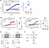 Figure 1
High CO2 elicits a triphasic Ca2+ response in ASM cells. (A–C) Intracellular Ca2+ dynamics were assessed in cultured mouse ASM cells expressing GCaMP6 (n = 3 biological replicates). (A) [Ca2+]i traces (left) and quantification of ΔF/F0 at 60 minutes (right) in the presence or absence of extracellular Ca2+. Cells were preincubated for 30 minutes with medium equilibrated to 5% CO2 (30–40 mmHg, pH 7.4), then exposed to high CO2 (60–80 or ~120 mmHg, pH 7.4) for 60 minutes. (B and C) Ca2+ influx in ASM cells treated with DMSO (vehicle) or 5 μM BTP-2 (B), or transfected with nontarget, Stim1, or Orai1 shRNA (C). Left: [Ca2+]i traces. Right: Quantification of ΔF/F0 at 60 minutes. Cells were preincubated in 5% CO2 medium for 30 minutes, exposed to high-CO2 medium for 30 minutes in the absence of extracellular Ca2+, then switched to 2.5 mM Ca2+ for an additional 30 minutes. (D) Representative Western blot (top) and quantification (bottom) of STIM1 and Orai1 in ASM cells exposed to 5% CO2 (Ctrl; 30–40 mmHg, pH 7.4) or high CO2 (~120 mmHg, pH 7.4) for 30 minutes (n = 4 biological replicates). (E) Schematic illustrating the proposed triphasic [Ca2+]i response in hypercapnic ASM cells, characterized by an initial transient decline, SR Ca2+ release, and SOCE activation. Data are presented as means ± SEM. Statistical analysis was performed by unpaired 2-tailed t test (B and D) or 1-way ANOVA with Tukey’s post hoc test (A and C). *P < 0.05, **P < 0.01. Pre-inc, preincubation; SR, sarcoplasmic reticulum; SOCE, store-operated Ca2+ entry.
When cells were exposed to high CO2 in Ca2+-free extracellular solution, the initial [Ca2+]i response, comprising a transient decline following a rapid elevation, was preserved; however, the sustained increase observed in the presence of extracellular Ca2+ was abolished (Figure 1A). These findings indicate that extracellular Ca2+ is required for the sustained phase of the response. Consistent with prior reports that hypercapnia stimulates InsP3R-mediated Ca2+ release from the SR (18, 19), administration of 2-APB, an inhibitor of InsP3R, eliminated the recovery of [Ca2+]i following its initial decline (Supplemental Figure 1). This result suggests that high-CO2-induced SR Ca2+ release underlies the secondary rise in [Ca2+]i. In contrast, ryanodine had no effect, indicating that ryanodine receptors are not involved in this process.
Because buffered hypercapnia is known to cause transient intracellular acidosis in various cell types (20, 21), we next examined whether intracellular pH changes contribute to the early [Ca2+]i decline. Exposure to high CO2 at extracellular pH 7.2 (mimicking respiratory acidosis) caused a sustained decrease in [Ca2+]i (Supplemental Figure 2A), coinciding with persistent intracellular acidosis throughout CO2 exposure (Supplemental Figure 2B). Similarly, transient intracellular acidification was observed under buffered hypercapnia, coinciding temporally with the initial [Ca2+]i decrease. These findings suggest that the early [Ca2+]i decline in response to hypercapnia is likely associated with intracellular acidification.
In many cell types, including smooth muscle cells, SOCE represents a primary mechanism of sustained Ca2+ influx (11). SOCE is mediated by Ca2+ release–activated calcium (CRAC) channels, including Orai1, which are activated when STIM1 senses SR Ca2+ depletion and translocates to SR–plasma membrane junctions (11). Increasing extracellular Ca2+ to 2.5 mM during hypercapnia enhanced the sustained [Ca2+]i elevation, which was abolished by the SOCE inhibitor BTP-2 in mouse ASM cells (Figure 1B). ASM cells stably expressing shRNAs targeting Stim1 or Orai1 also showed significant inhibition of high-CO2-induced Ca2+ influx (Figure 1C), confirming the functional requirement of STIM1-Orai1 engagement for SOCE. Notably, hypercapnia significantly upregulated STIM1 expression, whereas Orai1 and STIM2 expression remained unchanged (Figure 1D and Supplemental Figure 3), indicating selective regulation of STIM1 without compensatory induction of related isoforms under these conditions.
Collectively, these results demonstrate that hypercapnia elicits a triphasic [Ca2+]i response in ASM cells, comprising an initial acidification-associated decline, SR-dependent Ca2+ release, and sustained SOCE likely mediated through STIM1 activation (Figure 1E).
Hypercapnia activates ERK to promote SR Ca2+ depletion via InsP3R activation. Phosphorylation functions as a molecular switch that dynamically regulates InsP3R activity, controlling the rate and extent of SR Ca2+ store depletion (22). We previously demonstrated that hypercapnia activates ERK signaling in alveolar epithelial cells (23, 24). Consistent with these observations, exposure to high CO2 induced transient ERK activation in ASM cells (Figure 2A). ERK has been reported to phosphorylate InsP3R at optimal MAPK consensus sites (25), although the precise functional consequences of these modifications remain incompletely defined.
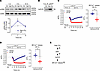 Figure 2
High CO2 activates ERK to promote SR Ca2+ depletion via InsP3R activation. Cultured ASM cells were incubated in control CO2 medium (Ctrl; 30–40 mmHg, pH 7.4) or exposed to buffered high CO2 (~120 mmHg, pH 7.4) for the indicated times. (A–C) Mouse ASM cells. (A) Representative Western blot (top) and quantification (bottom) of phosphorylated ERK (p-ERK) and total ERK (t-ERK) (n = 3 biological replicates). (B) Representative Western blot of proteins immunoprecipitated with anti–phospho-MAPK substrate (pXS*P) antibody from exposed cells. Blots were probed with anti-InsP3R1 antibody (n = 3 biological replicates). (C) [Ca2+]i traces (left) and quantification (right) of ΔF/F0 in cells pretreated with DMSO (vehicle) or U0126 (10 μM, 1 hour) and continuously exposed to high CO2 under Ca2+-free conditions (n = 3 biological replicates). (D) Human ASM cells. [Ca2+]i traces (left) and quantification (right) of ΔF/F0 in cells pretreated with DMSO (vehicle) or U0126 (10 μM, 1 hour) and continuously exposed to high CO2 under Ca2+-free conditions (n = 4 independent experimental replicates). Data are presented as means ± SEM. Statistical analysis was performed by unpaired 2-tailed t test (A, C, and D). (E) Schematic model illustrating ERK-mediated SR Ca2+ relesase via InsP3R activation during hypercapnia. *P < 0.05. InsP3R1, inositol 1,4,5-trisphosphate receptor 1; Pre-inc, preincubation; SR, sarcoplasmic reticulum.
To determine whether hypercapnia promotes SR Ca2+ depletion through ERK-dependent regulation of InsP3R, we examined MAPK-consensus serine phosphorylation using co-immunoprecipitation with an anti–phospho-MAPK/CDK substrate antibody that recognizes phosphorylated PXSP motifs. Under high-CO2 conditions, immunoreactive InsP3R species were enriched in phospho-PXSP immunoprecipitates from mouse ASM cells (Figure 2B), consistent with increased ERK-dependent phosphorylation of InsP3R.
Functionally, pharmacological inhibition of ERK signaling with the MEK inhibitor U0126 prevented hypercapnia-induced SR Ca2+ depletion in both mouse and human ASM cells (Figure 2, C and D). Together with the data shown in Supplemental Figure 1, these findings support a model in which hypercapnia activates ERK, leading to InsP3R phosphorylation and enhanced SR Ca2+ release (Figure 2E). This pathway provides a mechanistic trigger for STIM1-dependent SOCE.
Hypercapnia promotes STIM1 puncta formation via ERK-mediated phosphorylation. STIM1 phosphorylation promotes its translocation to puncta-like SR–plasma membrane junctions, facilitating SOCE via Orai1 channels (26–31). ERK-dependent phosphorylation at serine residues 575, 608, and 621 has been shown to enhance this process (26–28). We hypothesized that hypercapnia promotes STIM1 accumulation at junctional SR sites through ERK-mediated phosphorylation.
To test this hypothesis, we expressed STIM1-mCherry in mouse ASM cells and monitored subcellular localization by time-lapse single-cell imaging. High CO2 induced redistribution of STIM1 from a diffuse SR localization in resting cells to discrete puncta at the cell periphery (Figure 3A). Immunoprecipitated STIM1 from mouse ASM cells showed increased phosphorylation detected with an anti–phospho-MAPK/CDK substrate antibody, whereas mutation of Ser575/608/621 to alanine (S575A/S608A/S621A) or treatment with U0126 abolished this effect (Figure 3, B and C). U0126 also largely prevented high-CO2-induced STIM1 puncta formation in both mouse and human ASM cells (Figure 3, D and E). These results suggest that hypercapnia promotes STIM1 puncta formation through ERK-mediated phosphorylation (Figure 3F), in the context of SR Ca2+ depletion. Our findings indicate that ERK activation is required for coordinated upstream Ca2+ release and downstream STIM1 redistribution in hypercapnia ASM cells.
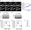 Figure 3
High CO2 promotes STIM1 puncta formation via ERK-mediated phosphorylation. Cultured ASM cells were incubated in control CO2 medium (Ctrl; 30–40 mmHg, pH 7.4) or exposed to buffered high CO2 (~120 mmHg, pH 7.4) for the indicated times. (A–D) Mouse ASM cells (n = 3 biological replicates). (A) Representative image (left) and time-course quantification (right) of mCherry-tagged STIM1 puncta formation. Cells were preincubated in Ctrl for 30 minutes, then exposed to high CO2. Scale bars: 50 μm. (B) Representative Western blot of proteins immunoprecipitated with anti-mCherry antibody from cells transfected with mCherry-tagged STIM1 wild type (WT STIM1) or STIM1S575A/S608A/S621A (Mutant STIM1). Blots were probed with anti–phospho-MAPK substrate (pXS*P) and -mCherry antibodies. (C) Representative Western blot of proteins immunoprecipitated with anti-STIM1 antibody from cells pretreated with DMSO or U0126 (10 μM, 1 hour) and continuously exposed to high CO2. Blots were probed with anti–phospho-MAPK substrate (pXS*P) and -STIM1 antibodies. (D) Quantification of STIM1 puncta in mouse ASM cells pretreated with DMSO (Vehicle) or U0126 (10 μM, 1 hour) and continuously exposed to high CO2. (E) Quantification of STIM1 puncta in human ASM cells under the same conditions (n = 3 independent experimental replicates). (F) Schematic model illustrating ERK-mediated STIM1 phosphorylation and puncta formation during hypercapnia. Data are presented as means ± SEM. Statistical analysis was performed by 1-way ANOVA with Dunnett’s (A) or Tukey’s post hoc test (D and E). *P < 0.05, **P < 0.01.
Hypercapnia increases STIM1 abundance via ERK/c-Fos signaling to enhance SOCE. A large-scale transcriptomic study identified transcription factors potentially regulating hypercapnia-responsive genes, including c-Fos (32), which can rapidly increase STIM1 expression in response to stress (33, 34). c-Fos activity is regulated by ERK-mediated phosphorylation at serine 374, which stabilizes nuclear c-Fos and enhances its transcriptional function (35). Given that STIM1 abundance determines the magnitude of SOCE (36, 37), we tested whether ERK regulates STIM1 expression via c-Fos during hypercapnia.
High CO2 caused transient ERK activation (Figure 2A) and increased nuclear c-Fos phosphorylation (Figure 4A). Silencing Fos with siRNA abrogated high-CO2-induced STIM1 upregulation (Figure 4B). Chromatin immunoprecipitation confirmed enhanced c-Fos binding to the Stim1 promoter under hypercapnia (Figure 4C). Pharmacological ERK inhibition with the MEK inhibitor U0126 prevented hypercapnia-induced c-Fos translocation and STIM1 protein abundance (Figure 4, D and E). We next examined whether hypercapnia-induced STIM1 activation facilitates its interaction with CRAC channels. Co-immunoprecipitation demonstrated increased STIM1-Orai1 association under hypercapnia, which was abolished by U0126 (Figure 4F). The ERK inhibition also attenuated high-CO2-induced elevations in [Ca2+]i (Figure 4G). These findings that hypercapnia activates the ERK/STIM1 pathway, increasing [Ca2+]i, were recapitulated in human cultured ASM cells (Figure 4, H and I). Together, our data indicate that ERK-dependent c-Fos activation increases STIM1 abundance and promotes STIM1-Orai1 interaction, sustaining SOCE during hypercapnia (Figure 4J).
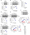 Figure 4
High CO2 increases STIM1 abundance via ERK/c-Fos signaling to enhance SOCE. Cultured ASM cells were incubated in control CO2 medium (Ctrl; 30–40 mmHg, pH 7.4) or exposed to buffered high CO2 (~120 mmHg, pH 7.4) for the indicated durations. (A–G) Mouse ASM cells. (A and B) Representative Western blot (top) and quantification (bottom) of phosphorylated c-Fosser374 (A; n = 4 biological replicates), and STIM1 in cells transfected with scramble (si-Scr) or Fos (si-Fos) siRNA (B; n = 3 biological replicates). (C) qPCR quantification of CUT&RUN assay showing c-Fos binding to the Stim1 promoter (n = 3 biological replicates). (D–G) Cells were preincubated with DMSO or U0126 (10 μM, 1 hour) and continuously exposed to the same treatment during high-CO2 exposure (n = 3 biological replicates). (D and E) Representative Western blot (top) and quantification (bottom) of c-Fos (D) and STIM1 (E). (F) Representative Western blot of proteins immunoprecipitated with anti-STIM1 antibody. Blots were probed with anti-STIM1 and -Orai1 antibodies. (G) [Ca2+]i traces (left) and quantification (right) of ΔF/F0. (H and I) Human ASM cells. Representative Western blot (top) and quantification (bottom) of STIM1 (H; n = 4 independent experimental replicates) and [Ca2+]i traces (left) and quantification (right) of ΔF/F0 (I; n = 3 independent experimental replicates) in cells pretreated with DMSO or U0126 (10 μM, 1 hour) and continuously exposed to the same treatment during high-CO2 exposure. (J) Schematic model illustrating ERK-dependent c-Fos activation, STIM1 upregulation, and enhanced SOCE during hypercapnia. Data are presented as means ± SEM. Statistical analysis was performed by unpaired 2-tailed t test (A, C, and I), 1-way ANOVA with Tukey’s post hoc test (B, D, E, and H), or Welch’s t test (G). *P < 0.05, **P < 0.01, ***P < 0.001. Pre-inc, preincubation.
Hypercapnia induces ERK-dependent STIM1 signaling required for ASM contraction. To examine the dynamics of hypercapnia-responsive signaling, we assessed the effects of elevated CO2 (~120 mmHg, extracellular pH 7.4) for up to 48 hours in cultured mouse ASM cells. This approach allows dissection of early mechanistic events triggered by elevated CO2 (3, 5, 8). Both [Ca2+]i and STIM1 protein abundance remained elevated for up to 6 hours of high-CO2 exposure (Figure 5, A and B), supporting sustained STIM1-dependent Ca2+ signaling.
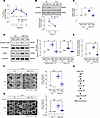 Figure 5
High CO2 induces ERK-dependent STIM1 signaling required for ASM contraction. Cultured mouse ASM cells or PCLSs were incubated in control CO2 medium (Ctrl; 30–40 mmHg, pH 7.4) or exposed to buffered high CO2 (~120 mmHg, pH 7.4) for the indicated times. (A and B) ASM cells. (A) [Ca2+]i measured using Fura-2 AM (n = 3 biological replicates). (B) Representative Western blot (top) and quantification (bottom) of STIM1 (n = 3 biological replicates). (C–E) ASM cells expressing non-targeting or Stim1 shRNA. (C) Calpain activity (n = 3 biological replicates). (D) Representative Western blots (left) and quantification (right) of nuclear cleaved caspase-7 (Casp7) and RhoA (n = 3 biological replicates). (E) Quantification of contraction induced by acetylcholine (ACh) (n = 3 biological replicates). (F and G) Representative images (left) and quantification (right) of ACh-induced airway contraction in PCLSs. (F) PCLSs obtained from Stim1-smKO (n = 3 biological replicates). Scale bars: 100 μm. (G) PCLSs obtained from C57BL/6J wild-type mice were pretreated with DMSO or 10 μM U0126 for 1 hour, followed by continuous exposure to Ctrl or high CO2 (n = 3–4 biological replicates). Scale bars: 200 μm. (H) Schematic illustrating hypercapnia-induced ASM contractility mediated via ERK/STIM1-dependent Ca2+ signaling pathways, thereby driving contractile responses. Data are presented as means ± SEM. Statistical analysis was performed by unpaired t test. (A and B) or 1-way ANOVA with Tukey’s post hoc test (C–G). *P < 0.05, **P < 0.01.
To determine the functional role of STIM1 in hypercapnia-induced ASM constriction, we examined hypercapnia-responsive signaling and acetylcholine-induced contractility in ASM cells expressing either non-targeting or Stim1-targeting shRNA. Consistent with prior findings (5), high CO2 increased calpain activation, nuclear caspase-7 cleavage, RhoA expression, and acetylcholine-induced contraction in cells expressing non-targeting shRNA, whereas these responses were abolished in Stim1-deficient cells (Figure 5, C–E).
These findings were further validated in PCLSs from C57BL/6J wild-type or smooth muscle–specific Stim1 knockout (Stim1-smKO) mice. High CO2 enhanced acetylcholine-induced airway contraction in wild-type PCLSs, which was abrogated by smooth muscle–specific Stim1 deletion (Figure 5F) or pharmacological ERK inhibition (Figure 5G). These small-airway PCLS data provide key functional evidence supporting the requirement for STIM1- and ERK-dependent signaling in hypercapnia-induced airway constriction. Together, these results indicate that ERK-dependent STIM1 signaling is a critical driver of hypercapnia-induced ASM contraction and airway responsiveness (Figure 5H).
Beyond contractility, hypercapnia may induce early phenotypic changes in ASM relevant to airway remodeling. We have recently reported that prolonged hypercapnia promotes ASM hypertrophy in mouse and human models (8). Here, we assessed α–smooth muscle actin (α-SMA) expression and proliferation in ASM cells under sustained hypercapnia. High CO2 increased α-SMA expression and impaired proliferation, effects that were prevented by Stim1 knockdown (Supplemental Figure 4). These data suggest that STIM1 contributes not only to acute Ca2+-dependent contractile responses but also to early remodeling-associated ASM phenotypic changes.
Collectively, these findings reveal a STIM1-dependent mechanistic link between elevated CO2 stress and airway dysfunction, suggesting that STIM1 plays a role in chronic airway disease.
Hypercapnia drives a biphasic STIM1 response across acute and chronic phases. To extend our mechanistic findings to clinically relevant conditions, we examined human ASM cells exposed to acute and prolonged hypercapnia (50–60 mmHg CO2, extracellular pH 7.4) and compared these responses with supraphysiological hypercapnia (~120 mmHg CO2, extracellular pH 7.4).
During the acute phase, clinically relevant hypercapnia induced activation of the ERK/c-Fos/STIM1 signaling axis (Figure 6, A and B), albeit with delayed kinetics compared with supraphysiological hypercapnia. Specifically, ERK phosphorylation was detected at 30 minutes after CO2 exposure (Figure 6A and Supplemental Figure 5A), whereas activation occurred within 5 minutes under 120 mmHg CO2 conditions (Figure 2A). These findings indicate that the magnitude of hypercapnic stress modulates signaling kinetics without altering the directionality of pathway activation.
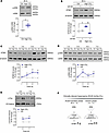 Figure 6
Hypercapnia drives a biphasic STIM1 response across acute and chronic phases. Cultured human ASM cells were incubated in control CO2 medium (Ctrl; 30–40 mmHg, pH 7.4) or exposed to buffered high CO2 (50–60 mmHg, pH 7.4) for the indicated durations. (A and B) Acute exposure phase (30 minutes). Representative Western blot (top) and quantification (bottom) of phosphorylated ERK (p-ERK; A), and STIM1 (B) in cells pretreated with DMSO or U0126 (10 μM, 1 hour) and maintained in the same treatment conditions during high-CO2 exposure (n = 3 independent experimental replicates). (C–E) Chronic exposure phase (up to 7 days). Representative Western blot (top) and quantification (bottom) of STIM1 (C; n = 4 independent experimental replicates), p-ERK (D; n = 4 independent experimental replicates), and nuclear c-Fos (E; n = 3 independent experimental replicates). (F) Schematic model illustrating the biphasic STIM1 response to clinically relevant hypercapnia, accompanied by temporally distinct ERK activation and persistent nuclear c-Fos localization. Data are presented as means ± SEM. Statistical analysis was performed by unpaired t test (A and C–E) or 1-way ANOVA with Tukey’s post hoc test (B). *P < 0.05, **P < 0.01. t-ERK, total ERK.
To determine whether STIM1 regulation is sustained over time, we next performed extended time-course analyses under clinically relevant hypercapnia. STIM1 protein abundance showed an initial increase (Figure 6B) followed by a transient normalization phase at 24 hours, and subsequently a secondary sustained increase during prolonged exposure (3–7 days) (Figure 6C). This biphasic pattern was accompanied by temporally distinct ERK activation (Figure 6D) and persistent nuclear c-Fos localization (Figure 6E). We also observed a similar directional regulation of STIM1 in mouse ASM cells exposed to clinically relevant chronic hypercapnia (Supplemental Figure 5B).
Collectively, these data demonstrate that hypercapnia induces a temporally mediated STIM1 response characterized by an early induction phase, transient normalization, and delayed secondary activation through ERK/c-Fos regulation (Figure 6F). This dynamic regulation provides a mechanistic framework linking acute hypercapnic signaling to chronic ASM dysfunction.
Gene-environment interaction between STIM1 variants and hypercapnia is associated with altered airway resistance in patients with COPD. As a disease-relevant context for hypercapnia-induced airway contractility, prior cohort studies demonstrated that airway resistance is higher in hypercapnic than in normocapnic patients with COPD and improves following noninvasive ventilation aimed at correcting hypercapnia (5). A recent study reported a trend linking STIM1 abundance with airflow limitation in COPD lung tissue (38). To test whether our mechanistic findings extend to humans, we interrogated publicly available datasets and analyzed 2 independent COPD cohorts.
Analysis of the COPD Cell Atlas (39) revealed that STIM1 expression is highly context and cell type dependent, with increased expression in specific epithelial lineages, while showing reduced expression in stromal populations, including smooth muscle cells, in explanted COPD lungs compared with controls (Figure 7A and Supplemental Figure 6). STIM1 expression has been shown to be lower in ASM cells associated with a differentiated contractile phenotype compared with a synthetic-like state, supporting tight regulation of STIM1 expression in relation to ASM cell state (40, 41). Consistent with these single-cell transcriptomic data, our in vitro experiments demonstrated lower STIM1 protein abundance in COPD-derived ASM cells compared with normal ASM cells (Figure 7B), suggesting that disease-associated differences in ASM phenotype may contribute to altered STIM1 expression.
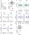 Figure 7
Interaction between hypercapnia and STIM1 variants influences airway obstruction in COPD. (A) STIM1 expression in smooth muscle cells from explanted COPD lungs and healthy controls, analyzed using the COPD Cell Atlas (GSE136831). Each dot represents the per-subject mean log-normalized STIM1 expression across smooth muscle cells. (B) Representative Western blot (top) and quantification (bottom) of STIM1 in ASM cells derived from normal or COPD lung tissue (n = 3 independent experimental replicates). (C) Genotype-specific STIM1 expression in 601 normal lung tissues, visualized using the GTEx eQTL calculator (https://www.gtexportal.org/home/testyourown). (D) Log (%sRtot, percentage of total specific resistance) in 26 normocapnic patients with chronic stable COPD from cohort study 2, grouped by minor allele zygosity (homozygous vs. heterozygous). (E) Log (%sRtot) in 74 patients with chronic stable COPD from cohort study 2, grouped by CO2 retention status (normocapnia [NC] vs. hypercapnia [HC]) and minor allele zygosity (homozygous vs. heterozygous). Main effect of CO2: P = 0.008. (F) Change (Δ) in log (%sRtot) in each genotype. Δ log (%sRtot) = log (%sRtot) from subjects with hypercapnia – averaged log (%sRtot) from normocapnic heterozygotes. (G) Representative Western blot (top) and quantification (bottom) of STIM1 in ASM cells from COPD donor exposed to control CO2 medium 30–40 mmHg, pH 7.4) or buffered high CO2 (HC; 50–60 mmHg, pH 7.4) for 7 days (n = 3 independent experimental replicates). Data are presented as means ± SEM. Statistical analysis was performed by unpaired (B, F, and G) or Welch’s (A and D) 2-tailed t test and 2-way ANOVA with post hoc uncorrected Fisher’s least significant difference test (E). Sample sizes and P values are indicated in the figure unless otherwise specified.
Consistent with these expression patterns, several noncoding STIM1 variants (rs1561876, rs3750994, rs3750996, rs3794050, and rs7934581) have been associated with altered STIM1 expression, with minor allele carriers showing reduced transcript levels in whole blood (42). Consistently, GTEx expression quantitative trait locus (eQTL) analysis identified rs1561876 and rs3794050 as loci influencing STIM1 expression in normal lung tissue, with minor alleles trending toward reduced expression (Figure 7C). Given their near-complete linkage disequilibrium (R2 = 0.981), we focused subsequent analyses on these two variants.
In cohort study 1, a case-control study of 118 European-ancestry subjects matched for age, sex, and smoking status, we first confirmed that potential confounders (age, sex, smoking status, BMI) did not influence the prevalence of rs1561876 and rs3794050 in 828 healthy European-ancestry individuals (Supplemental Figure 7 and Supplemental Table 1). Minor allele frequencies for both rs1561876 (adjusted OR: 9.85; P = 7.98 × 10⁻⁶) and rs3794050 (adjusted OR: 12.5; P = 6.26 × 10⁻⁶) were significantly higher in COPD patients than in healthy controls (Table 1), suggesting that these variants may contribute to reduced STIM1 expression in COPD.
In cohort study 2, we assessed the relationship between STIM1 variants and airway resistance in European-ancestry smokers with COPD carrying the minor alleles, stratified by CO2 retention status (Table 2). Disease severity did not differ significantly by genotype within either normocapnic or hypercapnic subgroups; however, as expected, severity overall was higher in the hypercapnic group (Table 2 and Supplemental Table 2). Among normocapnic individuals, homozygous carriers of the minor alleles showed a trend toward lower airway resistance (Figure 7D). Two-way ANOVA revealed a significant main effect of CO2 retention (F1,70 = 7.488, P = 0.008), indicating that hypercapnia increased airway resistance (Figure 7E). In contrast, the main effect of genotype was not significant (F1,70 = 1.104, P = 0.297), and no significant interaction was detected (F1,70 = 0.170, P = 0.682). Notably, the change in airway resistance (hypercapnia – normocapnia) did not differ between genotypes, suggesting that the beneficial association of the minor alleles under normocapnia was abolished under hypercapnic conditions (Figure 7F).
In vitro, exposure of ASM cells derived from COPD lung tissue to clinically relevant hypercapnia (50–60 mmHg, extracellular pH 7.4) (5, 8) for 7 days resulted in increased STIM1 abundance (Figure 7G). This sustained effect highlights the pathological relevance of STIM1 signaling and aligns with the clinical observation that the lower airway resistance associated with minor allele carriage under normocapnia was overridden by hypercapnia.
Taken together, these findings suggest that specific STIM1 variants may predispose individuals to altered airway contractility, potentially through modulation of gene expression, and that hypercapnia can exacerbate airway obstruction by overriding this genetic resilience, consistent with a gene-environment interaction in patients with COPD.


