
Research ArticleCell biologyGeneticsPulmonology
Open Access |  10.1172/jci.insight.199029
Polymerized Z-α-1 antitrypsin leads to lung injury in a murine model of emphysema
Maria Magallón Serrano,1 Nazli Khodayari,1 William Bowers,1 Edward P. Manning,2 Xinran Liu,3 Jungnam Lee,1 Tammy O. Flagg,1 Regina Oshins,1 Aidan Griffin,1 Sahil Patel,1 Jorge E. Lascano,1 Divay Chandra,4 Susan M. Majka,5 Irina Petrache,5,6 Mark L. Brantly,1,7 and Karina A. Serban1,7
1Department of Medicine, Division of Pulmonary, Critical Care, and Sleep Medicine, University of Florida, Gainesville, Florida, USA.
2Department of Medicine, Section of Pulmonary, Critical Care, and Sleep Medicine and
3Center for Cellular & Molecular Imaging, Electron Microscopy Core Facility, Yale University School of Medicine, New Haven, Connecticut, USA.
4Department of Medicine, University of Pittsburgh, Pittsburgh, Pennsylvania, USA.
5Department of Medicine, Division of Pulmonary, Critical Care, and Sleep Medicine, National Jewish Health, Denver, Colorado, USA.
6Department of Medicine, Division of Pulmonary, Critical Care, and Sleep Medicine, University of Colorado School of Medicine, University of Colorado, Aurora, Colorado, USA.
7Department of Molecular Genetics and Microbiology, University of Florida, Gainesville, Florida, USA.
Address correspondence to: Karina A. Serban, University of Florida, 1345 Center Drive, MSB – M330, Gainesville, Florida 32610, USA. Phone: 352.294.5116; Email: Karina.Serban@medicine.ufl.edu.
Find articles by Magallón Serrano, M. in: PubMed | Google Scholar
1Department of Medicine, Division of Pulmonary, Critical Care, and Sleep Medicine, University of Florida, Gainesville, Florida, USA.
2Department of Medicine, Section of Pulmonary, Critical Care, and Sleep Medicine and
3Center for Cellular & Molecular Imaging, Electron Microscopy Core Facility, Yale University School of Medicine, New Haven, Connecticut, USA.
4Department of Medicine, University of Pittsburgh, Pittsburgh, Pennsylvania, USA.
5Department of Medicine, Division of Pulmonary, Critical Care, and Sleep Medicine, National Jewish Health, Denver, Colorado, USA.
6Department of Medicine, Division of Pulmonary, Critical Care, and Sleep Medicine, University of Colorado School of Medicine, University of Colorado, Aurora, Colorado, USA.
7Department of Molecular Genetics and Microbiology, University of Florida, Gainesville, Florida, USA.
Address correspondence to: Karina A. Serban, University of Florida, 1345 Center Drive, MSB – M330, Gainesville, Florida 32610, USA. Phone: 352.294.5116; Email: Karina.Serban@medicine.ufl.edu.
Find articles by Khodayari, N. in: PubMed | Google Scholar
1Department of Medicine, Division of Pulmonary, Critical Care, and Sleep Medicine, University of Florida, Gainesville, Florida, USA.
2Department of Medicine, Section of Pulmonary, Critical Care, and Sleep Medicine and
3Center for Cellular & Molecular Imaging, Electron Microscopy Core Facility, Yale University School of Medicine, New Haven, Connecticut, USA.
4Department of Medicine, University of Pittsburgh, Pittsburgh, Pennsylvania, USA.
5Department of Medicine, Division of Pulmonary, Critical Care, and Sleep Medicine, National Jewish Health, Denver, Colorado, USA.
6Department of Medicine, Division of Pulmonary, Critical Care, and Sleep Medicine, University of Colorado School of Medicine, University of Colorado, Aurora, Colorado, USA.
7Department of Molecular Genetics and Microbiology, University of Florida, Gainesville, Florida, USA.
Address correspondence to: Karina A. Serban, University of Florida, 1345 Center Drive, MSB – M330, Gainesville, Florida 32610, USA. Phone: 352.294.5116; Email: Karina.Serban@medicine.ufl.edu.
Find articles by Bowers, W. in: PubMed | Google Scholar
1Department of Medicine, Division of Pulmonary, Critical Care, and Sleep Medicine, University of Florida, Gainesville, Florida, USA.
2Department of Medicine, Section of Pulmonary, Critical Care, and Sleep Medicine and
3Center for Cellular & Molecular Imaging, Electron Microscopy Core Facility, Yale University School of Medicine, New Haven, Connecticut, USA.
4Department of Medicine, University of Pittsburgh, Pittsburgh, Pennsylvania, USA.
5Department of Medicine, Division of Pulmonary, Critical Care, and Sleep Medicine, National Jewish Health, Denver, Colorado, USA.
6Department of Medicine, Division of Pulmonary, Critical Care, and Sleep Medicine, University of Colorado School of Medicine, University of Colorado, Aurora, Colorado, USA.
7Department of Molecular Genetics and Microbiology, University of Florida, Gainesville, Florida, USA.
Address correspondence to: Karina A. Serban, University of Florida, 1345 Center Drive, MSB – M330, Gainesville, Florida 32610, USA. Phone: 352.294.5116; Email: Karina.Serban@medicine.ufl.edu.
Find articles by Manning, E. in: PubMed | Google Scholar
1Department of Medicine, Division of Pulmonary, Critical Care, and Sleep Medicine, University of Florida, Gainesville, Florida, USA.
2Department of Medicine, Section of Pulmonary, Critical Care, and Sleep Medicine and
3Center for Cellular & Molecular Imaging, Electron Microscopy Core Facility, Yale University School of Medicine, New Haven, Connecticut, USA.
4Department of Medicine, University of Pittsburgh, Pittsburgh, Pennsylvania, USA.
5Department of Medicine, Division of Pulmonary, Critical Care, and Sleep Medicine, National Jewish Health, Denver, Colorado, USA.
6Department of Medicine, Division of Pulmonary, Critical Care, and Sleep Medicine, University of Colorado School of Medicine, University of Colorado, Aurora, Colorado, USA.
7Department of Molecular Genetics and Microbiology, University of Florida, Gainesville, Florida, USA.
Address correspondence to: Karina A. Serban, University of Florida, 1345 Center Drive, MSB – M330, Gainesville, Florida 32610, USA. Phone: 352.294.5116; Email: Karina.Serban@medicine.ufl.edu.
Find articles by
Liu, X.
in:
PubMed
|
Google Scholar
|

1Department of Medicine, Division of Pulmonary, Critical Care, and Sleep Medicine, University of Florida, Gainesville, Florida, USA.
2Department of Medicine, Section of Pulmonary, Critical Care, and Sleep Medicine and
3Center for Cellular & Molecular Imaging, Electron Microscopy Core Facility, Yale University School of Medicine, New Haven, Connecticut, USA.
4Department of Medicine, University of Pittsburgh, Pittsburgh, Pennsylvania, USA.
5Department of Medicine, Division of Pulmonary, Critical Care, and Sleep Medicine, National Jewish Health, Denver, Colorado, USA.
6Department of Medicine, Division of Pulmonary, Critical Care, and Sleep Medicine, University of Colorado School of Medicine, University of Colorado, Aurora, Colorado, USA.
7Department of Molecular Genetics and Microbiology, University of Florida, Gainesville, Florida, USA.
Address correspondence to: Karina A. Serban, University of Florida, 1345 Center Drive, MSB – M330, Gainesville, Florida 32610, USA. Phone: 352.294.5116; Email: Karina.Serban@medicine.ufl.edu.
Find articles by Lee, J. in: PubMed | Google Scholar
1Department of Medicine, Division of Pulmonary, Critical Care, and Sleep Medicine, University of Florida, Gainesville, Florida, USA.
2Department of Medicine, Section of Pulmonary, Critical Care, and Sleep Medicine and
3Center for Cellular & Molecular Imaging, Electron Microscopy Core Facility, Yale University School of Medicine, New Haven, Connecticut, USA.
4Department of Medicine, University of Pittsburgh, Pittsburgh, Pennsylvania, USA.
5Department of Medicine, Division of Pulmonary, Critical Care, and Sleep Medicine, National Jewish Health, Denver, Colorado, USA.
6Department of Medicine, Division of Pulmonary, Critical Care, and Sleep Medicine, University of Colorado School of Medicine, University of Colorado, Aurora, Colorado, USA.
7Department of Molecular Genetics and Microbiology, University of Florida, Gainesville, Florida, USA.
Address correspondence to: Karina A. Serban, University of Florida, 1345 Center Drive, MSB – M330, Gainesville, Florida 32610, USA. Phone: 352.294.5116; Email: Karina.Serban@medicine.ufl.edu.
Find articles by Flagg, T. in: PubMed | Google Scholar
1Department of Medicine, Division of Pulmonary, Critical Care, and Sleep Medicine, University of Florida, Gainesville, Florida, USA.
2Department of Medicine, Section of Pulmonary, Critical Care, and Sleep Medicine and
3Center for Cellular & Molecular Imaging, Electron Microscopy Core Facility, Yale University School of Medicine, New Haven, Connecticut, USA.
4Department of Medicine, University of Pittsburgh, Pittsburgh, Pennsylvania, USA.
5Department of Medicine, Division of Pulmonary, Critical Care, and Sleep Medicine, National Jewish Health, Denver, Colorado, USA.
6Department of Medicine, Division of Pulmonary, Critical Care, and Sleep Medicine, University of Colorado School of Medicine, University of Colorado, Aurora, Colorado, USA.
7Department of Molecular Genetics and Microbiology, University of Florida, Gainesville, Florida, USA.
Address correspondence to: Karina A. Serban, University of Florida, 1345 Center Drive, MSB – M330, Gainesville, Florida 32610, USA. Phone: 352.294.5116; Email: Karina.Serban@medicine.ufl.edu.
Find articles by Oshins, R. in: PubMed | Google Scholar
1Department of Medicine, Division of Pulmonary, Critical Care, and Sleep Medicine, University of Florida, Gainesville, Florida, USA.
2Department of Medicine, Section of Pulmonary, Critical Care, and Sleep Medicine and
3Center for Cellular & Molecular Imaging, Electron Microscopy Core Facility, Yale University School of Medicine, New Haven, Connecticut, USA.
4Department of Medicine, University of Pittsburgh, Pittsburgh, Pennsylvania, USA.
5Department of Medicine, Division of Pulmonary, Critical Care, and Sleep Medicine, National Jewish Health, Denver, Colorado, USA.
6Department of Medicine, Division of Pulmonary, Critical Care, and Sleep Medicine, University of Colorado School of Medicine, University of Colorado, Aurora, Colorado, USA.
7Department of Molecular Genetics and Microbiology, University of Florida, Gainesville, Florida, USA.
Address correspondence to: Karina A. Serban, University of Florida, 1345 Center Drive, MSB – M330, Gainesville, Florida 32610, USA. Phone: 352.294.5116; Email: Karina.Serban@medicine.ufl.edu.
Find articles by Griffin, A. in: PubMed | Google Scholar
1Department of Medicine, Division of Pulmonary, Critical Care, and Sleep Medicine, University of Florida, Gainesville, Florida, USA.
2Department of Medicine, Section of Pulmonary, Critical Care, and Sleep Medicine and
3Center for Cellular & Molecular Imaging, Electron Microscopy Core Facility, Yale University School of Medicine, New Haven, Connecticut, USA.
4Department of Medicine, University of Pittsburgh, Pittsburgh, Pennsylvania, USA.
5Department of Medicine, Division of Pulmonary, Critical Care, and Sleep Medicine, National Jewish Health, Denver, Colorado, USA.
6Department of Medicine, Division of Pulmonary, Critical Care, and Sleep Medicine, University of Colorado School of Medicine, University of Colorado, Aurora, Colorado, USA.
7Department of Molecular Genetics and Microbiology, University of Florida, Gainesville, Florida, USA.
Address correspondence to: Karina A. Serban, University of Florida, 1345 Center Drive, MSB – M330, Gainesville, Florida 32610, USA. Phone: 352.294.5116; Email: Karina.Serban@medicine.ufl.edu.
Find articles by Patel, S. in: PubMed | Google Scholar
1Department of Medicine, Division of Pulmonary, Critical Care, and Sleep Medicine, University of Florida, Gainesville, Florida, USA.
2Department of Medicine, Section of Pulmonary, Critical Care, and Sleep Medicine and
3Center for Cellular & Molecular Imaging, Electron Microscopy Core Facility, Yale University School of Medicine, New Haven, Connecticut, USA.
4Department of Medicine, University of Pittsburgh, Pittsburgh, Pennsylvania, USA.
5Department of Medicine, Division of Pulmonary, Critical Care, and Sleep Medicine, National Jewish Health, Denver, Colorado, USA.
6Department of Medicine, Division of Pulmonary, Critical Care, and Sleep Medicine, University of Colorado School of Medicine, University of Colorado, Aurora, Colorado, USA.
7Department of Molecular Genetics and Microbiology, University of Florida, Gainesville, Florida, USA.
Address correspondence to: Karina A. Serban, University of Florida, 1345 Center Drive, MSB – M330, Gainesville, Florida 32610, USA. Phone: 352.294.5116; Email: Karina.Serban@medicine.ufl.edu.
Find articles by Lascano, J. in: PubMed | Google Scholar
1Department of Medicine, Division of Pulmonary, Critical Care, and Sleep Medicine, University of Florida, Gainesville, Florida, USA.
2Department of Medicine, Section of Pulmonary, Critical Care, and Sleep Medicine and
3Center for Cellular & Molecular Imaging, Electron Microscopy Core Facility, Yale University School of Medicine, New Haven, Connecticut, USA.
4Department of Medicine, University of Pittsburgh, Pittsburgh, Pennsylvania, USA.
5Department of Medicine, Division of Pulmonary, Critical Care, and Sleep Medicine, National Jewish Health, Denver, Colorado, USA.
6Department of Medicine, Division of Pulmonary, Critical Care, and Sleep Medicine, University of Colorado School of Medicine, University of Colorado, Aurora, Colorado, USA.
7Department of Molecular Genetics and Microbiology, University of Florida, Gainesville, Florida, USA.
Address correspondence to: Karina A. Serban, University of Florida, 1345 Center Drive, MSB – M330, Gainesville, Florida 32610, USA. Phone: 352.294.5116; Email: Karina.Serban@medicine.ufl.edu.
Find articles by Chandra, D. in: PubMed | Google Scholar
1Department of Medicine, Division of Pulmonary, Critical Care, and Sleep Medicine, University of Florida, Gainesville, Florida, USA.
2Department of Medicine, Section of Pulmonary, Critical Care, and Sleep Medicine and
3Center for Cellular & Molecular Imaging, Electron Microscopy Core Facility, Yale University School of Medicine, New Haven, Connecticut, USA.
4Department of Medicine, University of Pittsburgh, Pittsburgh, Pennsylvania, USA.
5Department of Medicine, Division of Pulmonary, Critical Care, and Sleep Medicine, National Jewish Health, Denver, Colorado, USA.
6Department of Medicine, Division of Pulmonary, Critical Care, and Sleep Medicine, University of Colorado School of Medicine, University of Colorado, Aurora, Colorado, USA.
7Department of Molecular Genetics and Microbiology, University of Florida, Gainesville, Florida, USA.
Address correspondence to: Karina A. Serban, University of Florida, 1345 Center Drive, MSB – M330, Gainesville, Florida 32610, USA. Phone: 352.294.5116; Email: Karina.Serban@medicine.ufl.edu.
Find articles by
Majka, S.
in:
PubMed
|
Google Scholar
|
1Department of Medicine, Division of Pulmonary, Critical Care, and Sleep Medicine, University of Florida, Gainesville, Florida, USA.
2Department of Medicine, Section of Pulmonary, Critical Care, and Sleep Medicine and
3Center for Cellular & Molecular Imaging, Electron Microscopy Core Facility, Yale University School of Medicine, New Haven, Connecticut, USA.
4Department of Medicine, University of Pittsburgh, Pittsburgh, Pennsylvania, USA.
5Department of Medicine, Division of Pulmonary, Critical Care, and Sleep Medicine, National Jewish Health, Denver, Colorado, USA.
6Department of Medicine, Division of Pulmonary, Critical Care, and Sleep Medicine, University of Colorado School of Medicine, University of Colorado, Aurora, Colorado, USA.
7Department of Molecular Genetics and Microbiology, University of Florida, Gainesville, Florida, USA.
Address correspondence to: Karina A. Serban, University of Florida, 1345 Center Drive, MSB – M330, Gainesville, Florida 32610, USA. Phone: 352.294.5116; Email: Karina.Serban@medicine.ufl.edu.
Find articles by
Petrache, I.
in:
PubMed
|
Google Scholar
|
1Department of Medicine, Division of Pulmonary, Critical Care, and Sleep Medicine, University of Florida, Gainesville, Florida, USA.
2Department of Medicine, Section of Pulmonary, Critical Care, and Sleep Medicine and
3Center for Cellular & Molecular Imaging, Electron Microscopy Core Facility, Yale University School of Medicine, New Haven, Connecticut, USA.
4Department of Medicine, University of Pittsburgh, Pittsburgh, Pennsylvania, USA.
5Department of Medicine, Division of Pulmonary, Critical Care, and Sleep Medicine, National Jewish Health, Denver, Colorado, USA.
6Department of Medicine, Division of Pulmonary, Critical Care, and Sleep Medicine, University of Colorado School of Medicine, University of Colorado, Aurora, Colorado, USA.
7Department of Molecular Genetics and Microbiology, University of Florida, Gainesville, Florida, USA.
Address correspondence to: Karina A. Serban, University of Florida, 1345 Center Drive, MSB – M330, Gainesville, Florida 32610, USA. Phone: 352.294.5116; Email: Karina.Serban@medicine.ufl.edu.
Find articles by
Brantly, M.
in:
PubMed
|
Google Scholar
|
1Department of Medicine, Division of Pulmonary, Critical Care, and Sleep Medicine, University of Florida, Gainesville, Florida, USA.
2Department of Medicine, Section of Pulmonary, Critical Care, and Sleep Medicine and
3Center for Cellular & Molecular Imaging, Electron Microscopy Core Facility, Yale University School of Medicine, New Haven, Connecticut, USA.
4Department of Medicine, University of Pittsburgh, Pittsburgh, Pennsylvania, USA.
5Department of Medicine, Division of Pulmonary, Critical Care, and Sleep Medicine, National Jewish Health, Denver, Colorado, USA.
6Department of Medicine, Division of Pulmonary, Critical Care, and Sleep Medicine, University of Colorado School of Medicine, University of Colorado, Aurora, Colorado, USA.
7Department of Molecular Genetics and Microbiology, University of Florida, Gainesville, Florida, USA.
Address correspondence to: Karina A. Serban, University of Florida, 1345 Center Drive, MSB – M330, Gainesville, Florida 32610, USA. Phone: 352.294.5116; Email: Karina.Serban@medicine.ufl.edu.
Find articles by Serban, K. in: PubMed | Google Scholar
Published August 10, 2026 - More info
JCI Insight. 2026;11(15):e199029. https://doi.org/10.1172/jci.insight.199029.
© 2026 Serrano et al. This work is licensed under the Creative Commons Attribution 4.0 International License. To view a copy of this license, visit http://creativecommons.org/licenses/by/4.0/.


-
Results
Generation and phenotypic characterization of Z-AAT Serpina1Null murine model.
In individuals with AATD, SERPENA1 (serine protease inhibitor, clade A, member 1 cluster) gene encoding for normal serine protease inhibitor AAT, harbors a single point mutation, E342K (Figure 1A), resulting in mutated Z-AAT protein. Z-AAT is prone to polymerization within the hepatocytes and other epithelial cells embryologically derived from the gut endoderm, with some Z-AAT polymers being secreted into the blood stream (33). Therefore, we designed a 23-kb pMA-pHuman-Serpina1 construct (Figure 1B) that contains the entire human SERPENA1 gene under its endogenous hepatocyte and macrophage promoters plus 5 kB of the 5’ and 3 kb of the 3’ flanking genomic DNA sequences. The construct also contains the glutamic acid–to–lysine substitution at residue 366 (E366K, also designated 342; G to A; rs 28929474) (34). At the Jackson Laboratory, oocytes from Serpina1Null female mice were harvested and microinjected with our construct. Embryos were transferred into pseudo-pregnant females, and the transgenic litters (strain #412938) were backcrossed more than 3 time and maintained on a Serpina1Null background (Supplemental Figure 1; supplemental material available online with this article; https://doi.org/10.1172/jci.insight.199029DS1) to ensure germline transmission of the transgene. The offspring are viable and fertile and have a normal lifespan and gender distribution, although decreased litter productivity has been observed in Z-AAT Serpina1Null mating pairs. We confirmed their genotype by allelic discrimination assay (Figure 1C) and the somatic integration of our construct by measuring similar copy number of the transgene in various tissues: liver, leukocytes, lung, and AT2 cells (Figure 1D). Z-AAT Serpina1Null mice, carrying 5–6 copies of the transgene and having low AAT circulating levels (Figure 1G) were used for experiments.
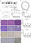 Figure 1
Generation and phenotypic characterization of Z-AAT Serpina1Null murine model. Graphic representation of (A) the human SERPENA1 gene on the chromosome 14, q arm, position q2.13. Note the SNP NC-000014.9:g.94378610 C>T (rs 28929474) in the exon V leading to the E [GAG] > K [AAG], E342K mutant AAT. (B) the 23-kb pMA-pHuman-Serpina1 construct harboring the glutamic acid to lysine substitution at residue 366 (E366K, also designated 342; G to A; rs 28929474) (C) Genotype identification of the Z-AAT Serpina1Null versus WT mice using SNP Genotyping TaqMan allelic discrimination assay. (D) Somatic integration of pMA-pHuman-Serpina1construct as demonstrated by equal, 5–6 copy number in the liver, lung, buffy coat and AT2 cells of Z-AAT Serpina1Null mice (E) Human SERPENA1 gene expression in the liver, lung, buffy coat, and AT2 cells of Z-AAT Serpina1Null mice relative to 18S control gene (F) Representative H&E, Periodic Acid Shiff globules after treatment with Diastase (PASD), Picro-Sirius Red (PSR) staining and α-smooth muscle actin (α-SMA) images of liver tissue from 4-mo old WT, Serpina1Null and Z-AAT Serpina1Null mice; scale 100 μm. Note increased PASD, PSR, and α-SMA staining (marked with *) in Z-AAT Serpina1Null mice. (G and H) Human AAT measured by nephelometry (G) and human Z-AAT polymers (H) measured by custom-made ELISA with specific 2C1 antibody (Hycult) against Z-AAT polymers in the plasma of 4-, 7- and greater than 12-month-old Z-AAT Serpina1Null versus Serpina1Null and WT mice. The red line, panel G represents the upper level (8 μM/mL) of serum AAT in Pi*ZZ individuals. Data are presented as mean ± SD, 1-way ANOVA, followed by Tukey’s multiple comparisons, #P < 0.05 versus 4-mo-old same genotype mice, *P < 0.05 versus same age WT mice.
Although the transgene is under human endogenous promoters, our Z-AAT Serpina1Null model recapitulated expected AAT expression pattern, with high Serpina1 expression in murine hepatocytes, followed by murine circulating monocytes and murine AT2 cells (Figure 1E). Murine H&E-stained liver tissue showed normal hepatocytes, sinusoids, and portal space’s morphology in WT, Serpina1Null, and Z-AAT Serpina1Null mice (Figure 1F), while 4-month-old Z-AAT Serpina1Null livers had accumulation of Periodic Acid Shiff globules after treatment with Diastase (PASD; Figure 1F), of collagen fibers on Picro-Sirius Red staining (Figure 1F), and of α-smooth muscle actin (α-SMA) expression (Figure 1F) suggestive of liver fibrogenesis. Moreover, the Z-AAT Serpina1Null mice present with low circulating AAT levels, as measured by nephelometry, like Pi*Z AATD individuals (Figure 1G), while expressing a constant high level of circuiting Z-AAT polymers across their lifespan (Figure 1H).
Z-AAT Serpina1Null mice develop spontaneous lung and systemic inflammation, vascular leak, and protease-antiprotease imbalance.
We measured baseline airway inflammation in the Z-AAT Serpina1Null mice kept in germ-free conditions and quantified total inflammatory cells recruited in the alveolar space using FACS gating strategy, as previously reported (6, 35) (Supplemental Figure 2A). Airway neutrophils (Figure 2A), and CD11b+ recruited monocytes (Figure 2B) were increased in 4-months, 7-months, and 12-months in Serpina1Null and Z-AAT Serpina1Null mice versus WT, control mice. Interestingly, neutrophils, CD11b+-recruited monocytes, and airway resident macrophages (Figure 2C) were significantly higher in 12-month-old Serpina1Null rather than Z-AAT Serpina1Null mice.
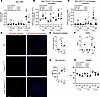 Figure 2
Z-AAT Serpina1Null mice develop lung, systemic inflammation, and protease-antiprotease imbalance. (A–C) Flow cytometry analysis of neutrophils (CD45+CD11b+Ly6G+), recruited monocytes (CD45+CD11b+Ly6G–F4/80+),and airway macrophages (CD45+CD11b– CD11c+SiglecF+). (A) neutrophils, (B) recruited monocytes, and (C) airway resident macrophages absolute counts in the BAL fluid of 4-, 7- and greater than 12-month-old Z-AAT Serpina1Null, Serpina1Null and WT mice. (D and E) Neutrophil elastase (NE) tissue abundance was measured using immunofluorescence and quantified using Image J plug-in. (D) Representative immunofluorescence images of 7-month-old WT, Serpina1Null and Z-AAT Serpina1Null mice; Neutrophil Elastase (NE, red), nuclei (blue, DAPI). scale bar: 100 μm. (E) Quantification of NE abundance reported as NE+ cells / number of nuclei; 5 fields / mouse, 3 mice per genotype were analyzed. (F) Plasma AAT level by nephelometry versus AAT inhibitory activity against NE by absorbance via NE activity assay kit. Note absence of total (open square) and functional AAT (black square) in 7-month-old Serpina1Null mice. Lower functional AAT was measured by absorbance via NE activity assay kit (◑) vs. total AAT (◐) measured by nephelometry in 7-month-old Z-AAT Serpina1Null mice (G) Albumin level (mg/mL) measured by ELISA in BAL fluid of 7-month-old Z-AAT Serpina1Null, Serpina1Null, and WT mice. (H) Harvest weight (g) of 4-, 7- and greater than 12-month-old Z-AAT Serpina1Null, Serpina1Null and WT mice. Data are presented as mean ± SD, 1-way ANOVA, followed by Tukey’s multiple comparisons or 2-way ANOVA with mixed-effects model (RELM), *P < 0.05 versus 4-mo old same genotype mice, #P < 0.05 versus same age WT mice, ^P < 0.05 versus same age Serpina1Null mice.
We next detected significantly higher neutrophil elastase by immunostaining in the lungs of Serpina1Null and Z-AAT Serpina1Null versus WT, control mice (Figure 2, D and E). Not only was neutrophil elastase abundance increased, but plasma antineutrophil elastase activity was decreased. Using the Anti Neutrophil Elastase Capacity Assay (ANEC) assay for functional AAT and nephelometry for total AAT, we detected in plasma of WT mice a higher capacity to inhibit human neutrophil elastase than in plasma of Z-AAT Serpina1Null mice, while the Serpina1Null mice had undetectable antielastase activity and total AAT (Figure 2F). The lung inflammation and imbalanced protease-antiprotease ratio was also associated with significantly higher vascular leak in the BALf of Serpina1Null and Z-AAT Serpina1Null versus WT, control mice, as measured by albumin extravasation (Figure 2G) and with alveolo-capillary unit morphological changes: thickened basement membrane, collagen accumulation, and fluid extravasation in the interstitial space (Supplemental Figure 3).
Lastly, the weight of 7- and 12-month-old Z-AAT Serpina1Null mice was lower than WT and Serpina1Null mice, possibly as a result of significant systemic inflammation (Figure 2H).
Z-AAT Serpina1Null mice develop spontaneous emphysema-like airspace enlargement.
To determine whether Z-AAT Serpina1Null mice develop spontaneous emphysema, we used functional and stereology markers of airspace enlargement. We measured pulmonary mechanics using flexiVent and alveolar architecture using lung morphometry. Brightfield images of representative H&E-stained tissue sections demonstrated normal lung morphology at 4-months old and airspace enlargement in 7- and 12-months old Serpina1Null and Z-AAT Serpina1Null versus WT, control mice (Figure 3, Ai-vi). We also measured higher inspiratory capacity (IC, Figure 3B) and static compliance (Cst, Figure 3C) in 7- and 12-month-old Serpina1Null and Z-AAT Serpina1Null versus WT, control mice.
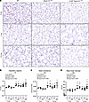 Figure 3
Z-AAT Serpina1Null mice develop spontaneous emphysema-like airspace enlargement. (A) Representative hematoxylin and eosin (H&E)-stained lung parenchyma sections from 4-, 7- and older than 12-month-old Z-AAT Serpina1Null, Serpina1Null versus WT mice, original magnification, × 10. (B and C) Murine lung physiology was measured using Flexivent. (B) Inspiratory capacity (IC, mL) and (C) static compliance (Cst, mL/cm H2O) of 4-, 7- and greater than 12-month-old Z-AAT Serpina1Null, Serpina1Null and WT mice. (D) Morphometry measured by Metamorph macro on low melting agarose–inflated and formalin-fixed lungs sampled using stereology methods. Mean linear intercept (MLI, μm, D) of 4-, 7- and older than 12-month-old Z-AAT Serpina1Null, Serpina1Null and WT mice. Data are presented as mean ± SD, 2-way ANOVA with mixed-effects model (RELM), #P < 0.05 versus 4-mo-old same genotype mice, *P < 0.05 versus same age WT mice, ^P < 0.05 versus same age Serpina1Null mice.
Following flexiVent testing the murine lungs were inflated with low melting agarose solution at constant pressure of 20 cm H2O and sectioned using stereology methods for unbiased estimation of alveolar number and alveolar surface. Quantitatively, the mean linear intercept (MLI) was significantly increased in 7- and 12-month-old Serpina1Null and Z-AAT Serpina1Null versus WT, control mice, with only 12-month-old Z-AAT Serpina1Null mice demonstrating progression of emphysema compared with Serpina1Null mice at the same age (Figure 3D). The higher static compliance and progressive airspace enlargement in 12-month-old Z-AAT Serpina1Null mice suggest that Z-AAT accumulation further worsens lung injury beyond lack of murine AAT, as in Serpina1Null mice.
Z-AAT polymer level in multiple lung compartments of Z-AAT Serpina1Null mice.
We next measured the level of Z-AAT polymers in various lung compartments, using the 2C1 monoclonal antibody that specifically detects naturally occurring human Z AAT polymers. We only measured polymerized Z-AAT in the BALf of Z-AAT Serpina1Null vs. Serpina1Nulland WT control mice, as early as at 4 months of age (Figure 4A and Supplemental Figure 4).
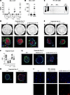 Figure 4
Z-AAT polymers accumulation in lung compartments of Z-AAT Serpina1Null mice. (A and B) Human Z-AAT polymers detected by ELISA using 2C1 antibody against Z-AAT polymers in BAL fluid of 4-, 7- and greater than 12-month-old Z-AAT Serpina1Null versus Serpina1Null and WT mice (A) and of healthy, never smokers (empty circles), Pi*Z AATD (black circles), and exsmoker Pi*Z AATD (larger black circles) individuals (B); Z-AAT polymer levels were normalized to epithelial lining fluid (ELF) volume imputed from BALf/plasma urea ratio and BAL volume. (C) Representative immunoblot of Z-AAT polymers (75–250 kDa, 2C1 antibody) of primary murine AT2 (EpCam+MHCII+) cell lysates isolated from Z-AAT Serpina1Null, Serpina1Null and WT mice that were greater than 12-month-old; polymerized AAT (heated M-AAT) served as positive control. (D–F) Primary murine AT2-derived organoids. Representative whole-well brightfield images (top) of Day 21 organoids derived from primary murine AT2 (EpCam+MHCII+) cells of greater than 12-month-old Z-AAT Serpina1Null, Serpina1Null, WT/Z-AAT, and WT mice cocultured with primary murine fibroblast in Matrigel (D and E). Organoid quantification (n = 4 wells from n = 3 mice / genotype) by CFE (F). Representative whole-mount immunofluorescent images of D21 organoids stained with anti-SPC (green), anti-RAGE (red, D and E, bottom), or polymerized Z-AAT (2C1, red, G and H) derived from AT2 of greater than 12-month-old Z-AAT Serpina1Null, Serpina1Null, WT/Z-AAT, and WT mice; scale bar: 100 μm (D and E, bottom; G and H) and 20 μm (D, top). (I) Representative immunofluorescence images of Cytospin slides from the BAL fluid of greater than 12-month-old Z-AAT Serpina1Null versus Serpina1Null and WT mice stained with 2C1 (red) and DAPI (blue), scale bar:100μm. Data are presented as mean ± SD, 1-way ANOVA, followed by Tukey’s multiple comparisons, or Mann-Whitney test. *P < 0.05 versus age-matched WT mice or healthy (H) individuals, ^ indicates versus age-matched Serpina1Null and Z-AAT Serpina1Null mice.
The Z-AAT polymer level in the BALf (26.6 ± 7.6 ng/mL) was lower than plasma level (77.5 ± 22 ng/mL) at every time point, including in greater than 12-month-old Z-AAT Serpina1Null mice (Figure 4A and Figure 1H). Our murine model recapitulates the findings seen in 6 of 17 AATD individuals with higher levels of Z-AAT polymers in the BALf (Figure 4B).
Accumulation and secretion of Z-AAT polymers in lung structural cells of individuals with AATD has been elusive and not described in previous murine models of AATD. We detect Z-AAT polymers by Western blotting in whole-cell lysates of unstimulated and nondifferentiated primary murine EpCam+MHCII+ AT2 cells (Figure 4C) isolated via FACS sorting from greater than 12-month-old Z-AAT Serpina1Null mice (Supplemental Figure 5). In a second experiment, FACS-sorted EpCam+MHCII+ AT2 cells from greater than 12-month-old Z-AAT Serpina1Null, Serpina1Null and WT mice (Supplemental Figure 2B) were cocultured with fetal murine fibroblasts in Matrigel in a 3D organoid model and allowed to proliferate and differentiate for 21 days. Brightfield images of day 21 organoids show lower number of organoids in Z-AAT Serpina1Null and Serpina1Null versus WT trans wells (Figure 4, D and E – top). During proliferation and differentiation, at day 21, the Serpina1Null and Z-AAT Serpina1Null organoids demonstrated lower proliferation capacity, as measured by colony forming units (CFE = organoid number / AT2 cells plated * 100, Figure 4F) and expressed less RAGE (Figure 4, D and E – bottom), suggestive of less AT2 proliferation potential and less AT2 to AT1 transition. Moreover, AT2 cells isolated from the Z-AAT mice on WT background (strain #037670) also showed lower CFE and decreased proliferation at day 21 in organoid culture versus AT2-derived organoids from WT littermates (Figure 4, E and F). These findings support an independent and detrimental role of Z-AAT polymers for AT2 proliferation and differentiation, even in presence of normal murine AAT levels as in Z-AAT mice on WT background (strain #037670).
By day 21 the WT-Z-AAT and Serpina1Null organoids secreted Z-AAT polymers, as detected by immunofluorescence using 2C1 monoclonal antibody (Figure 4, G and H, and Supplemental Figure 6A).
In a third experiment, after IgG panning AT2 cells isolated from greater than 12-month-old Z-AAT Serpina1Null, Serpina1Null, and WT mice were plated on 0.4 μm Corning inserts for 2D monolayer AT1-like differentiation, as previously published (36) (Supplemental Figure 7A). We detected patchy 2C1 staining, suggesting Z-AAT polymers production by day 9 in the Serpina1Null AT1-like monolayer (Supplemental Figure 7B).
Moreover, similar to recently published data in lung immune cells of individuals with AATD, we detected, using immunofluorescence, Z-AAT polymers within the airway macrophages of greater than 12-month-old Z-AAT Serpina1Null versus Serpina1Null and WT mice (Figure 4I and Supplemental Figure 6B).
Z-AAT polymers accumulation in AT2 cells is associated with mitochondria dysfunction and ER stress in Z-AAT Serpina1Null mice.
Within the hepatocytes of individuals with AATD and murine models of Z-AAT accumulation in the liver, Z-AAT polymerizes in the lumen of the ER, causing ER fragmentation, ER-stress, and activation of the unfolded protein response, resulting in oxidative stress and mitochondria dysfunction (14, 34).
To study ER morphology, mitochondria abundance, and localization we first evaluated the lungs of greater than 12-month-old Z-AAT Serpina1Null, Serpina1Null, and WT mice after fixation with chilled 4% paraformaldehyde / 2.5% glutaraldehyde and analyzed by transmission electron microscopy (TEM). We noticed dilated tubular smooth ER in the AT2 of old Z-AAT Serpina1Null versus Serpina1Null and WT mice (Supplemental Figure 3). We counted significantly higher number of mitochondria per AT2 surface area in Z-AAT Serpina1Null, Serpina1Null versus WT mice that were greater than 12-months old (Figure 5, A and B). The mitochondria were diffusely localized within the AT2 of Z-AAT Serpina1Null mice, while, in the AT2 of Serpina1Null and WT mice, they were localized primarily perinuclear. Moreover, mitochondria of Z-AAT Serpina1Null mice that were greater than 12-months old showed higher fusion and fission events, as demonstrated by white arrows in Figure 5A. Both Z-AAT Serpina1Null and Serpina1Null AT2 cells showed fewer lamellar body count (Supplemental Figure 8), but not significantly different than WT AT2 cells.
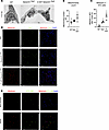 Figure 5
Z-AAT polymers accumulation in alveolar type-2 epithelial cells is associated with mitochondria dysfunction in Z-AAT Serpina1Null mice and AATD lung. (A) Representative TEM images of in situ murine AT2 of Z-AAT Serpina1Null, Serpina1Null and WT mice; scale bar: 2 μm. Note higher numbers of fused mitochondria (white arrows head), but unchanged number of surfactant-containing lamellar bodies (black star) in greater than 12-month-old Z-AAT Serpina1Null versus normal mitochondria (white arrow) Serpina1Null and WT mice. (B) Number of mitochondria per AT2 cell quantified by manual counting of 5 different TEM images per mouse. (C and D) Quantification of Mitofusin 2 (red) and pro-SPC (green) and representative immunofluorescence in lungs of 7-month-old Z-AAT Serpina1Null, Serpina1Null and WT mice. Colocalization Mitofusin 2 and pro-SPC (yellow) and quantified by ImageJ analysis of 5 different fields /slide from 3 different mice / genotype. Scale bar: 100 μm. (E) Representative immunofluorescence images of healthy, never smoker and AATD lungs stained for Mitofusin 2 (red, mitochondrial marker), HTII-280 (green), and DAPI (nuclei); scale bar: 100 μm. Data are presented as mean ± SD, 1-way ANOVA, followed by Tukey’s multiple comparisons, or Mann-Whitney test *P < 0.05 versus age-matched WT mice.
Mitofusin 2, a mitochondria outer membrane protein that controls fusion, was highly expressed in the AT2 cells (Figure 5C), murine lungs (Figure 5D) of greater than12-month-old Z-AAT Serpina1Null versus Serpina1Null and WT mice, and in the distal lung of individuals with AATD (Figure 5E).
The unstimulated, nondifferentiated AT2 cells from Z-AAT Serpina1Null mice also had significantly higher expression of ER stress, mitophagy, and autophagy markers, as demonstrated by higher expression of Gadd153 (ER stress, Figure 6A), Atf4 (unfolded protein response, Figure 6B), Pink1 (mitophagy, Figure 6C), Sesn3 (antioxidant and reactive oxygen species defense, Figure 6D), and of Sqstm1/p62 (impaired autophago-lysosomal degradation, Figure 6E) versus AT2 cells from Serpina1Null and WT mice.
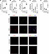 Figure 6
Z-AAT polymers accumulation in alveolar type-2 epithelial cells is associated with ER stress and autophagy in Z-AAT Serpina1Null mice and AATD lungs. (A–E) Expression of indicated genes by RT-qPCR in undifferentiated EpCam+MHCII+ AT2 cells isolated from greater than 7-month-old Z-AAT Serpina1Null, Serpina1Null and WT mouse lungs. Gadd153 (ER stress marker, A), Atf4 (unfolded protein response, B), Pink1 (mitophagy, C), Sesn3 (antioxidant and reactive oxygen species defense, D), and Sqstm1/p62 (autophagy marker, E). (F and G) Representative immunofluorescence images of healthy, never smoker, and AATD lungs stained for CHOP (red, ER stress marker, F), and LC3B (red, autophagy marker, G), Muc1 or HTII-280 (green), and DAPI (nuclei); scale bar: 100 μm. Note colocalization (yellow) between CHOP, and LC3B with AT2 cell markers. Data are presented as mean ± SD, t test, 1-way ANOVA, followed by Tukey’s multiple comparisons, or Mann-Whitney test *P < 0.05 versus age-matched WT mice.
Similarly, we found markers of ER stress and increased autophagy in the distal lung of individuals with AATD, as shown by increased colocalization between CHOP (Figure 6F) and LC3B (Figure 6G) with Muc1 or HT2-280 (AT2 marker), respectively, versus lungs of individuals who have never smoked.


