
Research ArticlePulmonologyVascular biology
Open Access |  10.1172/jci.insight.199743
Transplantation of endothelial progenitor cells attenuates pulmonary fibrosis via IFITM3-containing exosomes
Ying-Wei Lan,1 Jonathan Do,1 Wen Gao,1 Zicheng Deng,1 Xiaomei Xia,1 Enhong Li,1 Jillian Kash,1 Gautam Verma,1 Angara Sureshbabu,2 Kenneth S. Knox,3 Ross M. Bremner,2 Nicholas E. Banovich,4 Vladimir V. Kalinichenko,1,5 and Tanya V. Kalin1,6
1Phoenix Children’s Research Institute, Department of Child Health, University of Arizona College of Medicine–Phoenix, Phoenix, Arizona, USA.
2Norton Thoracic Institute, St. Joseph’s Hospital and Medical Center, Dignity Health, Phoenix, Arizona, USA.
3Department of Internal Medicine, Division of Pulmonary Medicine, University of Arizona College of Medicine–Phoenix, Phoenix, Arizona, USA.
4Division of Bioinnovation and Genome Sciences, The Translational Genomics Research Institute (TGen), Phoenix, Arizona, USA.
5Division of Neonatology and
6Center for Cancer and Blood Disorders, Phoenix Children’s Hospital, Phoenix, Arizona, USA.
Address correspondence to: Tanya V. Kalin, Phoenix Children’s Research Institute, Department of Child Health, University of Arizona College of Medicine–Phoenix, 475 N. 5th Street, Phoenix, Arizona 85004, USA. Phone: 602.827.2961; Email: tatianakalin@arizona.edu.
Find articles by Lan, Y. in: PubMed | Google Scholar
1Phoenix Children’s Research Institute, Department of Child Health, University of Arizona College of Medicine–Phoenix, Phoenix, Arizona, USA.
2Norton Thoracic Institute, St. Joseph’s Hospital and Medical Center, Dignity Health, Phoenix, Arizona, USA.
3Department of Internal Medicine, Division of Pulmonary Medicine, University of Arizona College of Medicine–Phoenix, Phoenix, Arizona, USA.
4Division of Bioinnovation and Genome Sciences, The Translational Genomics Research Institute (TGen), Phoenix, Arizona, USA.
5Division of Neonatology and
6Center for Cancer and Blood Disorders, Phoenix Children’s Hospital, Phoenix, Arizona, USA.
Address correspondence to: Tanya V. Kalin, Phoenix Children’s Research Institute, Department of Child Health, University of Arizona College of Medicine–Phoenix, 475 N. 5th Street, Phoenix, Arizona 85004, USA. Phone: 602.827.2961; Email: tatianakalin@arizona.edu.
Find articles by Do, J. in: PubMed | Google Scholar
1Phoenix Children’s Research Institute, Department of Child Health, University of Arizona College of Medicine–Phoenix, Phoenix, Arizona, USA.
2Norton Thoracic Institute, St. Joseph’s Hospital and Medical Center, Dignity Health, Phoenix, Arizona, USA.
3Department of Internal Medicine, Division of Pulmonary Medicine, University of Arizona College of Medicine–Phoenix, Phoenix, Arizona, USA.
4Division of Bioinnovation and Genome Sciences, The Translational Genomics Research Institute (TGen), Phoenix, Arizona, USA.
5Division of Neonatology and
6Center for Cancer and Blood Disorders, Phoenix Children’s Hospital, Phoenix, Arizona, USA.
Address correspondence to: Tanya V. Kalin, Phoenix Children’s Research Institute, Department of Child Health, University of Arizona College of Medicine–Phoenix, 475 N. 5th Street, Phoenix, Arizona 85004, USA. Phone: 602.827.2961; Email: tatianakalin@arizona.edu.
Find articles by Gao, W. in: PubMed | Google Scholar
1Phoenix Children’s Research Institute, Department of Child Health, University of Arizona College of Medicine–Phoenix, Phoenix, Arizona, USA.
2Norton Thoracic Institute, St. Joseph’s Hospital and Medical Center, Dignity Health, Phoenix, Arizona, USA.
3Department of Internal Medicine, Division of Pulmonary Medicine, University of Arizona College of Medicine–Phoenix, Phoenix, Arizona, USA.
4Division of Bioinnovation and Genome Sciences, The Translational Genomics Research Institute (TGen), Phoenix, Arizona, USA.
5Division of Neonatology and
6Center for Cancer and Blood Disorders, Phoenix Children’s Hospital, Phoenix, Arizona, USA.
Address correspondence to: Tanya V. Kalin, Phoenix Children’s Research Institute, Department of Child Health, University of Arizona College of Medicine–Phoenix, 475 N. 5th Street, Phoenix, Arizona 85004, USA. Phone: 602.827.2961; Email: tatianakalin@arizona.edu.
Find articles by Deng, Z. in: PubMed | Google Scholar
1Phoenix Children’s Research Institute, Department of Child Health, University of Arizona College of Medicine–Phoenix, Phoenix, Arizona, USA.
2Norton Thoracic Institute, St. Joseph’s Hospital and Medical Center, Dignity Health, Phoenix, Arizona, USA.
3Department of Internal Medicine, Division of Pulmonary Medicine, University of Arizona College of Medicine–Phoenix, Phoenix, Arizona, USA.
4Division of Bioinnovation and Genome Sciences, The Translational Genomics Research Institute (TGen), Phoenix, Arizona, USA.
5Division of Neonatology and
6Center for Cancer and Blood Disorders, Phoenix Children’s Hospital, Phoenix, Arizona, USA.
Address correspondence to: Tanya V. Kalin, Phoenix Children’s Research Institute, Department of Child Health, University of Arizona College of Medicine–Phoenix, 475 N. 5th Street, Phoenix, Arizona 85004, USA. Phone: 602.827.2961; Email: tatianakalin@arizona.edu.
Find articles by Xia, X. in: PubMed | Google Scholar
1Phoenix Children’s Research Institute, Department of Child Health, University of Arizona College of Medicine–Phoenix, Phoenix, Arizona, USA.
2Norton Thoracic Institute, St. Joseph’s Hospital and Medical Center, Dignity Health, Phoenix, Arizona, USA.
3Department of Internal Medicine, Division of Pulmonary Medicine, University of Arizona College of Medicine–Phoenix, Phoenix, Arizona, USA.
4Division of Bioinnovation and Genome Sciences, The Translational Genomics Research Institute (TGen), Phoenix, Arizona, USA.
5Division of Neonatology and
6Center for Cancer and Blood Disorders, Phoenix Children’s Hospital, Phoenix, Arizona, USA.
Address correspondence to: Tanya V. Kalin, Phoenix Children’s Research Institute, Department of Child Health, University of Arizona College of Medicine–Phoenix, 475 N. 5th Street, Phoenix, Arizona 85004, USA. Phone: 602.827.2961; Email: tatianakalin@arizona.edu.
Find articles by Li, E. in: PubMed | Google Scholar
1Phoenix Children’s Research Institute, Department of Child Health, University of Arizona College of Medicine–Phoenix, Phoenix, Arizona, USA.
2Norton Thoracic Institute, St. Joseph’s Hospital and Medical Center, Dignity Health, Phoenix, Arizona, USA.
3Department of Internal Medicine, Division of Pulmonary Medicine, University of Arizona College of Medicine–Phoenix, Phoenix, Arizona, USA.
4Division of Bioinnovation and Genome Sciences, The Translational Genomics Research Institute (TGen), Phoenix, Arizona, USA.
5Division of Neonatology and
6Center for Cancer and Blood Disorders, Phoenix Children’s Hospital, Phoenix, Arizona, USA.
Address correspondence to: Tanya V. Kalin, Phoenix Children’s Research Institute, Department of Child Health, University of Arizona College of Medicine–Phoenix, 475 N. 5th Street, Phoenix, Arizona 85004, USA. Phone: 602.827.2961; Email: tatianakalin@arizona.edu.
Find articles by Kash, J. in: PubMed | Google Scholar
1Phoenix Children’s Research Institute, Department of Child Health, University of Arizona College of Medicine–Phoenix, Phoenix, Arizona, USA.
2Norton Thoracic Institute, St. Joseph’s Hospital and Medical Center, Dignity Health, Phoenix, Arizona, USA.
3Department of Internal Medicine, Division of Pulmonary Medicine, University of Arizona College of Medicine–Phoenix, Phoenix, Arizona, USA.
4Division of Bioinnovation and Genome Sciences, The Translational Genomics Research Institute (TGen), Phoenix, Arizona, USA.
5Division of Neonatology and
6Center for Cancer and Blood Disorders, Phoenix Children’s Hospital, Phoenix, Arizona, USA.
Address correspondence to: Tanya V. Kalin, Phoenix Children’s Research Institute, Department of Child Health, University of Arizona College of Medicine–Phoenix, 475 N. 5th Street, Phoenix, Arizona 85004, USA. Phone: 602.827.2961; Email: tatianakalin@arizona.edu.
Find articles by Verma, G. in: PubMed | Google Scholar
1Phoenix Children’s Research Institute, Department of Child Health, University of Arizona College of Medicine–Phoenix, Phoenix, Arizona, USA.
2Norton Thoracic Institute, St. Joseph’s Hospital and Medical Center, Dignity Health, Phoenix, Arizona, USA.
3Department of Internal Medicine, Division of Pulmonary Medicine, University of Arizona College of Medicine–Phoenix, Phoenix, Arizona, USA.
4Division of Bioinnovation and Genome Sciences, The Translational Genomics Research Institute (TGen), Phoenix, Arizona, USA.
5Division of Neonatology and
6Center for Cancer and Blood Disorders, Phoenix Children’s Hospital, Phoenix, Arizona, USA.
Address correspondence to: Tanya V. Kalin, Phoenix Children’s Research Institute, Department of Child Health, University of Arizona College of Medicine–Phoenix, 475 N. 5th Street, Phoenix, Arizona 85004, USA. Phone: 602.827.2961; Email: tatianakalin@arizona.edu.
Find articles by Sureshbabu, A. in: PubMed | Google Scholar
1Phoenix Children’s Research Institute, Department of Child Health, University of Arizona College of Medicine–Phoenix, Phoenix, Arizona, USA.
2Norton Thoracic Institute, St. Joseph’s Hospital and Medical Center, Dignity Health, Phoenix, Arizona, USA.
3Department of Internal Medicine, Division of Pulmonary Medicine, University of Arizona College of Medicine–Phoenix, Phoenix, Arizona, USA.
4Division of Bioinnovation and Genome Sciences, The Translational Genomics Research Institute (TGen), Phoenix, Arizona, USA.
5Division of Neonatology and
6Center for Cancer and Blood Disorders, Phoenix Children’s Hospital, Phoenix, Arizona, USA.
Address correspondence to: Tanya V. Kalin, Phoenix Children’s Research Institute, Department of Child Health, University of Arizona College of Medicine–Phoenix, 475 N. 5th Street, Phoenix, Arizona 85004, USA. Phone: 602.827.2961; Email: tatianakalin@arizona.edu.
Find articles by Knox, K. in: PubMed | Google Scholar
1Phoenix Children’s Research Institute, Department of Child Health, University of Arizona College of Medicine–Phoenix, Phoenix, Arizona, USA.
2Norton Thoracic Institute, St. Joseph’s Hospital and Medical Center, Dignity Health, Phoenix, Arizona, USA.
3Department of Internal Medicine, Division of Pulmonary Medicine, University of Arizona College of Medicine–Phoenix, Phoenix, Arizona, USA.
4Division of Bioinnovation and Genome Sciences, The Translational Genomics Research Institute (TGen), Phoenix, Arizona, USA.
5Division of Neonatology and
6Center for Cancer and Blood Disorders, Phoenix Children’s Hospital, Phoenix, Arizona, USA.
Address correspondence to: Tanya V. Kalin, Phoenix Children’s Research Institute, Department of Child Health, University of Arizona College of Medicine–Phoenix, 475 N. 5th Street, Phoenix, Arizona 85004, USA. Phone: 602.827.2961; Email: tatianakalin@arizona.edu.
Find articles by Bremner, R. in: PubMed | Google Scholar
1Phoenix Children’s Research Institute, Department of Child Health, University of Arizona College of Medicine–Phoenix, Phoenix, Arizona, USA.
2Norton Thoracic Institute, St. Joseph’s Hospital and Medical Center, Dignity Health, Phoenix, Arizona, USA.
3Department of Internal Medicine, Division of Pulmonary Medicine, University of Arizona College of Medicine–Phoenix, Phoenix, Arizona, USA.
4Division of Bioinnovation and Genome Sciences, The Translational Genomics Research Institute (TGen), Phoenix, Arizona, USA.
5Division of Neonatology and
6Center for Cancer and Blood Disorders, Phoenix Children’s Hospital, Phoenix, Arizona, USA.
Address correspondence to: Tanya V. Kalin, Phoenix Children’s Research Institute, Department of Child Health, University of Arizona College of Medicine–Phoenix, 475 N. 5th Street, Phoenix, Arizona 85004, USA. Phone: 602.827.2961; Email: tatianakalin@arizona.edu.
Find articles by
Banovich, N.
in:
PubMed
|
Google Scholar
|

1Phoenix Children’s Research Institute, Department of Child Health, University of Arizona College of Medicine–Phoenix, Phoenix, Arizona, USA.
2Norton Thoracic Institute, St. Joseph’s Hospital and Medical Center, Dignity Health, Phoenix, Arizona, USA.
3Department of Internal Medicine, Division of Pulmonary Medicine, University of Arizona College of Medicine–Phoenix, Phoenix, Arizona, USA.
4Division of Bioinnovation and Genome Sciences, The Translational Genomics Research Institute (TGen), Phoenix, Arizona, USA.
5Division of Neonatology and
6Center for Cancer and Blood Disorders, Phoenix Children’s Hospital, Phoenix, Arizona, USA.
Address correspondence to: Tanya V. Kalin, Phoenix Children’s Research Institute, Department of Child Health, University of Arizona College of Medicine–Phoenix, 475 N. 5th Street, Phoenix, Arizona 85004, USA. Phone: 602.827.2961; Email: tatianakalin@arizona.edu.
Find articles by Kalinichenko, V. in: PubMed | Google Scholar
1Phoenix Children’s Research Institute, Department of Child Health, University of Arizona College of Medicine–Phoenix, Phoenix, Arizona, USA.
2Norton Thoracic Institute, St. Joseph’s Hospital and Medical Center, Dignity Health, Phoenix, Arizona, USA.
3Department of Internal Medicine, Division of Pulmonary Medicine, University of Arizona College of Medicine–Phoenix, Phoenix, Arizona, USA.
4Division of Bioinnovation and Genome Sciences, The Translational Genomics Research Institute (TGen), Phoenix, Arizona, USA.
5Division of Neonatology and
6Center for Cancer and Blood Disorders, Phoenix Children’s Hospital, Phoenix, Arizona, USA.
Address correspondence to: Tanya V. Kalin, Phoenix Children’s Research Institute, Department of Child Health, University of Arizona College of Medicine–Phoenix, 475 N. 5th Street, Phoenix, Arizona 85004, USA. Phone: 602.827.2961; Email: tatianakalin@arizona.edu.
Find articles by Kalin, T. in: PubMed | Google Scholar
Published October 8, 2026 - More info
JCI Insight. 2026;11(19):e199743. https://doi.org/10.1172/jci.insight.199743.
© 2026 Lan et al. This work is licensed under the Creative Commons Attribution 4.0 International License. To view a copy of this license, visit http://creativecommons.org/licenses/by/4.0/.
Received: September 2, 2025; Accepted: August 17, 2026


-
Results
Adult lung capEPCs localize within CAP1 and are decreased in fibrotic lungs of PF patients. Since KIT+FOXF1+ EPCs were originally identified in embryonic and neonatal lungs (22–24), we next determined whether these EPCs exist in adult lungs. We analyzed publicly available single-cell RNA sequencing (scRNA-seq) datasets of human lung tissue from 66 patients with PF and 48 unaffected donors (25). The samples from PF patients were stratified into 2 groups, “less fibrotic” and “more fibrotic,” based on the differentially affected areas of one lung used to prepare sequencing libraries (25). Endothelial cells were reclustered using EC-specific marker genes (26), identifying 5 distinct endothelial subclusters (Figure 1A and Supplemental Figure 1, A and B; supplemental material available online with this article; https://doi.org/10.1172/jci.insight.199743DS1). Consistent with previously published data (27), the percentage of CAP1, CAP2, arterial ECs, and venous ECs was decreased, while that of systemic venous ECs was increased, in PF lungs compared with donor lungs (Supplemental Figure 1C). Double-positive KIT+FOXF1+ EPCs (referred to as capEPCs hereafter) were mostly present within the CAP1 subcluster (Figure 1A) and had upregulated signaling pathways important for progenitor cells, including pluripotent stem cell differentiation, KIT, IGF-1, and FGF-1 signaling (Supplemental Figure 1D). The percentage of capEPCs was reduced in the lungs of PF patients, including both “less fibrotic” and “more fibrotic” PF, compared with donor lungs, suggesting that the loss of capEPCs is an early event in PF progression (Figure 1, B and C, and Supplemental Figure 1F). Next, we used immunostaining with CD31, FOXF1, and KIT antibodies and demonstrated that the abundance of capEPCs was decreased in the lungs of PF patients (Figure 1D). Compared with donor lungs, transcriptional signatures of capEPCs from PF lungs showed decreased gene signatures important for cell survival, proliferation, and progenitor functions, including NRF2, JAK-STAT, PDGFB, KIT, MAPK, and IGF-1 signaling pathways (Figure 1E and Supplemental Figure 1E). In contrast, profibrotic and proinflammatory pathways, such as TGF-β, IL-2, p53, and IL-6 pathways, were upregulated in fibrotic capEPCs (Figure 1E). Altogether, capEPCs are present in the human adult lung and belong to the CAP1 ECs, the number of capEPCs is decreased in lungs of PF patients, and their transcriptional signature is changed in PF.
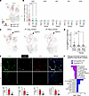 Figure 1
Lung capEPCs localize within CAP1 and are decreased in fibrotic lungs of PF patients. (A) UMAP visualization of EC subclusters (n = 37,752 cells) from donor and PF lungs (GSE227136), highlighting KIT+FOXF1+ cells (capEPCs) in red. (B) Quantification of FOXF1+KIT+ capEPCs across endothelial subtypes in donor, less fibrotic PF, and more fibrotic PF lungs. Data shown as the percentage of FOXF1+KIT+ cells among total ECs, calculated separately for each sample. Each dot represents an individual donor or PF patient sample. Horizontal lines indicate mean. Statistical significance was assessed using the Kruskal-Wallis test followed by pairwise Wilcoxon’s rank-sum tests with Benjamini-Hochberg correction. (C) Reduced abundance of capEPCs in fibrotic lungs. Left: UMAP visualization showing capEPCs (red dots) in donor, less fibrotic PF, and more fibrotic PF lungs. Dashed outlines indicate capEPC-enriched region. Right: Percentage of capEPCs among total ECs: donor (n = 44), less fibrotic PF (n = 39), and more fibrotic PF (n = 49). (D) Top: Representative immunofluorescence images of donor and PF lung sections stained for CD31 (green), FOXF1 (white), KIT (red), and DAPI (blue). Bottom: CD31+, FOXF1+, and KIT+ cells are shown as percentages of total DAPI+ nuclei, and capEPCs as a percentage of total CD31+ ECs, calculated per lung sample. PF lungs showed a significant reduction in capEPCs compared with donor lungs. n = 5 lungs/group. Scale bars: 100 μm. (E) Pathway enrichment analysis of genes differentially expressed in capEPCs from donor and PF lungs. Pathways downregulated or upregulated in PF capEPCs. Data presented as mean ± SD. *P < 0.05, **P < 0.01, ***P < 0.001, ****P < 0.0001. One-way ANOVA, Fisher’s least significant difference (LSD).
Mouse lung capEPCs localize within CAP1 and are decreased in lungs of bleomycin-treated mice. We next determined whether capEPCs existed in the adult mouse lung. Using publicly available scRNA-seq (28), we reclustered ECs to identify 4 distinct subclusters (Figure 2A and Supplemental Figure 2, A and B). Consistent with human data, the mouse capEPCs were identified within the CAP1 subcluster of ECs (Figure 2, A–C) and had upregulated signaling pathways important for progenitor cells (Supplemental Figure 2C). The abundance of capEPCs was decreased in the lungs of bleomycin-treated mice compared with control mouse lungs (Figure 2, B and C, and Supplemental Figure 2E). The numbers of capEPCs were decreased as early as day 7 after bleomycin injury and remained reduced during fibrotic stages (days 10–21) based on the analysis of the publicly available scRNA-seq dataset (GEO GSE141259) (29) (Supplemental Figure 2F). The decrease in the capEPC abundance correlated with decreased vascular gene module score and increased fibrosis gene module score (Supplemental Figure 2G). No changes in the numbers of KIT+CD45+ cells were shown between control and bleomycin-treated lungs (Supplemental Figure 2H). Consistent with human data, the transcriptional signature of capEPCs was changed during fibrotic remodeling in bleomycin-treated mice (Figure 2D). Specifically, capEPCs within fibrotic mouse lungs exhibited decreased signaling pathways important for cell survival, proliferation, and progenitor functions, including VEGF signaling, IGF signaling, PI3K/Akt signaling, and Ras signaling (Figure 2D). As in human capEPCs, profibrotic and proinflammatory signatures were increased in murine capEPCs from bleomycin-injured lungs (Figure 2D and Supplemental Figure 2D). Immunostaining of lung tissue sections with CD31, FOXF1, and KIT antibodies demonstrated that capEPCs were abundantly present in the normal mouse lung but decreased in the bleomycin-treated lungs (Figure 2E and Supplemental Figure 3). To quantify the percentage of capEPCs within lung ECs, we next used Foxf1-GFP reporter mice, in which GFP was knocked into the endogenous Foxf1 gene locus (24) (Figure 2F). Flow cytometry analysis demonstrated that the total number of lung ECs and the percentage of capEPCs within ECs were decreased in the bleomycin-treated lungs of Foxf1-GFP reporter mice compared with untreated control lungs at day 21 after 2 bleomycin administrations (Figure 2F). Altogether, capEPCs are present in the adult mouse lung and decreased after bleomycin injury. Strong similarities in capEPC signatures indicate common signaling mechanisms in human and mouse capEPCs during fibrotic lung remodeling.
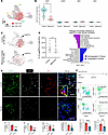 Figure 2
Mouse lung capEPCs localize within CAP1 and are decreased in lungs of bleomycin-treated mice. (A) UMAP visualization of lung EC subclusters from control and bleomycin-treated mouse lungs (GSE264151 and GSE264162), highlighting capEPCs (FOXF1+KIT+) in red. (B) Quantification of FOXF1+KIT+ capEPCs across endothelial subtypes in control and bleomycin-treated lungs. Data are shown as the percentage of FOXF1+KIT+ cells among total ECs within each subtype, calculated separately for each mouse lung. Each dot represents an individual mouse. Horizontal lines indicate the mean. Statistical significance was assessed using the Wilcoxon’s rank-sum test. (C) Reduced abundance of capEPCs following bleomycin injury. Left: UMAP visualization of FOXF1+KIT+ capEPCs (red dots) in control and bleomycin-treated lungs. Dashed outlines indicate the capEPC-enriched region. Right: Percentage of capEPCs among total ECs, calculated per mouse lung (control, n = 4; bleomycin, n = 3). Statistical significance was assessed using a linear model with dataset covariate adjustment. (D) Pathway enrichment analysis of capEPCs under fibrotic conditions, showing signaling pathways up- or downregulated following bleomycin treatment. (E) Top: Immunofluorescent staining of lung sections from control (n = 6) and bleomycin-treated mice (n = 8) for CD31 (green), FOXF1 (white), KIT (red), and DAPI (blue). Bottom: Quantification of CD31+, FOXF1+, and KIT+ cells expressed as a percentage of total DAPI+ nuclei, and FOXF1+KIT+ capEPCs expressed as a percentage of total CD31+ ECs, calculated per lung. Representative images from independent lungs are shown. Scale bars: 100 μm. (F) Flow cytometry analysis using Foxf1-GFP reporter mice (experimental design shown above) shows decreased percentage of capEPCs (CD117+Foxf1+ ECs) in bleomycin-treated lungs (n = 5 mice per group). Data are presented as mean ± SD. *P < 0.05, **P < 0.01, ****P < 0.0001. For E and F, unpaired 2-tailed t tests were used.
Transplantation of capEPCs improves mouse survival and lung functions in a preclinical model of PF. Since the number of capEPCs is decreased during PF and their transcriptomic signatures are conserved in humans and mice, we next tested whether the transplantation of donor capEPCs to recipient fibrotic lungs would have the therapeutic benefit. To lineage-label donor cells, we crossed Foxf1-GFP reporter mice with transgenic mice expressing ubiquitous tdTomato (Figure 3A). The double-transgenic donor mice were used for FACS sorting of FoxF1+KIT+ capEPCs from adult lungs. As a control for cell transplantation, we used adult FoxF1–KIT+ ECs that show no progenitor properties in neonatal lungs (24). The donor cells were transplanted intravenously to bleomycin-treated recipient mice that had already developed fibrosis and had shown measurable collagen deposition at day 8 after 2 intratracheal administrations of bleomycin (30) (Figure 3A). The other control included bleomycin-treated recipient mice that were mock-transplanted (treated with saline instead of cells).
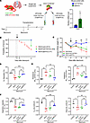 Figure 3
Transplantation of capEPCs improves survival and lung function in bleomycin-injured mice. (A) Top: Schematic of the FACS sorting strategy to isolate donor capEPCs (Foxf1+Kit+) and control Foxf1–Kit+ ECs from Foxf1-GFP;CAG-tdTomato reporter mice. Bottom: Experimental timeline. Right: qRT-PCR confirmed higher Foxf1 mRNA in capEPCs (n = 4). (B) Transplantation of capEPCs improved survival of bleomycin-injured mice. Numbers in parentheses indicate surviving mice/total mice per group. (C) Body weight recovery after bleomycin injury was improved in mice treated with capEPCs compared with mock-treated mice (BLM-mock) or mice treated with Foxf1–Kit+ ECs (BLM-Foxf1–Kit+). *Comparison with BLM-mock; #comparison with BLM-Foxf1–Kit+ ECs. (D) Arterial oxygen saturation on day 21 was increased after capEPC transplantation (control, n = 3; BLM-mock, n = 7; BLM-Foxf1–Kit+ ECs, n = 4; BLM-capEPCs, n = 4). (E) Lung resistance (Rrs) and elastance (Ers) measured by flexiVent show improved respiratory mechanics in capEPCs-treated mice (control, n = 3; BLM-mock, n = 5; BLM-Foxf1–Kit+ ECs, n = 4; BLM-capEPCs, n = 4). (F) Transplantation of capEPCs increased inspiratory capacity (IC), static compliance (Cst), and pressure-volume loop area, indicating enhanced lung compliance and elasticity (n = 3–5 mice per group). Data are presented as mean ± SD. Body weight recovery in C was analyzed by 2-way ANOVA with Greenhouse-Geisser correction (#P < 0.05, ##P < 0.01). Other comparisons were performed using 1-way ANOVA with Fisher’s LSD test. *P < 0.05, **P < 0.01, ***P < 0.001, ****P < 0.0001.
Mice transplanted with capEPCs exhibited markedly improved survival, with all mice surviving in this group, which was a better survival outcome than that of mock-transplanted mice or mice transplanted with the control FoxF1–KIT+ ECs (Figure 3B). We next assessed body weight changes in response to bleomycin treatment. Transplantation of capEPCs improved recovery of body weight loss in fibrotic mice by day 21 compared with both controls (Figure 3C). Furthermore, transplantation of capEPCs improved the peripheral arterial blood oxygenation measured by oximeter (Figure 3D), as well as lung mechanics measured by flexiVent (Figure 3, E and F). Respiratory system resistance (Rrs) was decreased, whereas lung inspiratory capacity and lung compliance were increased, in the capEPC-transplanted group compared with other bleomycin-injured groups (Figure 3, E and F). Thus, transplantation of capEPCs increases mouse survival, promotes body weight recovery, and improves respiratory functions after fibrotic bleomycin injury.
Transplantation of capEPCs attenuates fibrotic remodeling in bleomycin-treated mice. To further assess the therapeutic benefits of capEPC transplantation in a mouse model of PF, we compared fibrotic remodeling and collagen deposition in the lungs of bleomycin-injured mice transplanted with either capEPCs or control FoxF1–KIT+ ECs. Transplantation of capEPCs reduced Ashcroft score (Figure 4A) and decreased collagen deposition assessed by Masson’s trichrome staining (Figure 4B), Sirius red/fast green staining (Figure 4C), and immunostaining for αSMA (Figure 4D). Quantitative hydroxyproline assay demonstrated the decreased collagen levels in the lungs of capEPC-transplanted mice compared with controls (Figure 4E). Consistent with decreased fibrotic remodeling after capEPC transplantation, Col1a1, Col3a1, Fn1, and Acta2 mRNAs were reduced in total lung RNA (Figure 4, F–I). Moreover, capEPC transplantation successfully restored the Epcam and Cdh1 mRNAs compared with control treatments, suggesting improvement in the lung epithelium (Supplemental Figure 4A). The CTHRC1+ fibroblasts were decreased in the capEPC-treated group (Supplemental Figure 4B). No changes in the numbers of inflammatory cells were found in capEPC-transplanted and control lungs using flow cytometry analysis (Supplemental Figure 4C). Thus, transplantation of donor capEPCs effectively reduced fibrotic lung remodeling and collagen deposition in the bleomycin-injured recipient mice.
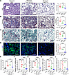 Figure 4
Transplantation of capEPCs attenuates fibrotic remodeling in bleomycin-treated mice. (A–D) Histological analysis of lungs collected 21 days after bleomycin injury shows reduced fibrotic remodeling following capEPC transplantation. Lung sections were stained with H&E (A), Masson’s trichrome (B), Sirius red/fast green (C), and immunofluorescence for αSMA (green) (D). Nuclei were counterstained with DAPI (blue). Right panels show quantitative analysis of Ashcroft score, trichrome-positive area, Sirius red–positive area, and αSMA-positive area. (BLM-mock, n = 10; BLM-Foxf1–Kit+ ECs, n = 6; BLM-capEPCs, n = 6.) Scale bars: 50 μm. (E) Hydroxyproline assay demonstrates reduced collagen deposition in lungs of capEPC-transplanted mice. n = 5–9 mice per group. (F–I) qRT-PCR analysis of total lung RNA reveals downregulation of fibrosis-associated genes in capEPC-transplanted mice. Actb was used for normalization. (Control group, n = 3; BLM-mock, n = 9; BLM-FOXF1–KIT+ ECs, n = 5; BLM-capEPCs. n = 7.) Data are presented as mean ± SD. One-way ANOVA with Fisher’s LSD test was used for all comparisons. *P < 0.05, **P < 0.01, ***P < 0.001, ****P < 0.0001.
Transplantation of capEPCs increases the number of lung ECs in bleomycin-injured mice. Since the number of capillary ECs is decreased in mouse and human fibrotic lungs (30), we examined EC numbers after capEPC transplantation. Immunofluorescent imaging and FACS analysis of lung tissue demonstrated an increase in the number of ECs in the lungs of bleomycin-injured mice transplanted with capEPCs (Figure 5, A and B). The mean fluorescence intensity of CD31 staining in ECs was increased in the capEPC-transplanted group, indicating that transplantation of capEPCs not only increased the number of CD31+ ECs but also increased CD31 cell surface expression in these cells (Figure 5B). Furthermore, more proliferating ECs and fewer apoptotic ECs were found in the lungs of bleomycin-injured mice transplanted with capEPCs (Supplemental Figure 5, A and B). Next, quantitative reverse transcription PCR (qRT-PCR) analysis of total lung tissue demonstrated that transplantation of capEPCs restored the expression of endothelial cell–specific Cdh5, Pecam1, and Kdr mRNAs as well as expression of Foxf1 (Figure 5C), which is consistent with the increased numbers of lung ECs (Figure 5, A and B).
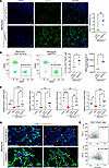 Figure 5
Transplantation of capEPCs increases the number of lung ECs in bleomycin-injured mice. (A) Left: Immunofluorescent staining of lung sections collected 21 days after bleomycin injury with CD31 (green) and DAPI (blue) shows increased endothelial coverage in capEPC-transplanted lungs compared with Foxf1–Kit+ EC–transplanted lungs. Right: Quantification of CD31+ area normalized to DAPI+ area (BLM-Foxf1–Kit+ ECs, n = 3; BLM-capEPCs, n = 4). Scale bars: 50 μm. (B) Flow cytometry analysis shows a higher percentage of ECs and increased CD31 mean fluorescence intensity of ECs in lungs of mice transplanted with capEPCs (BLM-Foxf1–Kit+ ECs, n = 3; BLM-capEPCs, n = 4). (C) qRT-PCR analysis of total lung RNA demonstrates increased Pecam1, Cdh5, Kdr, and Foxf1 mRNAs in capEPC-treated lungs. Actb was used for normalization (control, n = 3; BLM-mock, n = 9; BLM-Foxf1–Kit+ ECs, n = 5; BLM-capEPCs, n = 7). (D) Confocal imaging of lungs shows increased integration of donor tdTomato+ cells within CD31+ vasculature in the capEPC-treated group. Arrowheads indicate CD31+tdTomato+ double-positive cells; arrows mark tdTomato+ single-positive donor cells. Scale bars: 10 μm. (E) Flow cytometry analysis at day 13 after transplantation shows a higher percentage of tdTomato+ cells within lung ECs of capEPC-transplanted lungs (BLM-Foxf1–Kit+ ECs, n = 3; BLM-capEPCs, n = 7). Data are presented as mean ± SD. Unpaired 1-tailed t tests were used in A and B; 1-way ANOVA with Fisher’s LSD test was used in C. *P < 0.05, **P < 0.01, ***P < 0.001, ****P < 0.0001.
Finally, since the donor capEPCs were labeled with tdTomato, we assessed whether capEPCs engrafted into the lung endothelium of the bleomycin-injured mice. Immunostaining for CD31 and tdTomato showed a higher percentage of tdTomato+ capEPCs present within CD31+ ECs in comparison with the mice transplanted with control Foxf1–Kit+ ECs (Figure 5D). Flow cytometry analysis further supported these findings, detecting approximately 16% tdTomato+ ECs in the lungs of capEPC-transplanted mice 2 weeks after transplantation, compared with only 3% tdTomato+ ECs in the lungs of control mice (Figure 5E). These data indicate better engraftment efficiency of donor capEPCs to the recipient lung, compared with control Foxf1–Kit+ ECs.
Since the increased engraftment efficiency of donor capEPCs may still be insufficient to explain the robust therapeutic effect of capEPC transplantation, we next determined whether capEPCs mediate antifibrotic effects via paracrine signals to recipient cells.
IFITM3 is a direct transcriptional target of FOXF1. To identify molecular mechanisms underlying antifibrotic effects of capEPCs, we compared the transcriptome of capEPCs and control Foxf1–Kit+ ECs using the scRNA-seq dataset of adult mouse lungs. High Foxf1 mRNA levels in capEPCs and the absence of Foxf1 in control Foxf1–Kit+ ECs were confirmed using violin plot (Figure 6A). We next identified differentially expressed genes between capEPCs and control Foxf1–Kit+ ECs. One of the highly differentially expressed genes in capEPCs was interferon-induced transmembrane protein 3 (Ifitm3) (Figure 6B). The high level of Ifitm3 mRNA in capEPCs was confirmed by qRT-PCR using FACS-sorted capEPCs and Foxf1–Kit+ ECs from the lungs of Foxf1-GFP reporter mice (Figure 6C). Next, we used the human donor lung scRNA-seq dataset to compare IFITM3 expression in human capEPCs and FOXF1–KIT+ ECs. IFITM3 mRNA was increased in human capEPCs, correlating with FOXF1 expression (Figure 6D). In bronchoalveolar lavage fluid (BALF) from patients with idiopathic pulmonary fibrosis (IPF), IFITM3 protein levels were significantly reduced in comparison with healthy controls, as reported in a quantitative BALF proteomics study (31) (Figure 6E). Knockdown of Foxf1 in Kit+ ECs using siRNA decreased Ifitm3 mRNA and reduced angiogenesis in vitro (Figure 6, F and G). Finally, using a publicly available ChIP-seq dataset (32), we identified the FOXF1-binding region in the –267/+0 bp Ifitm3 promoter (Figure 6H). To functionally validate this FOXF1-DNA interaction, we cloned the Ifitm3 promoter region into a pGL4.23 luciferase reporter plasmid. In cotransfection experiments, the Foxf1 expression vector increased luciferase activity, whereas the empty luciferase vector had no effect (Figure 6I). In colocalization studies using both human and mouse lung tissue sections, we demonstrated that IFITM3 protein colocalized with capEPCs (Supplemental Figure 6, A and B). In IPF human and bleomycin-treated mouse lungs, the IFITM3 signal intensity and the capEPC abundance were markedly reduced (Supplemental Figure 6, A and B). Thus, IFITM3 is expressed in capEPCs and FOXF1 directly activates transcription of Ifitm3, indicating that the Ifitm3 gene is a direct transcriptional target of FOXF1.
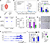 Figure 6
Ifitm3 is a direct transcriptional target of FOXF1. (A) scRNA-seq of adult mouse lungs shows Foxf1 mRNA in capEPCs (n = 312 cells) compared with Foxf1–Kit+ ECs (n = 70 cells). (B) Differentially expressed genes between capEPCs and Foxf1–Kit+ ECs show upregulation of Ifitm3 in capEPCs. (C) qRT-PCR of FACS-sorted lung ECs shows higher Foxf1 and Ifitm3 mRNA levels in capEPCs (n = 4). (D) Dot plot analysis of human lung scRNA-seq data (GSE227136) demonstrates coexpression of IFITM3 and FOXF1 in capEPCs. (E) Quantitative proteomic analysis of bronchoalveolar lavage fluid (BALF) from donor controls (n = 5) and patients with idiopathic pulmonary fibrosis (IPF; n = 4) demonstrates decreased IFITM3 protein levels in IPF BALF samples. Data were extracted and reanalyzed from a previously published BALF proteomics dataset (31). A 2-tailed Mann-Whitney test was used. (F and G) siRNA-mediated knockdown of Foxf1 in Kit+ lung ECs decreased Foxf1 and Ifitm3 mRNA expression (F; control, n = 4; siFoxf1, n = 3), and conditioned medium from these cells reduced endothelial tube formation in vitro (G; n = 4 per group). Scale bars in G: 100 μm. (H) ChIP-seq analysis (GSE77951) shows FOXF1 binding to the Ifitm3 promoter region. (I) Luciferase (Luc) assay demonstrates increased transcription from Ifitm3 promoter region (–267/+0 bp) in the presence of FOXF1 (n = 4). Data are presented as mean ± SD. Unpaired 2-tailed t tests were used. *P < 0.05, **P < 0.01, ****P < 0.0001.
IFITM3 improves EC functions but inhibits fibroblast activation. Next, we examined the effects of IFITM3 on lung ECs and fibroblasts in vitro. Mouse MFLM-91U cells were transfected with CMV-Ifitm3 plasmid to overexpress IFITM3 (Figure 7A). Conditioned medium from Ifitm3-overexpressing ECs increased endothelial tube formation in an in vitro assay (Figure 7B). In addition, treatment of human pulmonary artery endothelial cells (HPAECs) with recombinant IFITM3 (rIFITM3) protein significantly improved tube formation in vitro and increased the number of HPAECs in culture (Figure 7, C and D). These results were verified using human pulmonary microvascular endothelial cells (HPMECs), demonstrating that treatment with rIFITM3 increased angiogenesis and numbers of HPMECs in vitro (Supplemental Figure 6, C–E). In contrast to the activating effect of IFITM3 on ECs, the rIFITM3 treatment decreased proliferation and migration of CCD-19Lu fibroblasts (Figure 7, E and F) and inhibited profibrotic COL3A1, ACTA2, and VIM mRNAs in lung fibroblasts (Figure 7G). Thus, IFITM3 increased ECs’ pro-angiogenic properties but inhibited fibroblast activation in vitro.
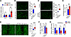 Figure 7
IFITM3 promotes EC functions but inhibits fibroblast activation. (A) qRT-PCR analysis confirms increased Ifitm3 expression in MFLM-91U cells transfected with CMV-Ifitm3 (n = 6). (B) Conditioned medium (CM) from IFITM3-overexpressing MFLM-91U cells enhances endothelial tube formation in vitro (n = 5). (C) Recombinant IFITM3 protein (rIFITM3; 100 ng/mL) promotes tube formation in cultured HPAECs (n = 7). (D) rIFITM3 increases HPAEC proliferation at 72 hours (n = 4). (E) Cell migration assay using a 2-well culture insert demonstrates reduced migration of CCD-19Lu fibroblasts following rIFITM3 treatment (n = 6 independent wells per group). Scale bars in B and C: 100 μm. Images in E were acquired using a 10× objective. (F) rIFITM3 suppresses fibroblast proliferation after 72 hours (n = 6). (G) qRT-PCR shows reduced expression of profibrotic COL3A1, ACTA2, and VIM after rIFITM3 treatment (control, n = 3; rIFITM3, n = 4 for each gene). Data are presented as mean ± SD. Unpaired 2-tailed t tests were used. *P < 0.05, **P < 0.01, ***P < 0.001, ****P < 0.0001.
IFITM3 is exported through an exosome-mediated pathway. Since IFITM3 lacks a signaling peptide for conventional protein secretion (33) (Supplemental Figure 6, F and G), and its export occurs through an exosome-mediated pathway (34), we tested the function of IFITM3 in exosomes. We generated an HPAEC cell line that expressed exogenous IFITM3 protein fused with an 11–amino acid HiBiT tag that possessed bioluminescence activity. Furthermore, the IFITM3-HiBiT fusion protein was expressed under control of doxycycline (Dox). Addition of Dox led to overexpression of exogenous IFITM3, which was detected by luminescence (Figure 8A). Endogenous IFITM3 in this cell line was inhibited by FOXF1-targeting siRNA (Figure 8A). The HiBiT tag enabled the detection of the IFITM3-HiBiT fusion protein in exosomes using the Nano-Glo HiBiT Extracellular Detection System. Exosomes were isolated from the conditioned medium of parental HPAECs or siFOXF1-transfected HPAECs in the presence or absence of Dox. Dynamic light scattering analysis confirmed that the isolated exosomes had an expected size distribution with a major peak intensity ranging between 94.50 ± 11.37 nm and 111.47 ± 21.88 nm, with no significant differences observed between groups (Figure 8B and Supplemental Table 1). Bioluminescence analysis of purified exosomes demonstrated increased bioluminescence activity in Dox-treated samples compared with exosomes from parental cells (control) or exosomes from cells without Dox, consistent with the presence of exogenous HiBiT-IFITM3 protein in exosomes after Dox treatment (Figure 8C).
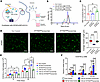 Figure 8
Treatment with IFITM3-containing exosomes improves EC functions but inhibits fibroblast activation. (A) Schematic diagram illustrating generation of ECs expressing exogenous IFITM3 fused to a HiBiT tag under doxycycline-inducible control. FOXF1 siRNA was used to suppress endogenous IFITM3 expression. Doxycycline (Dox) treatment induced HiBiT-IFITM3 expression, and conditioned media were collected for exosome isolation. Isolated exosomes were subsequently applied to endothelial cells (HPAECs) or fibroblasts (CCD-19Lu). (B) Size distribution of isolated exosomes measured by dynamic light scattering, showing similar particle profiles across groups (Exo Control, n = 3; IFITM3lo exosomes, n = 8; IFITM3hi exosomes, n = 5 independent exosome preparations). (C) Nano-Glo HiBiT extracellular detection assay shows increased HiBiT-IFITM3 signal in exosomes from Dox-induced cells (IFITM3hi exosomes; n = 4) compared with non-induced controls (IFITM3lo exosomes; n = 3). (D) Left: Endothelial tube formation assay in HPAECs shows impaired angiogenesis following treatment with IFITM3lo exosomes, while IFITM3hi exosomes restore tube-forming ability. Scale bars: 100 μm. Right: Quantification of vessel length (n = 12). Boxes indicate the 25th to 75th percentiles, center lines indicate the medians, and whiskers indicate the minimum and maximum values. (E) Cell proliferation assay using CCD-19Lu fibroblasts shows that IFITM3hi exosomes suppress TGF-β1–induced fibroblast proliferation at 24 and 48 hours, compared with IFITM3lo exosomes (n = 3). (F) qRT-PCR analysis demonstrates that treatment with IFITM3hi exosomes suppresses TGF-β1–induced expression of the profibrotic genes FN1, VIM, and COL3A1 in CCD-19Lu fibroblasts (control, n = 5; Exo Control + TGF-β1, n = 3; IFITM3lo exosomes + TGF-β1, n = 3; IFITM3hi exosomes + TGF-β1, n = 4 for each gene). Data are presented as mean ± SD. One-way ANOVA with Fisher’s LSD test was used for multiple comparisons. *P < 0.05, **P < 0.01, ***P < 0.001, ****P < 0.0001.
Treatment with IFITM3-containing exosomes improves EC functions but inhibits fibroblast activation. To test the functions of IFITM3 in exosomes, we purified exosomes from parental HPAECs (Exo Control) or siFOXF1-transfected HPAECs cultured without Dox (IFITM3lo exosomes) or with Dox (IFITM3hi exosomes). Endothelial cells were treated with these exosomes to test their effects in vitro. The IFITM3lo exosomes decreased tube formation in ECs compared with control exosomes as evidenced by reduced length of EC sprouts (Figure 8D). The tube formation in ECs was restored when the cells were treated with IFITM3hi exosomes (Figure 8D). We next tested the impact of IFITM3-containing exosomes on fibroblast activation. Consistent with published studies, addition of TGF-β1 to culture media increased human CCD-19Lu fibroblast proliferation as previously shown (35). IFITM3lo exosomes further increased TGF-β1–induced fibroblast proliferation at 48 hours after treatment. In contrast, IFITM3hi exosomes reduced fibroblast proliferation at both 24 and 48 hours after TGF-β1 treatment (Figure 8E). Furthermore, IFITM3hi exosomes decreased profibrotic FN1, VIM, and COL3A1 mRNAs in TGF-β1–activated fibroblasts (Figure 8F). Altogether, IFITM3-containing exosomes improved EC tube formation but inhibited TGF-β1–induced fibroblast activation in vitro.
Treatment with IFITM3-containing exosomes improves survival and lung functions in mouse model of lung fibrosis. To evaluate the therapeutic effects of IFITM3-containing exosomes in lung fibrosis, mice were intratracheally injected with IFITM3hi exosomes or control IFITM3lo exosomes on days 8 and 11 after 2 bleomycin administrations (Figure 9A). While mice treated with control exosomes exhibited continuous body weight loss throughout the study period, mice treated with IFITM3hi exosomes showed improved recovery of body weights (Figure 9A). Survival rates were also improved in the IFITM3hi exosome–treated group compared with the control group (Figure 9B). Luminescence analysis of lung tissues revealed higher HiBiT activity in the IFITM3hi exosome–treated group (Figure 9C), which was consistent with increased immunostaining of lung tissue sections using anti-HiBiT antibody and increased IFITM3 protein levels (Supplemental Figure 7A). Treatment with IFITM3hi exosomes restored peripheral blood oxygen saturation after bleomycin injury (Figure 9D). Lung mechanics measured by flexiVent showed significant improvement in the IFITM3hi exosome–treated group, including decreased elastance and resistance, and increased compliance and inspiratory capacity (Figure 9, E–H). Furthermore, treatment with IFITM3hi exosomes decreased lung fibrosis as shown by reduced hydroxyproline levels (Figure 9I), histological staining of lung tissue using H&E, Masson’s trichrome staining, and Sirius red/fast green (Figure 9J), and PDGFRA+ cells (Supplemental Figure 7B), Ashcroft score (Figure 9K), and qRT-PCR for fibrosis-associated mRNAs (Figure 9L). Finally, treatment with IFITM3hi exosomes increased abundance of lung pro-SPC+ epithelial cells (Supplemental Figure 7B). Altogether, intratracheal delivery of IFITM3-containing exosomes improved survival, body weight, and lung function and decreased PF in bleomycin-injured mice.
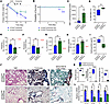 Figure 9
Treatment with IFITM3-containing exosomes decreases PF and improves survival in bleomycin-injured mice. (A) Treatment with IFITM3-containing (IFITM3hi) exosomes attenuated body weight loss in mice after bleomycin (BLM) injury. Exosomes were delivered on days 8 and 11 after BLM treatment. (B) Survival was improved in mice treated with IFITM3hi exosomes compared with mice treated with IFITM3lo exosomes. Numbers in parentheses indicate surviving mice/total mice per group. (C) HiBiT luminescence in lung lysates on day 15 post-bleomycin confirmed presence of IFITM3-containing exosomes in lung tissue (IFITM3lo, n = 4; IFITM3hi, n = 5). (D) Oxygen saturation was increased in mice treated with IFITM3hi exosomes (IFITM3lo, n = 4; IFITM3hi, n = 6). (E–H) FlexiVent measurements on day 15 showed that IFITM3hi exosomes improved elastance (Ers) (E), resistance (Rrs) (F), static compliance (Cst) (G), and inspiratory capacity (IC) (H). Red and black dotted lines represent the average values of the bleomycin and wild-type (WT) groups, respectively. (I) Hydroxyproline assay revealed reduced collagen deposition in mice treated with IFITM3hi exosomes (IFITM3lo, n = 4; IFITM3hi, n = 5). (J) Representative H&E, Masson’s trichrome, and Sirius red/fast green staining of lungs collected on day 15. Scale bars: 100 μm. (K) Quantification of Ashcroft scores and trichrome and Sirius red areas from J. (L) qRT-PCR showed reduced Col1a1, Col3a1, Fn1, and Vim in the IFITM3hi exosome–treated mice. Expression was normalized to Actb. Data are presented as mean ± SD. IFITM3hi exosome–treated mice, n = 5; IFITM3lo exosome–treated mice, n = 4. For box-and-whisker plots in D, I, and K, boxes indicate the 25th to 75th percentiles, center lines indicate the medians, and whiskers indicate the minimum and maximum values. Unpaired 2-tailed t tests were used in C–I, K, and L. **P < 0.01, ****P < 0.0001.


