
Research ArticleCell biologyMuscle biology
Open Access |  10.1172/jci.insight.203167
Histopathology and spatial transcriptomics jointly map myofiber-specific pathological programs in mTORC1-driven myopathy
Jer-En Hsu,1 Qingyang Zhao,1 Weiqiu Cheng,2 Hyun Min Kang,2 Susan V. Brooks,1 Myungjin Kim,1 and Jun Hee Lee1
1Department of Molecular & Integrative Physiology and
2Department of Biostatistics, University of Michigan, Ann Arbor, Michigan, USA.
Address correspondence to: Hyun-Min Kang, 1415 Washington Heights, Room 4632, SPH I Tower, Ann Arbor, Michigan 48109-2029, USA. Email: hmkang@umich.edu. Or to: Susan V. Brooks, 109 Zina Pitcher Place, 2029 BSRB, Ann Arbor, Michigan 48109-2200, USA. Email: svbrooks@umich.edu. Or to: Myungjin Kim, 109 Zina Pitcher Place, 3005 BSRB, Ann Arbor, Michigan 48109-2200, USA. Email: myungjin@umich.edu. Or to: Jun Hee Lee, 109 Zina Pitcher Place, 3019 BSRB, Ann Arbor, Michigan 48109-2200, USA. Email: leeju@umich.edu.
Find articles by Hsu, J. in: PubMed | Google Scholar
1Department of Molecular & Integrative Physiology and
2Department of Biostatistics, University of Michigan, Ann Arbor, Michigan, USA.
Address correspondence to: Hyun-Min Kang, 1415 Washington Heights, Room 4632, SPH I Tower, Ann Arbor, Michigan 48109-2029, USA. Email: hmkang@umich.edu. Or to: Susan V. Brooks, 109 Zina Pitcher Place, 2029 BSRB, Ann Arbor, Michigan 48109-2200, USA. Email: svbrooks@umich.edu. Or to: Myungjin Kim, 109 Zina Pitcher Place, 3005 BSRB, Ann Arbor, Michigan 48109-2200, USA. Email: myungjin@umich.edu. Or to: Jun Hee Lee, 109 Zina Pitcher Place, 3019 BSRB, Ann Arbor, Michigan 48109-2200, USA. Email: leeju@umich.edu.
Find articles by Zhao, Q. in: PubMed | Google Scholar
1Department of Molecular & Integrative Physiology and
2Department of Biostatistics, University of Michigan, Ann Arbor, Michigan, USA.
Address correspondence to: Hyun-Min Kang, 1415 Washington Heights, Room 4632, SPH I Tower, Ann Arbor, Michigan 48109-2029, USA. Email: hmkang@umich.edu. Or to: Susan V. Brooks, 109 Zina Pitcher Place, 2029 BSRB, Ann Arbor, Michigan 48109-2200, USA. Email: svbrooks@umich.edu. Or to: Myungjin Kim, 109 Zina Pitcher Place, 3005 BSRB, Ann Arbor, Michigan 48109-2200, USA. Email: myungjin@umich.edu. Or to: Jun Hee Lee, 109 Zina Pitcher Place, 3019 BSRB, Ann Arbor, Michigan 48109-2200, USA. Email: leeju@umich.edu.
Find articles by Cheng, W. in: PubMed | Google Scholar
1Department of Molecular & Integrative Physiology and
2Department of Biostatistics, University of Michigan, Ann Arbor, Michigan, USA.
Address correspondence to: Hyun-Min Kang, 1415 Washington Heights, Room 4632, SPH I Tower, Ann Arbor, Michigan 48109-2029, USA. Email: hmkang@umich.edu. Or to: Susan V. Brooks, 109 Zina Pitcher Place, 2029 BSRB, Ann Arbor, Michigan 48109-2200, USA. Email: svbrooks@umich.edu. Or to: Myungjin Kim, 109 Zina Pitcher Place, 3005 BSRB, Ann Arbor, Michigan 48109-2200, USA. Email: myungjin@umich.edu. Or to: Jun Hee Lee, 109 Zina Pitcher Place, 3019 BSRB, Ann Arbor, Michigan 48109-2200, USA. Email: leeju@umich.edu.
Find articles by Kang, H. in: PubMed | Google Scholar
1Department of Molecular & Integrative Physiology and
2Department of Biostatistics, University of Michigan, Ann Arbor, Michigan, USA.
Address correspondence to: Hyun-Min Kang, 1415 Washington Heights, Room 4632, SPH I Tower, Ann Arbor, Michigan 48109-2029, USA. Email: hmkang@umich.edu. Or to: Susan V. Brooks, 109 Zina Pitcher Place, 2029 BSRB, Ann Arbor, Michigan 48109-2200, USA. Email: svbrooks@umich.edu. Or to: Myungjin Kim, 109 Zina Pitcher Place, 3005 BSRB, Ann Arbor, Michigan 48109-2200, USA. Email: myungjin@umich.edu. Or to: Jun Hee Lee, 109 Zina Pitcher Place, 3019 BSRB, Ann Arbor, Michigan 48109-2200, USA. Email: leeju@umich.edu.
Find articles by
Brooks, S.
in:
PubMed
|
Google Scholar
|

1Department of Molecular & Integrative Physiology and
2Department of Biostatistics, University of Michigan, Ann Arbor, Michigan, USA.
Address correspondence to: Hyun-Min Kang, 1415 Washington Heights, Room 4632, SPH I Tower, Ann Arbor, Michigan 48109-2029, USA. Email: hmkang@umich.edu. Or to: Susan V. Brooks, 109 Zina Pitcher Place, 2029 BSRB, Ann Arbor, Michigan 48109-2200, USA. Email: svbrooks@umich.edu. Or to: Myungjin Kim, 109 Zina Pitcher Place, 3005 BSRB, Ann Arbor, Michigan 48109-2200, USA. Email: myungjin@umich.edu. Or to: Jun Hee Lee, 109 Zina Pitcher Place, 3019 BSRB, Ann Arbor, Michigan 48109-2200, USA. Email: leeju@umich.edu.
Find articles by Kim, M. in: PubMed | Google Scholar
1Department of Molecular & Integrative Physiology and
2Department of Biostatistics, University of Michigan, Ann Arbor, Michigan, USA.
Address correspondence to: Hyun-Min Kang, 1415 Washington Heights, Room 4632, SPH I Tower, Ann Arbor, Michigan 48109-2029, USA. Email: hmkang@umich.edu. Or to: Susan V. Brooks, 109 Zina Pitcher Place, 2029 BSRB, Ann Arbor, Michigan 48109-2200, USA. Email: svbrooks@umich.edu. Or to: Myungjin Kim, 109 Zina Pitcher Place, 3005 BSRB, Ann Arbor, Michigan 48109-2200, USA. Email: myungjin@umich.edu. Or to: Jun Hee Lee, 109 Zina Pitcher Place, 3019 BSRB, Ann Arbor, Michigan 48109-2200, USA. Email: leeju@umich.edu.
Find articles by Lee, J. in: PubMed | Google Scholar
Published August 20, 2026 - More info
JCI Insight. 2026;11(19):e203167. https://doi.org/10.1172/jci.insight.203167.
© 2026 Hsu et al. This work is licensed under the Creative Commons Attribution 4.0 International License. To view a copy of this license, visit http://creativecommons.org/licenses/by/4.0/.
Received: December 3, 2025; Accepted: August 12, 2026


-
Results
Seq-Scope enables combined analysis of histopathology and single-myofiber transcriptomics. We formerly generated a mouse model of unregulated mTORC1 hyperactivation by concomitantly deleting Tsc1 and Depdc5, thereby achieving maximal activation of both Rheb and Rag arms of mTORC1 signaling (Figure 1A). mTORC1 hyperactivation in skeletal muscle severely disrupted tissue homeostasis, leading to loss of muscle function associated with various pathologies, including basophilic fibers, enlarged fibers, and centrally nucleated fibers (Figure 1A; white, black, and green arrows, respectively). To capture the molecular changes underlying these phenotypes at the resolution of individual myofibers, we used Seq-Scope, a spatial transcriptomics platform with submicrometer resolution (average 0.5–0.7 μm), which we recently developed and optimized (30, 31). By aggregating transcripts according to the histology-based myofiber segmentation, a spatial transcriptome could be precisely assigned into individual myofibers. This approach preserved fiber morphology and spatial context while revealing and mapping transcriptomic diversity.
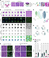 Figure 1
Seq-Scope enables histopathology-guided single-myofiber transcriptome analysis. (A) Schematic of the experimental workflow. Arrows indicate pathological phenotypes including basophilia (white), hypertrophy (black), and central nuclei (green). H&E histology, DAPI/WGA imaging, and Seq-Scope whole transcriptome analysis were performed on the same slide, which allowed precise segmentation and single-myofiber transcriptome analysis. Scale bar: 50 μm. (B) Cross-sections of control and mutant skeletal muscles examined by Seq-Scope. From top to bottom, images are displayed as H&E histology, DAPI/WGA staining, RNA density map from Seq-Scope transcriptome data, single-myofiber segmentation, and spatial myofiber type projection, according to the clusters identified in C. Tsc1fl/fl Depdc5fl/fl: control; Ckm-Cre Tsc1fl/fl Depdc5fl/fl: mutant. MF1: myofiber type I; MF2A: myofiber type IIa; MF2X: myofiber type IIx; MF2B: myofiber type IIb. Scale bars: 1 mm and 50 μm. (C) UMAP manifolds displaying the major myofiber types identified by multidimensional clustering. Datasets were integrated across mice. Each point (n = 8,616) represents an individual myofiber and is colored according to its corresponding myofiber type (center) or its originating muscle depot (lower right). (D) UMAP manifold colored by the expression levels of myosin heavy chain (Myh) isoforms that represent each myofiber type. (E) Pathological phenotypes observed in mTORC1 hyperactivated skeletal muscle. Left panel: centrally nucleated myofibers (arrows in the top row) and hypertrophic fibers (arrows in the bottom row). Right panel: transitional basophilic fibers (top row, scattered dark particles) and developed basophilic fibers (bottom row, dense and strip-like filling). Boxed areas are magnified on the right. Scale bars: 12 μm and 50 μm. (F) Quantification of pathological phenotype prevalence by muscle depot (left) or by myofiber type (right). “Other” includes myofibers from control muscles as well as morphologically normal fibers from mutant muscles. CTRL, control; KO, mutant.
In this study, we harvested EDL and SOL muscles from 4 Ckm-Cre Tsc1fl/fl Depdc5fl/fl and 4 Tsc1fl/fl Depdc5fl/fl male littermates at 10 weeks of age. A total of 16 cross-sections (2 muscles per mouse) were placed onto Seq-Scope arrays for spatial transcriptomic profiling. The exact same sections were subjected to H&E and DAPI/wheat germ agglutinin (WGA) imaging on the array surface prior to transcript capture and library generation (Figure 1B; first and second row, respectively). Sequencing of the Seq-Scope library yielded a total of 700 million unique reads that successfully aligned to the mm10 reference genome with valid spatial barcodes. The resulting RNA density map (Figure 1B, third row) revealed strong localization of transcripts at the periphery of myofibers, consistent with known muscle architecture. Using WGA imaging data and the Cellpose segmentation algorithm to delineate cellular membranes, we segmented 8,616 individual myofibers across 16 tissue sections (Figure 1B, fourth row, and Supplemental Figure 1A; supplemental material available online with this article; https://doi.org/10.1172/jci.insight.203167DS1). Data from each sample were processed independently and then integrated and clustered to assign myofiber identities. The data showed minimal batch effects, aside from the expected separation of SOL and EDL with their distinct myofiber compositions (Figure 1C and Supplemental Figure 1, B and C). Myofibers were also clearly distinguished by the expression of their corresponding myosin heavy chain isoforms (Figure 1D and Supplemental Figure 1D) and by structural and metabolic genes associated with oxidative (MF1/MF2A) or glycolytic (MF2B) types (Supplemental Figure 1E).
To systematically assess the histopathological features observed in mutant muscle, we quantified the prevalence of centrally nucleated fibers, enlarged fibers, and basophilic fibers across all sections. Centrally nucleated fibers were identified by detecting DAPI-positive nuclei located within a segmented myofiber and not overlapping the WGA-defined sarcolemmal boundary (Figure 1E). Enlarged fibers were identified from the myofiber segmentation output by measuring the cross-sectional area of each fiber. Because fiber area can vary with sectioning angle, enlargement was defined using a section-specific internal reference: mutant fibers with cross-sectional area greater than the mean area of morphologically healthy fibers were classified as enlarged (Figure 1E and Supplemental Figure 1F).
Basophilia was handled differently because it represents a histological pattern, defined by enhanced hematoxylin accumulation in the cytoplasm, that could not be captured reliably by a single intensity threshold alone. We therefore defined 2 prespecified morphology-based categories and performed blinded manual annotation. Transitional basophilic fibers were defined by scattered intracytoplasmic dark hematoxylin-positive particles (Figure 1E), whereas developed basophilic fibers were defined by dense, stripe-like, or broadly filled hematoxylin-positive staining within the myofiber (Figure 1E). These criteria were provided to an independent rater blinded to genotype for manual labeling. The reproducibility of these annotations was further validated by interrater agreement across 4 independent raters, as well as by quantification of intrafiber hematoxylin signal (Supplemental Figure 1, G–I).
Basophilic fibers were observed only in mutant EDL muscle, while enlarged fibers were restricted to mutant SOL muscle (Figure 1F and Supplemental Figure 1J). Fibers with central nuclei were also more frequent in mutant SOL muscle. When stratified by fiber type, we found that MF2X fibers showed the highest frequency of pathological features relative to other fiber types, suggesting that MF2X fibers exhibit the strongest pathological response to mTORC1 hyperactivation in this dataset (Figure 1F and Supplemental Figure 1J).
Identification of fiber type–specific responses to mTORC1 hyperactivation. The intrinsic differences among skeletal muscle fiber types suggest that myofibers may exhibit distinct transcriptional responses to sustained mTORC1 hyperactivation (1). To examine this question, we conducted differential gene expression analysis comparing transcriptomic signatures between control and mutant muscle within each myofiber type, both at the fiber level (Figure 2A and Supplemental Figure 2A) and at the biological replicate level, using pseudobulk analysis (Supplemental Figure 3A). Although many genes were commonly upregulated in response to mTORC1 hyperactivation, a number of genes showed myofiber type–specific expression patterns (Figure 2B). Importantly, phospho-RPS6 immunostaining co-registered with fiber-type markers showed that mTORC1 activation was broadly increased across mutant fiber types and was not restricted to the fiber types with the most prominent pathological changes (Supplemental Figure 2B).
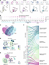 Figure 2
Identification of fiber type–specific responses to mTORC1 hyperactivation. (A) Schematic of the analytical workflow for fiber type–specific differential expression (DE) analysis comparing control (homeostasis) and mutant (hyperactivation) muscle. Upregulated genes determined by Wald’s test (adjusted P value < 0.05, log2FC > 1.5). (B) Representative expression patterns of genes that are exclusively upregulated in individual myofiber types, selected from a broader set (Supplemental Figure 2C). Exclusivity was determined by the Kruskal-Wallis test (adjusted P value < 0.05). Exclusively expressed genes were not identified in mutant MF1 fibers. (C) Venn diagram depicting the uniqueness and overlap of upregulated genes across different myofiber types, with numbers and percentages shown for both shared and unique gene sets. (D) Genes upregulated across all 4 fiber types (global response) were analyzed by gene ontology (GO) enrichment and visualized as a network. Each node represents a GO term, grouped by functional proximity within the enrichment hierarchy. Node size indicates the number of genes associated with the term, and node color reflects enrichment significance determined by 1-sided Fisher’s exact test (adjusted P value). (E) Genes selectively upregulated in specific fiber types were analyzed using compareGO. GO terms were grouped by functional proximity. Node size reflects the number of contributing genes, and node colors are shown as pie charts indicating the relative enrichment strength determined by 1-sided Fisher’s exact test (adjusted P value) contributed by each fiber type. (F) Genes globally upregulated across all fiber types and genes selectively upregulated in specific fiber types were mapped to enriched GO groups. The origin of each gene set is indicated by the stratum, and connecting threads represent gene-to-pathway relationships, colored by GO term identity.
We classified genes upregulated under mTORC1 hyperactivation into global response genes (expressed across all 4 fiber types, 25%), partially specific response genes (expressed in 2 or 3 fiber types, 49.2% in total), and genes specific for MF2A (5.6%, 7 genes), MF2X (4.0%, 5 genes), and MF2B (16.1%, 20 genes) fiber types (Figure 2C and Supplemental Figure 2, C and D). Notably, the MF1 transcriptome showed only global responses, without unique MF1-specific signatures in this analysis. Despite some differences between analytical frameworks, similar fiber type–associated patterns were observed in the mouse-level pseudobulk analysis (Supplemental Figure 3, A–H).
To systematically evaluate the functional annotations associated with both global and fiber type–specific signatures, we performed gene ontology (GO) enrichment analysis. Globally expressed genes were enriched in pathways related to ion transport regulation, muscle differentiation, cytoskeletal and synaptic remodeling, protein localization to membranes, and proteostasis (Figure 2D). These pathways overlap with proteostasis- and remodeling-related programs observed in our previous bulk RNA-Seq findings (6), supporting the reproducibility of these transcriptional features across different platforms.
In contrast, the present study also identified fiber type–specific transcriptional patterns associated with mTORC1 hyperactivation (Figure 2, B and E). Gene ontology clustering showed that MF2B myofibers exhibited relatively limited enrichment of oxidative stress- or detoxification-related GO terms compared with other fiber types. Instead, MF2B-associated genes were enriched for carbohydrate metabolic pathways, including Pkm, Phka1, Agl, and Pfkb3, consistent with preservation of a glycolytic transcriptional profile under mTORC1 hyperactivation. By contrast, MF2A myofibers showed enrichment of genes associated with stress-response and protein quality-control pathways, including Tfrc, Mt1, Psmd4, and Psmc3, along with regulators such as Igfbp5 and Sln. MF2X myofibers showed enrichment of remodeling-associated transcripts, including Asb15, Sparcl1, and Actc1, together with Ctsb and Gpx3, indicating a distinct structural and oxidative gene expression profile. Together, these results indicate an association between gene-level signatures and higher-order functional annotations (Figure 2F), supporting the conclusion that different fiber types exhibit distinct transcriptional programs associated with sustained mTORC1 hyperactivation.
Distinct mTORC1-associated states of type IIx myofibers in different muscles. MF2X represents the major fiber type consistently present in both the predominantly oxidative SOL muscle and the glycolytic EDL muscle, providing an opportunity to compare a shared fiber type across distinct muscle contexts (32). To investigate how MF2X fibers are altered under mTORC1 hyperactivation, we isolated a total of 1,447 MF2X myofibers from all tissue samples (Figure 3A, Supplemental Figure 1B, and Supplemental Figure 4A). Although control MF2X fibers from SOL and EDL were mixed in the UMAP manifold (Figure 3A), mTORC1-hyperactivated MF2X fibers separated into 2 distinct clusters corresponding to SOL- and EDL-derived MF2X fibers (Figure 3A). MF2X-SOL fibers showed strong enrichment for myofiber enlargement (Figure 3B), whereas MF2X-EDL fibers were preferentially associated with basophilia (Figure 3C and Supplemental Figure 1J).
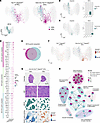 Figure 3
Distinct hyperactive mTORC1 responses of type IIx myofibers in different muscles. (A) UMAP visualization of myofiber data points separated as control (left) or mutant (right) groups, with muscle identity (EDL: red; SOL: blue) annotated within the original MF2X depot (lower left). Dotted arrows and shapes indicate the bidirectional shift of the MF2X subclusters in response to mTORC1 hyperactivation. (B and C) Enlarged fibers (B) and developed basophilic fibers (C) marked in the UMAP manifold (left). Proportions quantified from MF2X cluster of indicated muscles and presented in a bar graph (right). (D) Representative expression patterns of genes upregulated in response to mTORC1 hyperactivation in all MF2X fibers (MF2X global upregulation) or specifically in MF2X of EDL or MF2X of SOL, selected from a broader set in Supplemental Figure 4C. Exclusivity was determined using pairwise Wilcoxon’s tests (adjusted P value < 0.05). (E) Venn diagram depicting the uniqueness and overlap of genes upregulated in MF2X of SOL and MF2X of EDL. (F) Cumulative module score of muscle-specific upregulated genes displayed in integrated UMAP manifold with control (left) and mutant (right) myofiber data points separated. (G) Spatial images of representative mutant sections from each muscle. Boxed areas are magnified below (H&E). MF2X myofiber identity and cumulative module scores representing the axis of SOL- and EDL-specific responses are visualized. Scale bars: 100 μm and 500 μm. (H) GO enrichment network representing group of genes upregulated in response to mTORC1 hyperactivation in all MF2X fibers (global response) or specifically in MF2X of EDL (EDL-specific) or MF2X of SOL (SOL-specific). Pathways are grouped by functional similarity. Node size reflects the number of genes associated with each pathway, and node colors are shown as pie charts indicating the relative enrichment significance determined by 1-sided Fisher’s exact test (adjusted P values) contributed by each muscle-specific gene set. For the complete results with GO term names, see Supplemental Figure 4F.
Differential expression analysis comparing control and mutant MF2X-SOL or MF2X-EDL fibers revealed muscle context–associated transcriptional differences within the MF2X population (Supplemental Figure 4B). Among 216 genes highly upregulated under mTORC1 hyperactivation, 26 genes were classified as EDL specific and 72 genes were classified as SOL specific (Figure 3, D and E, and Supplemental Figure 4C). Gene score analysis showed that these SOL- and EDL-associated signatures were more prominent in mutant MF2X fibers than in control MF2X fibers (Figure 3F). Histology and spatial profiling further supported the separation between mutant SOL and EDL MF2X fibers at both the transcriptional and morphological levels (Figure 3G and Supplemental Figure 4, D and E).
We then performed gene set enrichment analysis using the SOL- and EDL-specific mTORC1-responsive genes (Figure 3H and Supplemental Figure 4F). Three categories of pathways emerged: those enriched in the global MF2X response (proteostasis and apoptosis, muscle contraction, myogenic development, and oxidative stress response), the SOL-specific MF2X response (cytoskeletal complex assembly and glucose homeostasis), and the EDL-specific MF2X response (ribonucleotide/purine metabolism and monosaccharide catabolism). Together, these findings indicate that, even within a shared fiber class, MF2X fibers exhibit muscle-specific transcriptional and histopathological programs under mTORC1 hyperactivation, supporting context-dependent divergence of type IIx myofibers.
Type IIx myofibers in SOL undergo hypertrophy with structural remodeling. Abnormal myofiber enlargement is a pathological hallmark observed in diverse muscle disorders and experimental models (1). Given that mTORC1 hyperactivation induced myofiber enlargement specifically in SOL muscles, we isolated and reanalyzed 2,972 SOL myofibers (Figure 4A). In mutant SOL muscles, enlarged fibers were mainly MF2X, and fiber-size analysis confirmed marked expansion of MF2X fibers compared with controls, whereas MF1 and most MF2A fibers were smaller (Figure 4, A–C, and Supplemental Figure 5A). These observations are consistent with IHC validation (Supplemental Figure 4E) and previously reported immunostaining quantification (8). We further noted a universal increase in Myh1 expression and decrease in Myh7 and Myh2 expression (Supplemental Figure 5B). The proportion of MF2X fibers in general was elevated in mutant SOL muscle (Supplemental Figure 5C). Importantly, this shift was observed consistently across independent batches, suggesting a potential fiber-type shift under sustained mTORC1 hyperactivation.
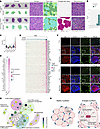 Figure 4
Type IIx myofibers in SOL undergo hypertrophy with structural remodeling. (A) Spatial images of SOL sections. Left: histology overview of 4 control sections (top) and 4 mutant sections (bottom) overlaid with fiber type images. Right: spatial images of magnified boxed area from corresponding overview, presented from left to right as histology, fiber types, enlarged fiber labels, and raw transcripts plotted as colored dots. Scale bars: 900 μm and 50 μm. (B) Prevalence of enlarged fibers in different myofiber clusters of SOL quantified and presented as a bar graph. (C) Size of fibers in different myofiber clusters of SOL calculated and presented as a violin plot. One pixel corresponds to ~0.57 μm2. (D) Expression of genes highly upregulated in enlarged fibers presented in a dot plot. (E) Correlation between fiber size and gene expression. Myofibers (x axis) are ordered from small to large. Positively correlated genes (y axis) are ranked by correlation strength. The expression level of the genes is colored based on z scores. RNA counts within a single fiber were normalized by fiber size to estimate RNA density. (F) Immunostaining of indicated proteins in control or mutant SOL muscle. WGA staining (green) outlines myofiber borders. Staining was performed on 2 serial sections (top and bottom). Boxed areas highlighting an enlarged fiber are magnified at the bottom. Representative images are shown from a single staining experiment. Scale bars: 20 μm and 50 μm. (G) GO enrichment network based on genes upregulated in enlarged fibers. Pathways grouped by functional similarity. Node size corresponds to the number of genes associated with each pathway, and node color reflects enrichment significance determined by one-sided Fisher’s exact test (adjusted P value). For the complete results with GO term names, see Supplemental Figure 5F. (H) Schematic diagram illustrating transcriptional signatures associated with pathological hypertrophy in mTORC1-activated type IIx myofibers in SOL.
In addition to exhibiting prominent hypertrophy, MF2X fibers also displayed more pronounced changes at the molecular level. Among the genes differentially expressed between control and mutant SOL tissues, MF2X fibers showed the strongest overall expression changes compared with other fiber types (Supplemental Figure 5D). We directly compared enlarged and nonenlarged fibers, enabling clearer identification of genes associated with mTORC1-driven hypertrophy (Figure 4D). These genes were generally expressed at low levels in non-hypertrophic fibers and showed strong positive correlations with myofiber size (Figure 4E and Supplemental Figure 5E).
Genes strongly correlated with fiber enlargement are classical autophagy regulators such as Sqstm1 (p62), Map1lc3a (LC3A), and Ubb, as well as proteasome or protease components such as Psmb5 and Ctsd, suggesting an association between myofiber hypertrophy and mTORC1 hyperactivation–driven proteostasis disruption on the transcriptional level. Genes involved in muscle growth and regeneration programs (e.g., Igfbp5, Mustn1, and Ankrd1) and structural genes associating with sarcomeric and cytoskeletal remodeling (e.g., Actc1, Myl4, Mylpf, Actn3, Tpm1, and Mybpc3) were also specifically upregulated in hypertrophic myofibers from mTORC1-hyperactivated muscles.
In addition, Uchl1, a ubiquitin C-terminal hydrolase linked to mTORC1 regulation, autophagy, and ubiquitin homeostasis in skeletal muscle (33, 34), was strongly correlated with fiber enlargement. Its elevated expression in enlarged fibers suggests a potential compensatory response to proteostasis disruption under sustained mTORC1 hyperactivation.
Importantly, the specific upregulation of p62, Igfbp5, Actc1, and Uchl1 in hypertrophic myofibers was confirmed through IHC staining of their protein products (Figure 4F). This coordinated program, combining impaired proteostasis, persistent growth signaling, and cytoskeletal remodeling, was reflected in our GO network analysis (Figure 4G and Supplemental Figure 5F). Together, these transcriptional results suggested a potential connection of the molecular changes to the enlargement myopathy observed in SOL muscle (Figure 4H).
Central nuclei are prevalent across various types of myofibers. The presence of centrally located nuclei in skeletal myofibers is a well-recognized histopathological feature, commonly associated with regeneration, myopathies, and various muscle stress conditions (35, 36). Histological analysis revealed a higher proportion of centrally nucleated myofibers in mutant SOL compared with mutant EDL muscles (Figure 1F and Supplemental Figure 5G). Although no strong fiber-type specificity was detected, oxidative fibers had a subtle tendency to show more frequent central nuclei compared with glycolytic fibers (Supplemental Figure 5, G and H). To assess whether central nuclei regions display distinct transcriptional features, we segmented 430 central nuclei from 2 mutant EDL and 2 mutant SOL muscles using combined H&E, DAPI, and WGA staining. Nuclei were dilated by 2, 4, or 6 μm to capture sufficient transcripts, and their transcriptomes were compared with 24-μm hexagon profiles from the 4 selected sections to generate a spatial sn-like dataset (Supplemental Figure 5, I and J). The analysis did not reveal strong enrichment for a specific gene or pathway, likely reflecting that the central nuclei feature could happen for a variety of different myofibers without representing a specific pathway. However, we were able to identify several zinc finger proteins, such as Zfp93 and Zfp429, which were substantially enriched in central nuclei regions and may be involved in positioning the nucleus during the injury (Supplemental Figure 5K).
Basophilic fibers are associated with increased RNA and metabolism-related signatures. Basophilic fibers represent a distinctive histological phenotype often associated with increased RNA content and muscle stress responses (37). Here, we isolated 48 developed basophilic fibers and 387 transitional basophilic fibers based on their histological patterns (Figure 5, A and B). Most basophilic fibers were restricted to MF2X fibers in mutant EDL muscle (Figure 5C and Supplemental Figure 6A). Consistent with the histological interpretation of basophilia, both transitional and developed basophilic fibers showed increased RNA content in the spatial transcriptomic dataset (Figure 5, D and E, and Supplemental Figure 6B), as well as increased RNA signal by RNA in situ hybridization (Supplemental Figure 6C).
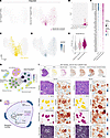 Figure 5
Basophilic fibers are associated with increased RNA and metabolism-related transcriptional signatures. (A) Integrated UMAP manifold, colored with transitional (pink) and developed (red) basophilic fibers, shown separately for control (left) and mutant (right) groups. (B) The prevalence of basophilia was quantified for each fiber-type cluster. (C) UMAP manifold displaying MF2X myofibers originating from EDL muscles (yellow). (D) UMAP manifold displaying RNA density (size-normalized RNA count) of each myofiber. (E) Violin plot visualizing the distribution of size-normalized RNA counts for transitional (pink) and developed (red) basophilic fibers as well as other fibers (gray). (F) Dot plot visualizing the expression of genes upregulated in basophilic fibers. (G) GO enrichment network analysis based on genes specifically upregulated in basophilic fibers. Pathways grouped by functional similarity. Node size corresponds to the number of genes associated with each pathway, and node color reflects enrichment significance determined by 1-sided Fisher’s exact test (adjusted P value). For the complete results with GO term names, see Supplemental Figure 6D. (H) Schematic diagram illustrating transcriptional signatures associated with mTORC1-induced basophilia in type IIx fibers in EDL. (I) Spatial images of a representative section from mutant EDL. Top: overview images presented from left to right as H&E histology, MF2X identity, basophilic states, and cumulative score maps for the following genes: lipid handling score (Fabp3, Acadl, Oxct1, Mfn2, Chpt1, Gpx4, and Ckmt2); respiration score (Mb, Cox genes, and Atp genes, see F); mitochondrial gene score (mitochondrial genome-encoded genes, see F); nucleotide synthesis score (Idh2, Ogdh, Mdh1, Mdh2, Ldhb, Mpc2, and Nmrk2). Bottom: representative basophilic fibers magnified in 2 views, corresponding to the boxes above in the H&E image. Raw transcripts are shown in colored dots as indicated in insets. Crosses and dotted borders highlight representative MF2X fibers. Scale bars: 300 μm and 50 μm. For the images from other mutant EDL sections, see Supplemental Figure 6A.
Through differential expression analysis, we identified a set of transcripts enriched in basophilic fibers, including genes associated with lipid metabolism, mitochondrial function, oxidative metabolism, and nucleotide metabolism (Figure 5F). GO pathway enrichment analysis showed strong representation of pathways related to lipid metabolism, mitochondrial transport, the TCA cycle, electron transport, and nucleotide synthesis (Figure 5G and Supplemental Figure 6D). These findings identify metabolism-related transcriptional signatures associated with myofiber basophilia.
Consistent with this pattern, multiple fatty acid– and lipid metabolism–related genes, including Fabp3, Acadl, Oxct1, Mfn2, Chpt1, and Gpx4, were prominently enriched in basophilic fibers (Figure 5F). Basophilic fibers also showed elevated expression of oxidative metabolism–associated genes, including Mb, multiple Cox subunits, Atp genes, and several mitochondrially encoded transcripts. This oxidative signature was further supported by increased COX activity in basophilic regions of mutant EDL muscle (Supplemental Figure 6E).
Enrichment of nucleotide metabolism–associated pathways was also observed in basophilic fibers (Figure 5, F and G). Several TCA cycle–, redox-, and nucleotide metabolism–related genes, including Idh2, Ogdh, Mdh1, Mdh2, Ldhb, Mpc2, and Nmrk2, were elevated in this population. Together, these transcriptional signatures indicate that lipid-, oxidative-, and nucleotide metabolism–related gene expression is associated with the increased RNA content of basophilic fibers (Figure 5H).
To illustrate these associations within a histological context, Fabp3, Mb, and Mdh1 were highlighted as representative genes associated with lipid handling, oxidative respiration, and nucleotide metabolism, respectively (Figure 5I, raw transcript view). In parallel, selected genes corresponding to these 3 gene sets were combined into cumulative scores, and their relative expression levels were assessed and visualized across the tissue (Figure 5I, Supplemental Figure 6A, and Supplemental Figure 6F). Taken together, our results indicate that basophilic MF2X fibers in mutant EDL muscle represent a distinct mTORC1-associated transcriptional state marked by increased RNA content and elevated expression of genes related to lipid, oxidative, and nucleotide metabolism.
Type IIb fibers in EDL exhibit transcriptional heterogeneity under mTORC1 hyperactivation. Type IIb fibers (MF2B), typically defined as the most glycolytic and fast-contracting population, have recently been shown to exhibit transcriptional variation under different contexts (38). Here, we identified transcriptional heterogeneity among MF2B fibers that was masked by aggressive batch correction during integration. This MF2B variation was initially observed in nonintegrated EDL clusters (Supplemental Figure 7, A–D). Since mutant samples showed greater variability across biological replicates, potentially reflecting biological heterogeneity at the 10-week time point, we first analyzed 2 mutant batches (KO-M1 and KO-M3) that integrated well even without batch correction (Figure 6, A and B). We then used these samples as reference datasets to define integration anchors and map all 4 mutant replicates into the same shared space (Supplemental Figure 8, A–C).
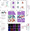 Figure 6
Type IIb fibers in EDL exhibit 3 distinct transcriptional states without gross morphological alterations. Myofibers from EDL muscles were subset and analyzed to reveal MF2B heterogeneity. (A) UMAP manifold showed MF2B clusters in selected control and mutant EDL sections (n = 2). Control MF2B cluster and non-MF2B clusters are labeled in dark gray and light gray, respectively. For full analysis, see Supplemental Figures 7 and 8. (B) Spatial images of selected mutant EDL sections. Left: H&E histology. Middle: MF2B fiber subtype map, according to the clustering presented in A. Right: Spatial map of a cumulative score created with the expression level of mitochondrial genome-encoded genes. Boxed areas in H&E images are magnified in F and G. Scale bar: 500 μm. (C) Expression of genes upregulated in each KO-MF2B subtype. (D) Mitochondrial gene cumulative score compared between control and mutant MF2B subtypes. (E) Frequency of basophilic fibers in control and mutant MF2B subtypes. (F) Representative views of MF2B subtype-2 with histology (H&E), fiber-subtype map, and mitochondrial gene cumulative score map. Crosses indicate representative subtype-2 myofibers. Scale bar: 50 μm. (G) Representative views of MF2B subtype-3 with histology (H&E), fiber-subtype map, and raw transcript plots of indicated genes. Crosses and dotted borders highlight representative subtype-3 myofibers. Scale bar: 50 μm.(H) Immunostaining of indicated proteins in control and mutant EDL muscles. WGA staining (green) delineates myofiber membranes. Boxed areas are magnified at the bottom. Representative images are shown from a single staining experiment. Scale bars: 20 μm and 50 μm. (I) Schematic diagram illustrating differential transcriptional states of MF2B fibers under sustained mTORC1 hyperactivation.
From these analyses, we identified 3 transcriptionally distinct MF2B subgroups under mTORC1 hyperactivation: MF2B subtype-1 (58% of mutant MF2B), which showed increased expression of transcripts resembling the shared mTORC1 hyperactivation signature; MF2B subtype-2 (22%), which showed increased expression of mitochondria-associated transcripts; and MF2B subtype-3 (20%), which was characterized by increased expression of developmental and remodeling-associated genes, including Actc1 and Myl4 (Figure 6C and Supplemental Figure 8D). These MF2B subgroups were represented across all 4 biological replicates (Supplemental Figure 7, D and E, and Supplemental Figure 8, E and F). All mutant subtypes exhibited elevated RNA content compared with controls, indicating altered transcriptional states under mTORC1 hyperactivation (Supplemental Figure 7F). Despite these transcriptomic differences, all MF2B subtypes maintained their fiber-type identity by robustly expressing the characteristic MF2B myosin isoform Myh4 (Supplemental Figure 7B and Supplemental Figure 8G).
The elevated expression of mitochondria-associated transcripts, including Atp and Cox genes, in MF2B subtype-2 suggested a distinct mitochondrial transcript signature (Figure 6C). To quantify this feature, we generated a composite mitochondrial transcript score based on detectable mitochondrially encoded genes. This analysis showed that MF2B subtype-2 exhibited a relatively high mitochondrial transcript score, comparable to basophilic MF2X fibers within EDL muscle (Figure 6, C and D, Supplemental Figure 7, G and H, and Supplemental Figure 8H). However, unlike basophilic MF2X fibers, MF2B subtype-2 fibers largely maintained normal morphology (Figure 6, E and F). These findings indicate that a subset of MF2B fibers exhibits increased mitochondria-associated gene expression without altered myosin isoform identity or overt pathological morphology under mTORC1 hyperactivation.
MF2B subtype-3, marked by developmental and remodeling-associated transcripts, did not display obvious morphological phenotypes (Figure 6, E and G). Spatial transcript mapping and quantitative analyses supported the elevated expression of Actc1 and Myl4 in this subgroup (Figure 6G, Supplemental Figure 7I, and Supplemental Figure 8I). Co-immunostaining of ACTC1 and MYL4 revealed both overlapping and nonoverlapping fibers (Figure 6H), suggesting that this transcriptional state may involve multiple downstream regulatory patterns.
Together, these transcriptional findings indicate that type IIb fibers exhibit distinct transcriptional states under mTORC1 hyperactivation while maintaining their fiber-type identity (Figure 6I). These results highlight previously unrecognized heterogeneity within a fiber type that has traditionally been considered relatively uniform.
mTORC1 hyperactivation drives macrophage-fibroblast accumulation in SOL muscle. Aside from myofibers or myocytes, many non-myocytic cell types, including fibroblasts, endothelial cells, and immune cells, reside within skeletal muscle, where they provide essential support for tissue maintenance and homeostasis. To investigate how these populations are altered under mTORC1 hyperactivation, we profiled non-myocytes using 24-μm diameter, flat-to-flat width, hexagon spatial gridding. This analysis identified diverse cell types and tissue-associated structures, including macrophages, fibroblasts, neuromuscular junctions, and erythrocytes (RBCs) (Figure 7, A and B).
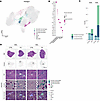 Figure 7
mTORC1 hyperactivation drives macrophage-fibroblast accumulation in SOL muscle. Non-myocytes were identified in skeletal muscle using 24 μm nonoverlapping hexagon gridding. (A) UMAP manifold of the hexagonal grid dataset displaying non-myocyte clusters, conducted without batch correction. (B) Expression of non-myocyte markers in different cell types. (C) Quantification of macrophage and fibroblast cell-type prevalence in control and mutant mice. (D) Spatial images showing H&E histology and cluster maps in representative muscle sections from each group. Boxed areas are magnified. Transcript expressions are displayed as colored dots as indicated Scale bars: 50 μm and 500 μm.
Resting non-myocytic cell types were broadly represented across batches (Supplemental Figure 9, A and B), but macrophage- and fibroblast-associated clusters were preferentially enriched in mutant SOL muscle (Figure 7C and Supplemental Figure 9C). The macrophage-associated cluster strongly expressed Apoe, consistent with a tissue-resident macrophage-like signature, whereas the activated fibroblast-associated cluster, separated from Gsn-expressing interstitial fibroblasts, expressed Mgp, Thbs4, Col1a1, and Col3a1, along with relatively low Gsn expression (Figure 7B and Supplemental Figure 9D). These observations suggest localized inflammatory and fibrotic remodeling in mutant SOL muscle, where histopathological features such as myofiber enlargement and central nuclei were also enriched. Macrophage- and fibroblast-associated clusters were frequently observed in close spatial proximity (Figure 7D). Consistent with this spatial transcriptomic pattern, immunofluorescence staining showed increased F4/80-positive macrophage signal and COL1A1-positive fibrotic signal in mutant SOL muscle, with enrichment in interstitial regions rather than diffuse distribution across myofibers (Supplemental Figure 9E). These findings support the presence of localized macrophage- and fibroblast-associated remodeling regions in mutant SOL muscle, consistent with prior studies showing that macrophage-fibroblast interactions can contribute to fibrotic tissue remodeling (39–41).
Overall, these results show that mTORC1 hyperactivation in myofibers is accompanied by a muscle context–dependent non-myocyte remodeling program. The preferential enrichment of macrophage- and fibroblast-associated clusters in mutant SOL, together with their close spatial proximity to each other and to regions of myofiber pathology, supports the presence of localized inflammatory and fibrotic remodeling in this muscle. Because the analysis was performed using 24-μm spatial hexagon bins at a single time point, these data do not establish the temporal order or causal direction between myofiber pathology and non-myocyte remodeling. Nevertheless, they identify a spatially organized macrophage-fibroblast–associated tissue state that is selectively linked to the mutant SOL microenvironment under sustained mTORC1 hyperactivation.


