
Research ArticleCardiologyVascular biology
Open Access |  10.1172/jci.insight.203575
GLS1 governs vascular smooth muscle cell phenotypic switching and aortic dissection via glutamate metabolism
Wei Xie,1 Chen Ning,2 Chen Lu,1 Dongjin Wang,3 Shuang Zhao,2 Tianyu Song,4 and Hailong Cao1
1Department of Cardiac Surgery, Zhongda Hospital, School of Medicine, Southeast University, Nanjing, Jiangsu, China.
2School of Pharmacy, Nanjing Medical University, Nanjing, Jiangsu, China.
3Department of Cardiac Surgery, Nanjing Drum Tower Hospital, Affiliated Hospital of Medical School, Nanjing University, Nanjing, Jiangsu, China.
4School of Medicine, Nanjing University of Chinese Medicine, Nanjing, Jiangsu, China.
Address correspondence to: Hailong Cao, Department of Cardiac Surgery, Zhongda Hospital, School of Medicine, Southeast University, No. 87 Dingjiaqiao, Gulou District, Nanjing, Jiangsu Province 210009, China. Phone: 86.25.83262261; Email: hailongcao_zd@163.com. Or to: Tianyu Song, School of Medicine, Nanjing University of Chinese Medicine, No. 138 Xianlin Avenue, Qixia District, Nanjing, Jiangsu Province 210023, China. Phone: 86.15250966190; Email: tianyusong@njucm.edu.cn. Or to: Shuang Zhao, School of Pharmacy, Nanjing Medical University, No. 101 Longmian Avenue, Jiangning District, Nanjing, Jiangsu 211166, China. Phone: 86.15720800551; Email: zhaoshuang@njmu.edu.cn.
Authorship note: WX and CN contributed equally to this work.
Find articles by Xie, W. in: PubMed | Google Scholar
1Department of Cardiac Surgery, Zhongda Hospital, School of Medicine, Southeast University, Nanjing, Jiangsu, China.
2School of Pharmacy, Nanjing Medical University, Nanjing, Jiangsu, China.
3Department of Cardiac Surgery, Nanjing Drum Tower Hospital, Affiliated Hospital of Medical School, Nanjing University, Nanjing, Jiangsu, China.
4School of Medicine, Nanjing University of Chinese Medicine, Nanjing, Jiangsu, China.
Address correspondence to: Hailong Cao, Department of Cardiac Surgery, Zhongda Hospital, School of Medicine, Southeast University, No. 87 Dingjiaqiao, Gulou District, Nanjing, Jiangsu Province 210009, China. Phone: 86.25.83262261; Email: hailongcao_zd@163.com. Or to: Tianyu Song, School of Medicine, Nanjing University of Chinese Medicine, No. 138 Xianlin Avenue, Qixia District, Nanjing, Jiangsu Province 210023, China. Phone: 86.15250966190; Email: tianyusong@njucm.edu.cn. Or to: Shuang Zhao, School of Pharmacy, Nanjing Medical University, No. 101 Longmian Avenue, Jiangning District, Nanjing, Jiangsu 211166, China. Phone: 86.15720800551; Email: zhaoshuang@njmu.edu.cn.
Authorship note: WX and CN contributed equally to this work.
Find articles by Ning, C. in: PubMed | Google Scholar
1Department of Cardiac Surgery, Zhongda Hospital, School of Medicine, Southeast University, Nanjing, Jiangsu, China.
2School of Pharmacy, Nanjing Medical University, Nanjing, Jiangsu, China.
3Department of Cardiac Surgery, Nanjing Drum Tower Hospital, Affiliated Hospital of Medical School, Nanjing University, Nanjing, Jiangsu, China.
4School of Medicine, Nanjing University of Chinese Medicine, Nanjing, Jiangsu, China.
Address correspondence to: Hailong Cao, Department of Cardiac Surgery, Zhongda Hospital, School of Medicine, Southeast University, No. 87 Dingjiaqiao, Gulou District, Nanjing, Jiangsu Province 210009, China. Phone: 86.25.83262261; Email: hailongcao_zd@163.com. Or to: Tianyu Song, School of Medicine, Nanjing University of Chinese Medicine, No. 138 Xianlin Avenue, Qixia District, Nanjing, Jiangsu Province 210023, China. Phone: 86.15250966190; Email: tianyusong@njucm.edu.cn. Or to: Shuang Zhao, School of Pharmacy, Nanjing Medical University, No. 101 Longmian Avenue, Jiangning District, Nanjing, Jiangsu 211166, China. Phone: 86.15720800551; Email: zhaoshuang@njmu.edu.cn.
Authorship note: WX and CN contributed equally to this work.
Find articles by Lu, C. in: PubMed | Google Scholar
1Department of Cardiac Surgery, Zhongda Hospital, School of Medicine, Southeast University, Nanjing, Jiangsu, China.
2School of Pharmacy, Nanjing Medical University, Nanjing, Jiangsu, China.
3Department of Cardiac Surgery, Nanjing Drum Tower Hospital, Affiliated Hospital of Medical School, Nanjing University, Nanjing, Jiangsu, China.
4School of Medicine, Nanjing University of Chinese Medicine, Nanjing, Jiangsu, China.
Address correspondence to: Hailong Cao, Department of Cardiac Surgery, Zhongda Hospital, School of Medicine, Southeast University, No. 87 Dingjiaqiao, Gulou District, Nanjing, Jiangsu Province 210009, China. Phone: 86.25.83262261; Email: hailongcao_zd@163.com. Or to: Tianyu Song, School of Medicine, Nanjing University of Chinese Medicine, No. 138 Xianlin Avenue, Qixia District, Nanjing, Jiangsu Province 210023, China. Phone: 86.15250966190; Email: tianyusong@njucm.edu.cn. Or to: Shuang Zhao, School of Pharmacy, Nanjing Medical University, No. 101 Longmian Avenue, Jiangning District, Nanjing, Jiangsu 211166, China. Phone: 86.15720800551; Email: zhaoshuang@njmu.edu.cn.
Authorship note: WX and CN contributed equally to this work.
Find articles by Wang, D. in: PubMed | Google Scholar
1Department of Cardiac Surgery, Zhongda Hospital, School of Medicine, Southeast University, Nanjing, Jiangsu, China.
2School of Pharmacy, Nanjing Medical University, Nanjing, Jiangsu, China.
3Department of Cardiac Surgery, Nanjing Drum Tower Hospital, Affiliated Hospital of Medical School, Nanjing University, Nanjing, Jiangsu, China.
4School of Medicine, Nanjing University of Chinese Medicine, Nanjing, Jiangsu, China.
Address correspondence to: Hailong Cao, Department of Cardiac Surgery, Zhongda Hospital, School of Medicine, Southeast University, No. 87 Dingjiaqiao, Gulou District, Nanjing, Jiangsu Province 210009, China. Phone: 86.25.83262261; Email: hailongcao_zd@163.com. Or to: Tianyu Song, School of Medicine, Nanjing University of Chinese Medicine, No. 138 Xianlin Avenue, Qixia District, Nanjing, Jiangsu Province 210023, China. Phone: 86.15250966190; Email: tianyusong@njucm.edu.cn. Or to: Shuang Zhao, School of Pharmacy, Nanjing Medical University, No. 101 Longmian Avenue, Jiangning District, Nanjing, Jiangsu 211166, China. Phone: 86.15720800551; Email: zhaoshuang@njmu.edu.cn.
Authorship note: WX and CN contributed equally to this work.
Find articles by Zhao, S. in: PubMed | Google Scholar
1Department of Cardiac Surgery, Zhongda Hospital, School of Medicine, Southeast University, Nanjing, Jiangsu, China.
2School of Pharmacy, Nanjing Medical University, Nanjing, Jiangsu, China.
3Department of Cardiac Surgery, Nanjing Drum Tower Hospital, Affiliated Hospital of Medical School, Nanjing University, Nanjing, Jiangsu, China.
4School of Medicine, Nanjing University of Chinese Medicine, Nanjing, Jiangsu, China.
Address correspondence to: Hailong Cao, Department of Cardiac Surgery, Zhongda Hospital, School of Medicine, Southeast University, No. 87 Dingjiaqiao, Gulou District, Nanjing, Jiangsu Province 210009, China. Phone: 86.25.83262261; Email: hailongcao_zd@163.com. Or to: Tianyu Song, School of Medicine, Nanjing University of Chinese Medicine, No. 138 Xianlin Avenue, Qixia District, Nanjing, Jiangsu Province 210023, China. Phone: 86.15250966190; Email: tianyusong@njucm.edu.cn. Or to: Shuang Zhao, School of Pharmacy, Nanjing Medical University, No. 101 Longmian Avenue, Jiangning District, Nanjing, Jiangsu 211166, China. Phone: 86.15720800551; Email: zhaoshuang@njmu.edu.cn.
Authorship note: WX and CN contributed equally to this work.
Find articles by Song, T. in: PubMed | Google Scholar
1Department of Cardiac Surgery, Zhongda Hospital, School of Medicine, Southeast University, Nanjing, Jiangsu, China.
2School of Pharmacy, Nanjing Medical University, Nanjing, Jiangsu, China.
3Department of Cardiac Surgery, Nanjing Drum Tower Hospital, Affiliated Hospital of Medical School, Nanjing University, Nanjing, Jiangsu, China.
4School of Medicine, Nanjing University of Chinese Medicine, Nanjing, Jiangsu, China.
Address correspondence to: Hailong Cao, Department of Cardiac Surgery, Zhongda Hospital, School of Medicine, Southeast University, No. 87 Dingjiaqiao, Gulou District, Nanjing, Jiangsu Province 210009, China. Phone: 86.25.83262261; Email: hailongcao_zd@163.com. Or to: Tianyu Song, School of Medicine, Nanjing University of Chinese Medicine, No. 138 Xianlin Avenue, Qixia District, Nanjing, Jiangsu Province 210023, China. Phone: 86.15250966190; Email: tianyusong@njucm.edu.cn. Or to: Shuang Zhao, School of Pharmacy, Nanjing Medical University, No. 101 Longmian Avenue, Jiangning District, Nanjing, Jiangsu 211166, China. Phone: 86.15720800551; Email: zhaoshuang@njmu.edu.cn.
Authorship note: WX and CN contributed equally to this work.
Find articles by Cao, H. in: PubMed | Google Scholar
Published April 23, 2026 - More info
JCI Insight. 2026;11(11):e203575. https://doi.org/10.1172/jci.insight.203575.
© 2026 Xie et al. This work is licensed under the Creative Commons Attribution 4.0 International License. To view a copy of this license, visit http://creativecommons.org/licenses/by/4.0/.
Received: December 15, 2025; Accepted: April 8, 2026


-
Results
Decreased GLS1 in VSMCs is associated with AD. To identify candidate genes potentially involved in VSMC phenotypic switching in AD, we integrated 2 transcriptomic datasets from AD patients (Gene Expression Omnibus [GEO] GSE52093 and GSE98770) and cross-referenced them with mitochondrial metabolism–related gene sets (MitoCarta3.0, ref. 14). As shown by Venn diagram, GLS1, ACADL, ALDH2, and ALDH1B1 were consistently shared across the datasets, identifying them as key candidate genes linking mitochondria-related metabolism and VSMC phenotypic switching to AD (Figure 1A). Notably, ACADL and ALDH2 have been previously associated with aortic aneurysm/dissection (7, 15), and ALDH1B1 shares high sequence/structure conservation with ALDH2 (16). Given that the precise role of GLS1 in AD remains undefined, we here sought to investigate its impact on VSMC phenotypic switching and AD pathogenesis.
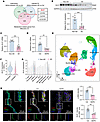 Figure 1
Expression of GLS1 in VSMCs is decreased in AD. (A) Venn diagram showing the 4 overlap genes (GLS1, glutaminase 1; ACADL, long-chain acylase A dehydrogenase; ALDH2, aldehyde dehydrogenase 2; ALDH1B1, aldehyde dehydrogenase 1 family member B1) between differentially expressed genes (DEGs) in human AD database (GSE52093, GSE98770) and metabolic-related genes from Mitocarta3. (B) Western blot analysis of GLS1 expression in aorta from non-AD groups and AD patients. (C) mRNA expression of GLS1 in aorta from non-AD groups and AD patients was detected by real-time qPCR. (D) Three-week-old male C57BL/6 mice were treated with β-aminopropionitrile (BAPN) in drinking water (1 g/kg/d) for 4 weeks. mRNA expression of Gls1 in mouse aorta was detected by real-time qPCR. (E) Uniform manifold approximation and projection (UMAP) visualization of single cells from human aortic tissues (GSE213740). Cells were partitioned into 10 major lineages: T cell, fibroblast, VSMC (contractile), VSMC (synthetic), monocyte, endothelial cell, macrophage, myofibroblast, epithelial cell, and B cell. (F) GLS1 expression among distinct cellular populations between non-AD groups and AD patients. (G) GLS1 expression among distinct cell types between non-AD groups and AD patients. (H) Three-week-old male C57BL/6 mice were treated with BAPN in drinking water (1 g/kg/d) for 4 weeks. Immunofluorescence staining for GLS1 (red), α-SMA (green), and DAPI (blue) in mouse aorta. Scale bars: 100 μm. Data are presented as mean ± SEM. Statistical analysis was performed using unpaired, 2-tailed Student’s t test (B–D, G, and H). **P < 0.01, ***P < 0.001.
Transcriptomic analysis of AD patient datasets (GSE52093 and GSE98770) revealed reduced GLS1 expression in AD tissues compared with controls (Supplemental Figure 1, A–D; supplemental material available online with this article; https://doi.org/10.1172/jci.insight.203575DS1). Importantly, we verified that the aortic GLS1 protein levels were significantly decreased in AD patients (Figure 1B). Meanwhile, the mRNA levels of GLS1 were reduced in aortic tissues from both AD patients and β-aminopropionitrile–induced (BAPN-induced) AD mice (Figure 1, C and D). To determine GLS1 expression patterns within the vascular wall, we analyzed a single-cell RNA sequencing dataset of AD patients (GSE213740). Uniform manifold approximation and projection (UMAP) visualization identified 10 major cell lineages (Figure 1E). The 2 separate populations of VSMCs were identified as contractile and synthetic phenotype, respectively, according the VSMC phenotypic marker genes (Supplemental Figure 1, E–G). Notably, we observed that expression of GLS1 was obviously decreased in the synthetic VSMCs (Figure 1F). Also, it was highly expressed in VSMCs from non-AD subjects and exhibited a striking reduction in AD patients (Figure 1G). Immunofluorescence also demonstrated decreased GLS1 in the tunica media of aortic tissues from mouse AD models (Figure 1H). These results suggested that the downregulation of GLS1 in VSMCs may participate in AD development.
GLS1 regulates VSMC phenotypic switching in vitro. In physiological conditions, VSMCs maintain a differentiated contractile phenotype. However, they dedifferentiate and convert to a synthetic phenotype in response to pathological stimuli, a process recognized as a pivotal mechanism in AD pathogenesis (17). Next, we used PDGF-BB to stimulate human aortic smooth muscle cells (HASMCs) to induce VSMC dedifferentiation and found that GLS1 was decreased upon PDGF-BB stimulation (Figure 2A). To determine whether GLS1 participated in VSMC dedifferentiation, we used siRNA to knock down GLS1 in HASMCs (Supplemental Figure 2A), followed by PDGF-BB treatment. The contractile-related genes (TAGLN and ACTA2) and synthetic-related gene (OPN) in HASMCs were detected, and the results showed that GLS1 knockdown further inhibited the mRNA levels of TAGLN and ACTA2, and promoted OPN mRNA levels upon PDGF-BB stimulation (Figure 2B). Meanwhile, Western blot analysis revealed that GLS1 knockdown further inhibited TAGLN expression and enhanced OPN expression in PDGF-BB–treated HASMCs (Supplemental Figure 2B). Also, in situ zymography revealed that knockdown of GLS1 promoted the MMP activities induced by PDGF-BB (Figure 2C). To further evaluate the role of GLS1 during VSMC phenotypic switching, we used adenovirus to overexpress GLS1 in HASMCs (Supplemental Figure 3A). We found that the VSMC phenotypic switching induced by PDGF-BB could be improved by GLS1 overexpressed in HASMCs, as evidenced by increased TAGLN and ACTA2, and decreased OPN mRNA levels upon GLS1 overexpression (Figure 3A), and similar results were obtained by Western blot analysis (Supplemental Figure 3B). GLS1 overexpression also inhibited MMP activities in the condition of PDGF-BB treatment (Figure 3B). Collectively, these findings demonstrated that downregulation of GLS1 promotes VSMC dedifferentiation into a synthetic phenotype, and GLS1 overexpression attenuates pathological VSMC phenotypic switching in vitro.
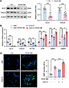 Figure 2
GLS1 knockdown promotes phenotypic switching of VSMCs in response to PDGF-BB. (A) Human aortic smooth muscle cells (HASMCs) were treated with PDGF-BB. Western blot analysis of GLS1 and TAGLN expression. (B and C) HASMCs were transfected with siRNA against GLS1 (siGLS1) or negative control (siNC), and then treated with PDGF-BB. (B) The mRNA expression of GLS1, TAGLN, ACTA2, OPN, MMP2, and MMP9 in HASMCs was detected by real-time qPCR. (C) Immunofluorescence images of in situ zymography (DQ gelatin) in HASMCs. MMP activity (green) was quantified by immunofluorescence intensity. Scale bars: 50 μm. (Blue, DAPI.) Data are presented as mean ± SEM. Statistical analysis was performed using unpaired, 2-tailed Student’s t test (A), or 1-way ANOVA followed by Tukey’s multiple-comparison test (B and C). *P < 0.05, **P < 0.01, ***P < 0.001.
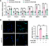 Figure 3
GLS1 overexpression preserves the contractile phenotype of VSMCs in response to PDGF-BB. (A and B) HASMCs were infected with adenovirus containing empty vector (ad-Vector) or GLS1-encoding plasmids (ad-GLS1), and then treated with PDGF-BB. (A) The mRNA expression of GLS1, TAGLN, ACTA2, OPN, MMP2, and MMP9 in HASMCs was detected by real-time qPCR. (B) Immunofluorescence images of in situ zymography (DQ gelatin) in HASMCs. MMP activity (green) was quantified by immunofluorescence intensity. Scale bars: 50 μm. (Blue: DAPI.) Data are presented as mean ± SEM. Statistical analysis was performed using 1-way ANOVA followed by Tukey’s multiple-comparison test (A and B). **P < 0.01, ***P < 0.001.
VSMC-specific GLS1 overexpression ameliorates BAPN-induced AD. To assess the role of GLS1 in VSMCs during AD, we constructed lentivirus carrying empty vector (Lenti-Vector) or Gls1 overexpression plasmid (Lenti-Gls1) containing 2 loxP sites that can be recognized by Cre recombinase. Then, 3-week-old male TaglnCre/+ mice received intravenous injections of either Lenti-Vector or Lenti-Gls1. One week after injection, AD was induced by administration of BAPN in drinking water for 4 weeks (Figure 4A). The expression of exogenous GLS1 in aortic VSMCs was verified by immunofluorescence staining with FLAG (Supplemental Figure 4A). Notably, we found that delivery of Lenti-Gls1 could attenuate AD lesion induced by BAPN (Figure 4B), and significantly decreased the incidence of aortic dissection and rupture (Figure 4C). Hematoxylin and eosin (H&E) staining showed reduced aortic dilatation in Lenti-Gls1 mice fed by BAPN, while elastic Verhoeff–van Gieson (EVG) staining also demonstrated that aortic media degeneration was improved by delivery of Lenti-Gls1 (Figure 4D). Meanwhile, overexpression of GLS1 significantly mitigated the dilatation of thoracic aorta diameter induced by BAPN (Figure 4E). The mRNA levels of VSMC phenotypic switching–related genes showed that delivery of Lenti-Gls1 could reverse the reduced expression of contractile-related genes (Tagln and Acta2) and enhanced expression of synthetic-related gene (Opn, Mmp2, and Mmp9) in aorta from BAPN-fed mice (Figure 4F). These data suggested that GLS1 overexpression could improve VSMC phenotypic switching and protect against AD in vivo.
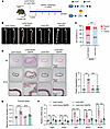 Figure 4
Overexpression of GLS1 in VSMCs attenuates AD in BAPN-treated mice. (A) Three-week-old male TaglnCre/+ mice were intravenously injected with lentivirus containing control vector or reverse Gls1 sequence with 2 loxP sites. After 1 week, BAPN (1 g/kg/d) was given in drinking water for 4 weeks. (B) Representative morphology of aortae from mice in the control group (n = 5) and the BAPN-treated group (n = 10). Scale bars: 5 mm. (C) The incidence of aortic dissection and rupture in BAPN-treated mice (n = 10). (D) Hematoxylin and eosin (H&E) and elastic Verhoeff–van Gieson (EVG) staining of aorta from control or BAPN-treated mice, and quantifications of elastic fiber breaks. Scale bars: 500 μm (top and middle panels), 100 μm (bottom panels). (E) Quantification of maximal diameters measured ex vivo of aorta from control and BAPN-treated mice. (F) The mRNA expression of Tagln, Acta2, Opn, Mmp2, and Mmp9 in aorta from control and BAPN-treated mice was detected by real-time qPCR. Data are presented as mean ± SEM. Statistical analysis was performed using Fisher’s exact test (C), or 1-way ANOVA followed by Tukey’s multiple-comparison test (E and F). ***P < 0.001, ****P < 0.0001.
GLS1 deficiency in VSMCs aggravates AD in BAPN-treated mice. To assess the effect of GLS1 deficiency in VSMCs during AD, we used smooth muscle cell–specific Gls1-knockout mice (Gls1SMKO) and their littermate controls (TaglnCre/+ and Gls1fl/fl). Five-week-old male TaglnCre/+, Gls1fl/fl, and Gls1SMKO mice were given BAPN in drinking water for 4 weeks to induce AD (Figure 5A). Notably, GLS1 deficiency markedly exacerbated BAPN-induced AD lesions (Figure 5B) and significantly increased the incidence of aortic dissection and rupture (Figure 5C). H&E staining revealed more severe aortic dilatation in Gls1SMKO mice exposed to BAPN, while EVG staining demonstrated pronounced aortic media degeneration in comparison with controls (Figure 5D). Moreover, GLS1 deficiency significantly increased thoracic aortic diameters after BAPN treatment (Figure 5E). Analysis of VSMC phenotypic switching–related genes showed that GLS1 deficiency further suppressed the expression of contractile markers (Tagln and Acta2) and enhanced the expression of synthetic markers (Opn, Mmp2, and Mmp9) in aortae from BAPN-fed mice (Figure 5F). These data indicate that GLS1 deficiency promotes VSMC phenotypic switching and aggravates AD in vivo.
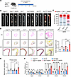 Figure 5
GLS1 deficiency in VSMCs aggravates AD in BAPN-treated mice. (A) Five-week-old male TaglnCre/+, Gls1fl/fl , and Gls1SMKO mice were given BAPN (0.5 g/kg/d) in drinking water for 4 weeks. (B) Representative morphology of aortae from TaglnCre/+, Gls1fl/fl, and Gls1SMKO mice under control (n = 5) conditions or after BAPN treatment (n = 10). Scale bars: 5 mm. (C) The incidence of aortic dissection and rupture in BAPN-treated mice (n = 10). (D) H&E and EVG staining of aorta from TaglnCre/+, Gls1fl/fl, and Gls1SMKO mice under control conditions or after BAPN-treatment, and quantifications of elastic fiber breaks. Scale bars: 500 μm (top and middle panels), 100 μm (bottom panels). (E) Quantification of maximal aortic diameters measured ex vivo in TaglnCre/+, Gls1fl/fl, and Gls1SMKO mice under control conditions or after BAPN treatment. (F) The mRNA expression of Tagln, Acta2, Opn, Mmp2, and Mmp9 in aortic tissues from TaglnCre/+, Gls1fl/fl, and Gls1SMKO mice under control conditions or after BAPN treatment was detected by real-time qPCR. Data are presented as mean ± SEM. Statistical analysis was performed using Fisher’s exact test (C), or 1-way ANOVA followed by Tukey’s multiple-comparison test (E and F). *P < 0.05, **P < 0.01, ***P < 0.001.
GLS1-mediated glutamate metabolism regulates PI3K/AKT signaling to modulate VSMC phenotypic switching. To further investigate the underlying regulatory mechanism for GLS1-related VSMC phenotypic switching, we performed RNA sequencing in HASMCs with GLS1 overexpression by adenovirus with or without PDGF-BB treatment to scan the potential genes regulated by GLS1. As shown by Venn diagram, 174 genes appeared to be differently expressed both in ad-Vector+PDGF-BB versus ad-Vector and in ad-GLS1+PDGF-BB versus ad-Vector+PDGF-BB (Figure 6A). Kyoto Encyclopedia of Genes and Genomes (KEGG) enrichment analysis revealed the 174 differentially expressed genes (DEGs) mainly involved in vascular smooth muscle contraction and PI3K/AKT signaling (Figure 6B), indicating that GLS1 may regulate VSMC phenotypic switching via the PI3K/AKT pathway.
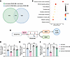 Figure 6
GLS1 regulates glutamine metabolism during VSMC phenotypic switching. (A) RNA sequencing was performed on HASMCs infected with adenovirus containing empty vector (ad-Vector) or GLS1-encoding plasmids (ad-GLS1) and then treated with PDGF-BB. Venn diagram showing the overlap DEGs between ad-Vector+PDGF-BB versus ad-Vector and ad-GLS1+PDGF-BB versus ad-Vector+PDGF-BB (n = 3). (B) KEGG enrichment analysis of these overlap DEGs. (C) Schematic diagram of GLS1-related metabolic pathway for glutamine. (D–F) HASMCs were infected with adenovirus containing empty vector (ad-Vector) or GLS1-encoding plasmids (ad-GLS1), and then treated with PDGF-BB. (D) The content of glutamate in HASMCs. (E) The content of glutathione (GSH) in HASMCs. (F) The content of α-ketoglutarate (αKG) in HASMCs. Data are presented as mean ± SEM. Statistical analysis was performed using 1-way ANOVA followed by Tukey’s multiple-comparison test (D–F). *P < 0.05, **P < 0.01, ***P < 0.001.
GLS1 plays an essential role by catalyzing the hydrolysis of glutamine to glutamate, a central metabolic intermediate with multifaceted functions (18). Glutamate serves as a precursor for α-ketoglutarate, which enters the TCA cycle to support mitochondrial ATP generation; and for glutathione synthesis, a critical antioxidant defense mechanism against oxidative stress (19, 20) (Figure 6C). Notably, mitochondrial dysfunction and disrupted glutathione homeostasis synergistically increase reactive oxygen species (ROS) accumulation. Elevated ROS levels can subsequently activate the PI3K/AKT signaling pathway (21). Therefore, we hypothesized that GLS1 may participate in the metabolism of glutamate to regulate PI3K/AKT signaling, and thereby modulate VSMC phenotypic switching. For verification, we assessed the cellular glutamate content and found that the glutamate level in HASMCs was reduced after PDGF-BB stimulation, which was increased by GLS1 overexpression (Figure 6D). Meanwhile, the levels of glutamate, glutathione, and α-ketoglutarate were suppressed upon PDGF-BB incubation, while overexpression of GLS1 restored the levels of glutathione and α-ketoglutarate to baseline values (Figure 6, E and F). Moreover, mitochondrial stress test showed that ad-GLS1 transfection effectively improved PDGF-BB–impaired oxidative phosphorylation in VSMCs, protecting against mitochondrial dysfunction (Figure 7A). MitoSOX staining revealed that GLS1 overexpression mitigated the mitochondrial oxidative stress induced by PDGF-BB (Figure 7B). In parallel, the PDGF-BB–induced phosphorylation of PI3K, AKT, and mTOR was significantly attenuated by GLS1 overexpression (Figure 7C). These findings collectively demonstrated that GLS1 promotes the glutamate-derived glutathione and α-ketoglutarate contents to suppress ROS-related activation of the PI3K/AKT pathway, thereby improving VSMC phenotypic switching under pathological conditions.
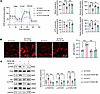 Figure 7
GLS1 modulates mitochondrial function and PI3K/AKT/mTOR signaling during VSMC phenotypic switching. (A–C) HASMCs were infected with adenovirus containing empty vector (ad-Vector) or GLS1-encoding plasmids (ad-GLS1), and then treated with PDGF-BB. (A) Summarized oxygen consumption rate (OCR) tracings in HASMCs. Basal OCR was measured, followed by sequential injections of 1 μM oligomycin, 1.5 μM FCCP, and a mixture of 2 μM rotenone and 2 μM antimycin A. (B) Mitochondrial reactive oxygen species were detected by MitoSOX staining in HASMCs. Scale bar: 50 μm. (C) Western blot analysis of phosphorylated and total PI3K, AKT, and mTOR levels in HASMCs. Data are presented as mean ± SEM. Statistical analysis was performed using 1-way ANOVA followed by Tukey’s multiple-comparison test (B and C). *P < 0.05, **P < 0.01, ***P < 0.001.
The expression of GLS1 is regulated by RARα. To further unravel the regulatory mechanism of GLS1 downregulation in the context of VSMC phenotypic switching, we integrated the prediction of GLS1 promoter–binding transcription factors with the AnimalTFDB database and the DEGs from transcriptomic data of AD patients (GSE52093 and GSE98770). Retinoic acid receptor-α (RARα) was identified as a transcriptional regulator of GLS1 (Figure 8A). We found that RARα expression was enhanced both in aortas from AD patients (Figure 8B) and in PDGF-BB–stimulated HASMCs (Supplemental Figure 5A). Next, we assessed the transcriptional effect of RARα on GLS1 by constructing luciferase reporter gene plasmids with mutation of the 3 potential binding sites of RARα from the JASPAR database (Figure 8C). The result of luciferase reporter gene assay revealed that mutation of transcription factor binding site 2 (TFBS2) effectively reversed the transcriptional inhibition of GLS1 that resulted from RARα overexpression in both HEK293 cells and HASMCs (Figure 8D and Supplemental Figure 6A), suggesting that RARα should bind to the –1,770 to –1,754 bp of the GLS1 promoter region to inhibit its transcription. Furthermore, the binding of RARα to the GLS1 promoter was significantly enhanced upon PDGF-BB stimulation in HASMCs (Supplemental Figure 6B). Then, we used siRNA to knock down RARα to evaluate the effect of RARα on VSMC phenotypic switching (Supplemental Figure 7A). The quantitative PCR (qPCR) results showed that inhibition of RARα by siRNA could promote the expression of GLS1 in HASMCs, while the decreased ACTA2 and TAGLN and increased OPN mRNA levels were reversed by RARα knockdown in PDGF-BB–treated HASMCs (Figure 8E). Meanwhile, Western blot analysis yielded consistent results (Supplemental Figure 7B). Also, MMP activities induced by PDGF-BB were inhibited by RARα knockdown (Figure 8F). These data suggested that RARα inhibition could improve VSMC phenotypic switching through increasing GLS1 expression.
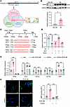 Figure 8
Transcription of GLS1 is regulated by the transcription factor RARα. (A) Prediction of GLS1 promoter–binding transcription factors by AnimalTFDB and using the region from 2,000 bp upstream to 100 bp downstream of the GLS1 transcription start site (TSS) as the promoter region. Venn diagram of DEGs in human aortic dissection samples relative to normal controls from the NCBI GEO database (GSE52093 and GSE98770). (B) Western blot analysis of RARα expression in the aorta from non-AD groups and AD patients. (C) The construction of wild-type (WT) and 3 mutated luciferase reporter gene plasmids for GLS1 promoter by mutation of 3 potential binding sites of RARα predicted by JASPAR. (D) Relative luciferase activity in HEK293 cells of luciferase reporter constructs containing GLS1 promoter or its mutants transfected along with pRL-TK (internal control plasmid) followed by transfection with RARα-encoding plasmid. (E and F) HASMCs were transfected with siRNA against RARα (siRARα) or negative control (siNC), and then treated with PDGF-BB. (E) The mRNA expression of RARα, GLS1, TAGLN, ACTA2, and OPN in HASMCs was detected by real-time qPCR. (F) Immunofluorescence images of in situ zymography (DQ gelatin) in HASMCs. MMP activity (green) was quantified by immunofluorescence intensity. Scale bars: 50 μm. (Blue: DAPI.) Data are presented as mean ± SEM. Statistical analysis was performed using unpaired, 2-tailed Student’s t test (B), or 1-way ANOVA followed by Tukey’s multiple-comparison test (D–F). *P < 0.05, ***P < 0.001.
The RARα inhibitor AR7 protects against BAPN-induced AD. We next evaluated whether pharmacological targeting of the RARα/GLS1 axis could attenuate AD. Given the lack of available GLS1 agonists, we used AR7, a RARα inhibitor, to evaluate whether pharmacological blockade of RARα exhibits a protective role in AD. Three-week-old mice were fed with BAPN water and injected intraperitoneally with AR7 (10 mg/kg/d) for 4 weeks. We found that AR7 treatment mitigated the AD lesion (Figure 9A) and lowered the incidence of aortic dissection and rupture in mice fed with BAPN (Figure 9B). H&E and EVG staining showed that AR7 administration reduced aortic dilatation and improved media degeneration in the BAPN-induced mouse AD model (Figure 9C). AR7 also mitigated the maximal thoracic aorta diameter induced by BAPN (Figure 9D). The mRNA levels of VSMC phenotypic switching–related genes showed that AR7 treatment could reverse the reduced expression of Tagln and Acta2 and enhanced expression of Opn in aorta from BAPN-fed mice (Figure 9E). Taken together, these data demonstrated that RARα inhibition via AR7 may represent a potential therapeutic strategy for AD.
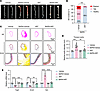 Figure 9
The RARα inhibitor AR7 attenuates AD in BAPN-treated mice. (A–E) Three-week-old male C57BL/6 mice were treated with BAPN in drinking water (1 g/kg/d) with or without intraperitoneal injection of AR7 (10 mg/kg/d) for 4 weeks. (A) Representative morphology of aortae from mice in the control group (n = 5) and the BAPN-treated group (n = 10). Scale bars: 5 mm. (B) The incidence of aortic dissection and rupture in BAPN-treated mice (n = 10). (C) H&E and EVG staining of aorta from control or BAPN-treated mice. Scale bars: 500 μm (top and middle panels), 100 μm (bottom panels). (D) Quantification of maximal diameters measured ex vivo of aorta from control and BAPN-treated mice. (E) The mRNA expression of Tagln, Acta2, and Opn in aorta from control and BAPN-treated mice was detected by real-time qPCR. Data are presented as mean ± SEM. Statistical analysis was performed using Fisher’s exact test (B), or 1-way ANOVA followed by Tukey’s multiple-comparison test (D and E). **P < 0.01, ***P < 0.001, ****P < 0.0001.


