
Research ArticleHematologyMetabolism
Open Access |  10.1172/jci.insight.203777
Direct pharmacological targeting of asparagine synthetase to overcome resistance to L-asparaginase in ALL therapy
Rodney Claude,1,2,3 Sankalp Srivastava,1,3 Kirk A. Staschke,2,3 Carlos Mellado-Fritz,4 Shaoxiong Chen,5 Lei Liu,1 Minghua Zhong,1 Harish Kothandaraman,6 Nadia A. Lanman,6,7 Utpal Davé,8 Sandeep Batra,9 Jiehao Zhou,10 Yue Fang,11 Chi Zhang,11 Reuben Kapur,1,3 Jing Fan,4 Ronald C. Wek,2,3 and Ji Zhang1,2,3
1Herman B. Wells Center for Pediatric Research,
2Department of Biochemistry, Molecular Biology, and Pharmacology, and
3Melvin and Bren Simon Comprehensive Cancer Center, Indiana University School of Medicine, Indianapolis, Indiana, USA.
4Morgridge Institute for Research & Department of Medical Microbiology and Immunology, University of Wisconsin-Madison, Madison, Wisconsin, USA.
5Department of Pathology, Indiana University School of Medicine, Indianapolis, Indiana, USA.
6Purdue University Institute for Cancer Research, and
7Department of Comparative Pathobiology, Purdue University, West Lafayette, Indiana, USA.
8Department of Medicine, Indiana University School of Medicine, Indianapolis, Indiana, USA.
9Riley Hospital for Children at Indiana University Health, Indianapolis, Indiana, USA.
10Department of Laboratory Medicine and Pathology, Mayo Clinic Arizona, Phoenix, Arizona, USA.
11Department of Medical and Molecular Genetics, Indiana University School of Medicine, Indianapolis, Indiana, USA.
Address correspondence to: Ji Zhang, Herman B. Wells Center for Pediatric Research, Indiana University School of Medicine, 1044 W Walnut Street, Room-170, Indianapolis, Indiana 46202, USA. Phone: 317.274.2134; Email: jzh1@iu.edu.
Authorship note: RC and SS contributed equally to this work.
Find articles by Claude, R. in: PubMed | Google Scholar
1Herman B. Wells Center for Pediatric Research,
2Department of Biochemistry, Molecular Biology, and Pharmacology, and
3Melvin and Bren Simon Comprehensive Cancer Center, Indiana University School of Medicine, Indianapolis, Indiana, USA.
4Morgridge Institute for Research & Department of Medical Microbiology and Immunology, University of Wisconsin-Madison, Madison, Wisconsin, USA.
5Department of Pathology, Indiana University School of Medicine, Indianapolis, Indiana, USA.
6Purdue University Institute for Cancer Research, and
7Department of Comparative Pathobiology, Purdue University, West Lafayette, Indiana, USA.
8Department of Medicine, Indiana University School of Medicine, Indianapolis, Indiana, USA.
9Riley Hospital for Children at Indiana University Health, Indianapolis, Indiana, USA.
10Department of Laboratory Medicine and Pathology, Mayo Clinic Arizona, Phoenix, Arizona, USA.
11Department of Medical and Molecular Genetics, Indiana University School of Medicine, Indianapolis, Indiana, USA.
Address correspondence to: Ji Zhang, Herman B. Wells Center for Pediatric Research, Indiana University School of Medicine, 1044 W Walnut Street, Room-170, Indianapolis, Indiana 46202, USA. Phone: 317.274.2134; Email: jzh1@iu.edu.
Authorship note: RC and SS contributed equally to this work.
Find articles by Srivastava, S. in: PubMed | Google Scholar
1Herman B. Wells Center for Pediatric Research,
2Department of Biochemistry, Molecular Biology, and Pharmacology, and
3Melvin and Bren Simon Comprehensive Cancer Center, Indiana University School of Medicine, Indianapolis, Indiana, USA.
4Morgridge Institute for Research & Department of Medical Microbiology and Immunology, University of Wisconsin-Madison, Madison, Wisconsin, USA.
5Department of Pathology, Indiana University School of Medicine, Indianapolis, Indiana, USA.
6Purdue University Institute for Cancer Research, and
7Department of Comparative Pathobiology, Purdue University, West Lafayette, Indiana, USA.
8Department of Medicine, Indiana University School of Medicine, Indianapolis, Indiana, USA.
9Riley Hospital for Children at Indiana University Health, Indianapolis, Indiana, USA.
10Department of Laboratory Medicine and Pathology, Mayo Clinic Arizona, Phoenix, Arizona, USA.
11Department of Medical and Molecular Genetics, Indiana University School of Medicine, Indianapolis, Indiana, USA.
Address correspondence to: Ji Zhang, Herman B. Wells Center for Pediatric Research, Indiana University School of Medicine, 1044 W Walnut Street, Room-170, Indianapolis, Indiana 46202, USA. Phone: 317.274.2134; Email: jzh1@iu.edu.
Authorship note: RC and SS contributed equally to this work.
Find articles by Staschke, K. in: PubMed | Google Scholar
1Herman B. Wells Center for Pediatric Research,
2Department of Biochemistry, Molecular Biology, and Pharmacology, and
3Melvin and Bren Simon Comprehensive Cancer Center, Indiana University School of Medicine, Indianapolis, Indiana, USA.
4Morgridge Institute for Research & Department of Medical Microbiology and Immunology, University of Wisconsin-Madison, Madison, Wisconsin, USA.
5Department of Pathology, Indiana University School of Medicine, Indianapolis, Indiana, USA.
6Purdue University Institute for Cancer Research, and
7Department of Comparative Pathobiology, Purdue University, West Lafayette, Indiana, USA.
8Department of Medicine, Indiana University School of Medicine, Indianapolis, Indiana, USA.
9Riley Hospital for Children at Indiana University Health, Indianapolis, Indiana, USA.
10Department of Laboratory Medicine and Pathology, Mayo Clinic Arizona, Phoenix, Arizona, USA.
11Department of Medical and Molecular Genetics, Indiana University School of Medicine, Indianapolis, Indiana, USA.
Address correspondence to: Ji Zhang, Herman B. Wells Center for Pediatric Research, Indiana University School of Medicine, 1044 W Walnut Street, Room-170, Indianapolis, Indiana 46202, USA. Phone: 317.274.2134; Email: jzh1@iu.edu.
Authorship note: RC and SS contributed equally to this work.
Find articles by Mellado-Fritz, C. in: PubMed | Google Scholar
1Herman B. Wells Center for Pediatric Research,
2Department of Biochemistry, Molecular Biology, and Pharmacology, and
3Melvin and Bren Simon Comprehensive Cancer Center, Indiana University School of Medicine, Indianapolis, Indiana, USA.
4Morgridge Institute for Research & Department of Medical Microbiology and Immunology, University of Wisconsin-Madison, Madison, Wisconsin, USA.
5Department of Pathology, Indiana University School of Medicine, Indianapolis, Indiana, USA.
6Purdue University Institute for Cancer Research, and
7Department of Comparative Pathobiology, Purdue University, West Lafayette, Indiana, USA.
8Department of Medicine, Indiana University School of Medicine, Indianapolis, Indiana, USA.
9Riley Hospital for Children at Indiana University Health, Indianapolis, Indiana, USA.
10Department of Laboratory Medicine and Pathology, Mayo Clinic Arizona, Phoenix, Arizona, USA.
11Department of Medical and Molecular Genetics, Indiana University School of Medicine, Indianapolis, Indiana, USA.
Address correspondence to: Ji Zhang, Herman B. Wells Center for Pediatric Research, Indiana University School of Medicine, 1044 W Walnut Street, Room-170, Indianapolis, Indiana 46202, USA. Phone: 317.274.2134; Email: jzh1@iu.edu.
Authorship note: RC and SS contributed equally to this work.
Find articles by Chen, S. in: PubMed | Google Scholar
1Herman B. Wells Center for Pediatric Research,
2Department of Biochemistry, Molecular Biology, and Pharmacology, and
3Melvin and Bren Simon Comprehensive Cancer Center, Indiana University School of Medicine, Indianapolis, Indiana, USA.
4Morgridge Institute for Research & Department of Medical Microbiology and Immunology, University of Wisconsin-Madison, Madison, Wisconsin, USA.
5Department of Pathology, Indiana University School of Medicine, Indianapolis, Indiana, USA.
6Purdue University Institute for Cancer Research, and
7Department of Comparative Pathobiology, Purdue University, West Lafayette, Indiana, USA.
8Department of Medicine, Indiana University School of Medicine, Indianapolis, Indiana, USA.
9Riley Hospital for Children at Indiana University Health, Indianapolis, Indiana, USA.
10Department of Laboratory Medicine and Pathology, Mayo Clinic Arizona, Phoenix, Arizona, USA.
11Department of Medical and Molecular Genetics, Indiana University School of Medicine, Indianapolis, Indiana, USA.
Address correspondence to: Ji Zhang, Herman B. Wells Center for Pediatric Research, Indiana University School of Medicine, 1044 W Walnut Street, Room-170, Indianapolis, Indiana 46202, USA. Phone: 317.274.2134; Email: jzh1@iu.edu.
Authorship note: RC and SS contributed equally to this work.
Find articles by Liu, L. in: PubMed | Google Scholar
1Herman B. Wells Center for Pediatric Research,
2Department of Biochemistry, Molecular Biology, and Pharmacology, and
3Melvin and Bren Simon Comprehensive Cancer Center, Indiana University School of Medicine, Indianapolis, Indiana, USA.
4Morgridge Institute for Research & Department of Medical Microbiology and Immunology, University of Wisconsin-Madison, Madison, Wisconsin, USA.
5Department of Pathology, Indiana University School of Medicine, Indianapolis, Indiana, USA.
6Purdue University Institute for Cancer Research, and
7Department of Comparative Pathobiology, Purdue University, West Lafayette, Indiana, USA.
8Department of Medicine, Indiana University School of Medicine, Indianapolis, Indiana, USA.
9Riley Hospital for Children at Indiana University Health, Indianapolis, Indiana, USA.
10Department of Laboratory Medicine and Pathology, Mayo Clinic Arizona, Phoenix, Arizona, USA.
11Department of Medical and Molecular Genetics, Indiana University School of Medicine, Indianapolis, Indiana, USA.
Address correspondence to: Ji Zhang, Herman B. Wells Center for Pediatric Research, Indiana University School of Medicine, 1044 W Walnut Street, Room-170, Indianapolis, Indiana 46202, USA. Phone: 317.274.2134; Email: jzh1@iu.edu.
Authorship note: RC and SS contributed equally to this work.
Find articles by Zhong, M. in: PubMed | Google Scholar
1Herman B. Wells Center for Pediatric Research,
2Department of Biochemistry, Molecular Biology, and Pharmacology, and
3Melvin and Bren Simon Comprehensive Cancer Center, Indiana University School of Medicine, Indianapolis, Indiana, USA.
4Morgridge Institute for Research & Department of Medical Microbiology and Immunology, University of Wisconsin-Madison, Madison, Wisconsin, USA.
5Department of Pathology, Indiana University School of Medicine, Indianapolis, Indiana, USA.
6Purdue University Institute for Cancer Research, and
7Department of Comparative Pathobiology, Purdue University, West Lafayette, Indiana, USA.
8Department of Medicine, Indiana University School of Medicine, Indianapolis, Indiana, USA.
9Riley Hospital for Children at Indiana University Health, Indianapolis, Indiana, USA.
10Department of Laboratory Medicine and Pathology, Mayo Clinic Arizona, Phoenix, Arizona, USA.
11Department of Medical and Molecular Genetics, Indiana University School of Medicine, Indianapolis, Indiana, USA.
Address correspondence to: Ji Zhang, Herman B. Wells Center for Pediatric Research, Indiana University School of Medicine, 1044 W Walnut Street, Room-170, Indianapolis, Indiana 46202, USA. Phone: 317.274.2134; Email: jzh1@iu.edu.
Authorship note: RC and SS contributed equally to this work.
Find articles by Kothandaraman, H. in: PubMed | Google Scholar
1Herman B. Wells Center for Pediatric Research,
2Department of Biochemistry, Molecular Biology, and Pharmacology, and
3Melvin and Bren Simon Comprehensive Cancer Center, Indiana University School of Medicine, Indianapolis, Indiana, USA.
4Morgridge Institute for Research & Department of Medical Microbiology and Immunology, University of Wisconsin-Madison, Madison, Wisconsin, USA.
5Department of Pathology, Indiana University School of Medicine, Indianapolis, Indiana, USA.
6Purdue University Institute for Cancer Research, and
7Department of Comparative Pathobiology, Purdue University, West Lafayette, Indiana, USA.
8Department of Medicine, Indiana University School of Medicine, Indianapolis, Indiana, USA.
9Riley Hospital for Children at Indiana University Health, Indianapolis, Indiana, USA.
10Department of Laboratory Medicine and Pathology, Mayo Clinic Arizona, Phoenix, Arizona, USA.
11Department of Medical and Molecular Genetics, Indiana University School of Medicine, Indianapolis, Indiana, USA.
Address correspondence to: Ji Zhang, Herman B. Wells Center for Pediatric Research, Indiana University School of Medicine, 1044 W Walnut Street, Room-170, Indianapolis, Indiana 46202, USA. Phone: 317.274.2134; Email: jzh1@iu.edu.
Authorship note: RC and SS contributed equally to this work.
Find articles by
Lanman, N.
in:
PubMed
|
Google Scholar
|

1Herman B. Wells Center for Pediatric Research,
2Department of Biochemistry, Molecular Biology, and Pharmacology, and
3Melvin and Bren Simon Comprehensive Cancer Center, Indiana University School of Medicine, Indianapolis, Indiana, USA.
4Morgridge Institute for Research & Department of Medical Microbiology and Immunology, University of Wisconsin-Madison, Madison, Wisconsin, USA.
5Department of Pathology, Indiana University School of Medicine, Indianapolis, Indiana, USA.
6Purdue University Institute for Cancer Research, and
7Department of Comparative Pathobiology, Purdue University, West Lafayette, Indiana, USA.
8Department of Medicine, Indiana University School of Medicine, Indianapolis, Indiana, USA.
9Riley Hospital for Children at Indiana University Health, Indianapolis, Indiana, USA.
10Department of Laboratory Medicine and Pathology, Mayo Clinic Arizona, Phoenix, Arizona, USA.
11Department of Medical and Molecular Genetics, Indiana University School of Medicine, Indianapolis, Indiana, USA.
Address correspondence to: Ji Zhang, Herman B. Wells Center for Pediatric Research, Indiana University School of Medicine, 1044 W Walnut Street, Room-170, Indianapolis, Indiana 46202, USA. Phone: 317.274.2134; Email: jzh1@iu.edu.
Authorship note: RC and SS contributed equally to this work.
Find articles by Davé, U. in: PubMed | Google Scholar
1Herman B. Wells Center for Pediatric Research,
2Department of Biochemistry, Molecular Biology, and Pharmacology, and
3Melvin and Bren Simon Comprehensive Cancer Center, Indiana University School of Medicine, Indianapolis, Indiana, USA.
4Morgridge Institute for Research & Department of Medical Microbiology and Immunology, University of Wisconsin-Madison, Madison, Wisconsin, USA.
5Department of Pathology, Indiana University School of Medicine, Indianapolis, Indiana, USA.
6Purdue University Institute for Cancer Research, and
7Department of Comparative Pathobiology, Purdue University, West Lafayette, Indiana, USA.
8Department of Medicine, Indiana University School of Medicine, Indianapolis, Indiana, USA.
9Riley Hospital for Children at Indiana University Health, Indianapolis, Indiana, USA.
10Department of Laboratory Medicine and Pathology, Mayo Clinic Arizona, Phoenix, Arizona, USA.
11Department of Medical and Molecular Genetics, Indiana University School of Medicine, Indianapolis, Indiana, USA.
Address correspondence to: Ji Zhang, Herman B. Wells Center for Pediatric Research, Indiana University School of Medicine, 1044 W Walnut Street, Room-170, Indianapolis, Indiana 46202, USA. Phone: 317.274.2134; Email: jzh1@iu.edu.
Authorship note: RC and SS contributed equally to this work.
Find articles by Batra, S. in: PubMed | Google Scholar
1Herman B. Wells Center for Pediatric Research,
2Department of Biochemistry, Molecular Biology, and Pharmacology, and
3Melvin and Bren Simon Comprehensive Cancer Center, Indiana University School of Medicine, Indianapolis, Indiana, USA.
4Morgridge Institute for Research & Department of Medical Microbiology and Immunology, University of Wisconsin-Madison, Madison, Wisconsin, USA.
5Department of Pathology, Indiana University School of Medicine, Indianapolis, Indiana, USA.
6Purdue University Institute for Cancer Research, and
7Department of Comparative Pathobiology, Purdue University, West Lafayette, Indiana, USA.
8Department of Medicine, Indiana University School of Medicine, Indianapolis, Indiana, USA.
9Riley Hospital for Children at Indiana University Health, Indianapolis, Indiana, USA.
10Department of Laboratory Medicine and Pathology, Mayo Clinic Arizona, Phoenix, Arizona, USA.
11Department of Medical and Molecular Genetics, Indiana University School of Medicine, Indianapolis, Indiana, USA.
Address correspondence to: Ji Zhang, Herman B. Wells Center for Pediatric Research, Indiana University School of Medicine, 1044 W Walnut Street, Room-170, Indianapolis, Indiana 46202, USA. Phone: 317.274.2134; Email: jzh1@iu.edu.
Authorship note: RC and SS contributed equally to this work.
Find articles by Zhou, J. in: PubMed | Google Scholar
1Herman B. Wells Center for Pediatric Research,
2Department of Biochemistry, Molecular Biology, and Pharmacology, and
3Melvin and Bren Simon Comprehensive Cancer Center, Indiana University School of Medicine, Indianapolis, Indiana, USA.
4Morgridge Institute for Research & Department of Medical Microbiology and Immunology, University of Wisconsin-Madison, Madison, Wisconsin, USA.
5Department of Pathology, Indiana University School of Medicine, Indianapolis, Indiana, USA.
6Purdue University Institute for Cancer Research, and
7Department of Comparative Pathobiology, Purdue University, West Lafayette, Indiana, USA.
8Department of Medicine, Indiana University School of Medicine, Indianapolis, Indiana, USA.
9Riley Hospital for Children at Indiana University Health, Indianapolis, Indiana, USA.
10Department of Laboratory Medicine and Pathology, Mayo Clinic Arizona, Phoenix, Arizona, USA.
11Department of Medical and Molecular Genetics, Indiana University School of Medicine, Indianapolis, Indiana, USA.
Address correspondence to: Ji Zhang, Herman B. Wells Center for Pediatric Research, Indiana University School of Medicine, 1044 W Walnut Street, Room-170, Indianapolis, Indiana 46202, USA. Phone: 317.274.2134; Email: jzh1@iu.edu.
Authorship note: RC and SS contributed equally to this work.
Find articles by Fang, Y. in: PubMed | Google Scholar
1Herman B. Wells Center for Pediatric Research,
2Department of Biochemistry, Molecular Biology, and Pharmacology, and
3Melvin and Bren Simon Comprehensive Cancer Center, Indiana University School of Medicine, Indianapolis, Indiana, USA.
4Morgridge Institute for Research & Department of Medical Microbiology and Immunology, University of Wisconsin-Madison, Madison, Wisconsin, USA.
5Department of Pathology, Indiana University School of Medicine, Indianapolis, Indiana, USA.
6Purdue University Institute for Cancer Research, and
7Department of Comparative Pathobiology, Purdue University, West Lafayette, Indiana, USA.
8Department of Medicine, Indiana University School of Medicine, Indianapolis, Indiana, USA.
9Riley Hospital for Children at Indiana University Health, Indianapolis, Indiana, USA.
10Department of Laboratory Medicine and Pathology, Mayo Clinic Arizona, Phoenix, Arizona, USA.
11Department of Medical and Molecular Genetics, Indiana University School of Medicine, Indianapolis, Indiana, USA.
Address correspondence to: Ji Zhang, Herman B. Wells Center for Pediatric Research, Indiana University School of Medicine, 1044 W Walnut Street, Room-170, Indianapolis, Indiana 46202, USA. Phone: 317.274.2134; Email: jzh1@iu.edu.
Authorship note: RC and SS contributed equally to this work.
Find articles by
Zhang, C.
in:
PubMed
|
Google Scholar
|
1Herman B. Wells Center for Pediatric Research,
2Department of Biochemistry, Molecular Biology, and Pharmacology, and
3Melvin and Bren Simon Comprehensive Cancer Center, Indiana University School of Medicine, Indianapolis, Indiana, USA.
4Morgridge Institute for Research & Department of Medical Microbiology and Immunology, University of Wisconsin-Madison, Madison, Wisconsin, USA.
5Department of Pathology, Indiana University School of Medicine, Indianapolis, Indiana, USA.
6Purdue University Institute for Cancer Research, and
7Department of Comparative Pathobiology, Purdue University, West Lafayette, Indiana, USA.
8Department of Medicine, Indiana University School of Medicine, Indianapolis, Indiana, USA.
9Riley Hospital for Children at Indiana University Health, Indianapolis, Indiana, USA.
10Department of Laboratory Medicine and Pathology, Mayo Clinic Arizona, Phoenix, Arizona, USA.
11Department of Medical and Molecular Genetics, Indiana University School of Medicine, Indianapolis, Indiana, USA.
Address correspondence to: Ji Zhang, Herman B. Wells Center for Pediatric Research, Indiana University School of Medicine, 1044 W Walnut Street, Room-170, Indianapolis, Indiana 46202, USA. Phone: 317.274.2134; Email: jzh1@iu.edu.
Authorship note: RC and SS contributed equally to this work.
Find articles by Kapur, R. in: PubMed | Google Scholar
1Herman B. Wells Center for Pediatric Research,
2Department of Biochemistry, Molecular Biology, and Pharmacology, and
3Melvin and Bren Simon Comprehensive Cancer Center, Indiana University School of Medicine, Indianapolis, Indiana, USA.
4Morgridge Institute for Research & Department of Medical Microbiology and Immunology, University of Wisconsin-Madison, Madison, Wisconsin, USA.
5Department of Pathology, Indiana University School of Medicine, Indianapolis, Indiana, USA.
6Purdue University Institute for Cancer Research, and
7Department of Comparative Pathobiology, Purdue University, West Lafayette, Indiana, USA.
8Department of Medicine, Indiana University School of Medicine, Indianapolis, Indiana, USA.
9Riley Hospital for Children at Indiana University Health, Indianapolis, Indiana, USA.
10Department of Laboratory Medicine and Pathology, Mayo Clinic Arizona, Phoenix, Arizona, USA.
11Department of Medical and Molecular Genetics, Indiana University School of Medicine, Indianapolis, Indiana, USA.
Address correspondence to: Ji Zhang, Herman B. Wells Center for Pediatric Research, Indiana University School of Medicine, 1044 W Walnut Street, Room-170, Indianapolis, Indiana 46202, USA. Phone: 317.274.2134; Email: jzh1@iu.edu.
Authorship note: RC and SS contributed equally to this work.
Find articles by Fan, J. in: PubMed | Google Scholar
1Herman B. Wells Center for Pediatric Research,
2Department of Biochemistry, Molecular Biology, and Pharmacology, and
3Melvin and Bren Simon Comprehensive Cancer Center, Indiana University School of Medicine, Indianapolis, Indiana, USA.
4Morgridge Institute for Research & Department of Medical Microbiology and Immunology, University of Wisconsin-Madison, Madison, Wisconsin, USA.
5Department of Pathology, Indiana University School of Medicine, Indianapolis, Indiana, USA.
6Purdue University Institute for Cancer Research, and
7Department of Comparative Pathobiology, Purdue University, West Lafayette, Indiana, USA.
8Department of Medicine, Indiana University School of Medicine, Indianapolis, Indiana, USA.
9Riley Hospital for Children at Indiana University Health, Indianapolis, Indiana, USA.
10Department of Laboratory Medicine and Pathology, Mayo Clinic Arizona, Phoenix, Arizona, USA.
11Department of Medical and Molecular Genetics, Indiana University School of Medicine, Indianapolis, Indiana, USA.
Address correspondence to: Ji Zhang, Herman B. Wells Center for Pediatric Research, Indiana University School of Medicine, 1044 W Walnut Street, Room-170, Indianapolis, Indiana 46202, USA. Phone: 317.274.2134; Email: jzh1@iu.edu.
Authorship note: RC and SS contributed equally to this work.
Find articles by Wek, R. in: PubMed | Google Scholar
1Herman B. Wells Center for Pediatric Research,
2Department of Biochemistry, Molecular Biology, and Pharmacology, and
3Melvin and Bren Simon Comprehensive Cancer Center, Indiana University School of Medicine, Indianapolis, Indiana, USA.
4Morgridge Institute for Research & Department of Medical Microbiology and Immunology, University of Wisconsin-Madison, Madison, Wisconsin, USA.
5Department of Pathology, Indiana University School of Medicine, Indianapolis, Indiana, USA.
6Purdue University Institute for Cancer Research, and
7Department of Comparative Pathobiology, Purdue University, West Lafayette, Indiana, USA.
8Department of Medicine, Indiana University School of Medicine, Indianapolis, Indiana, USA.
9Riley Hospital for Children at Indiana University Health, Indianapolis, Indiana, USA.
10Department of Laboratory Medicine and Pathology, Mayo Clinic Arizona, Phoenix, Arizona, USA.
11Department of Medical and Molecular Genetics, Indiana University School of Medicine, Indianapolis, Indiana, USA.
Address correspondence to: Ji Zhang, Herman B. Wells Center for Pediatric Research, Indiana University School of Medicine, 1044 W Walnut Street, Room-170, Indianapolis, Indiana 46202, USA. Phone: 317.274.2134; Email: jzh1@iu.edu.
Authorship note: RC and SS contributed equally to this work.
Find articles by
Zhang, J.
in:
PubMed
|
Google Scholar
|
Published June 4, 2026 - More info
JCI Insight. 2026;11(14):e203777. https://doi.org/10.1172/jci.insight.203777.
© 2026 Claude et al. This work is licensed under the Creative Commons Attribution 4.0 International License. To view a copy of this license, visit http://creativecommons.org/licenses/by/4.0/.
Received: December 19, 2025; Accepted: May 28, 2026


-
Results
Genetic inactivation of GCN2 alone does not delay leukemia onset in a KrasG12D-driven mouse T-ALL model. Previous studies indicate that GCN2 is activated under asparagine-depleted conditions to alleviate amino acid stress through ATF4-dependent induction of ASNS (6, 8). To address its role in ALL, we employed a mouse T-ALL model driven by KrasG12D activated by a T cell–specific Cre recombinase (Lck-Cre) (Figure 1A) (13, 14). Mice expressing Lck-Cre developed aggressive T-ALL within 16 weeks with 100% penetrance, characterized by enlarged thymus and spleen (Figure 1B), and increased CD4+CD8+ double-positive cells in the peripheral blood (PBL) and spleen (Figure 1C). Immunohistochemical (IHC) staining for p-GCN2 (T899), a marker of GCN2 activation, revealed regional positivity in the thymus, suggesting a potential role for GCN2 in leukemogenesis (Figure 1D). In addition, leukemic thymus tissues exhibited elevated expression of oncogenic molecules NOTCH-1 and c-MYC (Figure 1E), both critical for T-ALL pathogenesis (15). Furthermore, we also observed increased phosphorylation of eIF2α (S51), and upregulation of ATF4 and ASNS in leukemic compared with normal thymus (Figure 1E), implicating activation of an upstream eIF2α kinase. Among the candidate kinases, PERK and GCN2 are known to contribute to amino acid stress adaptation in MYC-driven cancers (16). While PERK primarily responds to endoplasmic reticulum (ER) stress (7), its phosphorylation at Thr982 — a marker of its activation — was minimally detected in leukemic thymus tissues (Supplemental Figure 1A; supplemental material available online with this article; https://doi.org/10.1172/jci.insight.203777DS1), suggesting PERK is not a major driver of eIF2α phosphorylation in this model. These results prompted us to further evaluate the role of GCN2 in T-ALL initiation and progression.
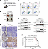 Figure 1
GCN2 is not required for leukemogenesis. (A) Schematic diagram of T-ALL mice that were established by a KrasG12D mutation knocked into the endogenous locus of the Kras gene. The loxP-flanked STOP codon was deleted only when Cre was expressed under the T cell–specific Lck promoter. (B) Representative images of thymus and spleen, collected at 16 weeks from KrasG12D mice with or without Cre. (C) CD4+/CD8+ cell profiles were assessed by flow cytometry analysis in spleen, peripheral blood (PBL), and thymus of KrasG12D mice with or without Cre. (D) IHC staining of p-GCN2 (T899) in thymus from KrasG12D mice with or without Cre. Images from 2 representative mice of each genotype are shown. (E) Western blot analysis was used to measure c-MYC, NOTCH-1, p-eIF2α (S51), ATF4, and ASNS protein in thymus from KrasG12D mice, with or without Cre. NOTCH-1 and ATF4 blots were run at a separate time, and protein lysates for the ATF4 blot were prepared from independent animals. (F) KrasG12D;Lck-Cre T-ALL mice were crossed with Gcn2–/– mice. Kaplan-Meier curve comparing survival between the Gcn2+/+ and Gcn2–/– mice. Statistical comparison between the 2 groups was done using the log-rank (Mantel-Cox) test.
We crossed KrasG12D;Lck-Cre mice with Gcn2–/– animals to generate GCN2-deficient T-ALL mice. Gcn2-germline-knockout mice develop normally unless fed on amino acid–restricted food, confirming its central role in amino acid sensing (17). Kaplan-Meier survival analysis revealed no significant difference in leukemia onset or overall survival between Gcn2+/+ and Gcn2–/– T-ALL mice (Figure 1F). We did not observe differences in spleen or thymus weights, nor in splenic CD4+/CD8+ cell profiles, between genotypes (Supplemental Figure 1, B and C). Western blot analysis of thymic tissues showed minimal PERK activation only in one sample, though ASNS expression trended lower in Gcn2–/– leukemias (Supplemental Figure 1D). To determine whether this effect was intrinsic to leukemia cells, we generated a secondary T-ALL model by transplanting preleukemic bone marrow from 7-week-old KrasG12D;Lck-Cre;Gcn2+/+ or Gcn2–/– mice into lethally irradiated WT recipients (Supplemental Figure 1E). At this preleukemic stage, donor bone marrow displayed a normal CD4+/CD8+ cell profile (Supplemental Figure 1F). This secondary model exhibited a shorter latency, yet no significant difference in leukemia onset was observed between genotypes (Supplemental Figure 1G). Control mice lacking the KrasG12D allele did not develop leukemia (Supplemental Figure 1G). Collectively, these data indicate that GCN2 is dispensable for leukemia initiation and progression in this KrasG12D-driven T-ALL model.
GCN2 deficiency sensitizes ALL cells to asparagine depletion. To assess the impact of GCN2 loss on asparagine depletion, we established primary T-ALL cell lines from Gcn2+/+ and Gcn2–/– leukemias. All lines proliferate normally in asparagine-containing RPMI medium (Figure 2A). However, upon treatment with pegylated L-asparaginase (PEG), Gcn2–/– cells failed to proliferate and underwent cell death, while Gcn2+/+ cells remained viable and proliferating (Figure 2, B and C). Western blotting analysis confirmed that Gcn2–/– cells failed to induce ATF4 and ASNS protein following PEG treatment (Figure 2D), and the failure of ASNS protein induction was due to mRNA expression (Figure 2E).
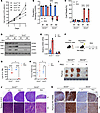 Figure 2
GCN2 deficiency sensitizes ALL cells to asparagine depletion. (A) Gcn2+/+ and Gcn2–/– murine leukemic lines were cultured in RPMI media and cell numbers were recorded over a 3-day period. (B and C) Gcn2+/+ and Gcn2–/– T-ALL cells from panel A were treated with pegylated L-asparaginase (PEG) (0.01 IU/mL) or untreated (UT) over a 3-day period. Population doublings and cell viability were measured on day 3. Statistical significance was determined using 2-way ANOVA. ****P < 0.0001. (D) Gcn2+/+ and Gcn2–/– T-ALL mouse cell lines were treated with PEG for 16 hours to collect cell lysates. Western blot was used to detect GCN2, ASNS, and ATF4 proteins. (E) RNA was isolated from representative Gcn2+/+ and Gcn2–/– mouse T-ALL cells treated with or without PEG for 16 hours. qPCR was used to measure Asns mRNA levels. (F) Schematic illustration of secondary T-ALL model. Seven-week-old preleukemic bone marrow cells from KrasG12D;Gcn2+/+ and KrasG12D;Gcn2–/– mice were transplanted into lethally irradiated WT recipients. Mice received 2 doses of PEG at 53 and 60 days after transplantation (arrows). Mice were euthanized on day 74 after transplantation. (G and H) Lymphocyte percentages in peripheral blood and thymus weights from F were recorded at the experimental endpoint (n = 3 per group). Statistical significance was determined using 2-way ANOVA (G) and multiple unpaired t test (H). *P < 0.05; ****P < 0.0001. (I) Images of representative thymus from mice in F. (J) Representative image of H&E-stained mouse spleen from F. (K) Representative IHC staining for ASNS in thymus tissues from mice in I.
To assess the impact of GCN2 deficiency in response to L-asparaginase treatment in vivo, we employed our secondary T-ALL model with Gcn2+/+ and Gcn2–/– preleukemic bone marrow transplantation. Mice received 2 doses of PEG 1 week apart, and analysis was performed 2 weeks after the second dose (Figure 2F). PEG treatment resulted in a greater reduction in peripheral lymphocyte percentage and thymus size in Gcn2–/– than Gcn2+/+ mice (Figure 2, G–I). Hematoxylin and eosin (H&E) staining revealed decreased leukemic infiltration in Gcn2–/– spleen (Figure 2J). IHC staining of ASNS showed strong induction in Gcn2+/+ but only modest induction in Gcn2–/– thymic tissues (Figure 2K). These findings demonstrate that GCN2 is required for ASNS induction during asparagine depletion, and its loss sensitizes leukemia cells to L-asparaginase both in vitro and in vivo.
A small-molecule inhibitor of GCN2 sensitizes ALL cells to asparagine depletion therapy. We next tested whether pharmacological inhibition of GCN2 phenocopied genetic deletion of GCN2. Treatment of Gcn2+/+ T-ALL lines with GCN2iB (6), a small-molecule inhibitor of GCN2, induced cell death only in combination with PEG (Figure 3A). Consistent with our prediction, GCN2iB blocked ATF4 and ASNS induction after PEG treatment, which correlated with increased PARP cleavage (Figure 3B). The suppression of ASNS protein induction by GCN2iB is attributable to inhibition of its mRNA induction (Figure 3C). In human T-ALL lines, GCN2iB similarly suppressed ASNS induction following asparagine depletion in KOPT-K1 and HPB-ALL cells, but not in Jurkat cells, which expressed high basal ASNS (Figure 3D). Correspondingly, GCN2iB suppressed proliferation in KOPT-K1 and HPB-ALL cells, but not Jurkat cells, only when asparagine was removed from the media (Figure 3E). GCN2iB inhibited eIF2α phosphorylation and ATF4 induction in KOPT-K1 and HPB-ALL cells within 4 hours of asparagine depletion (Supplemental Figure 2A), preceding ASNS induction. At 16 hours after asparagine depletion, GCN2iB suppressed both ASNS mRNA and protein expression (Supplemental Figure 2B). These results suggest that GCN2iB can effectively suppress the induction of ASNS in ALL cells expressing low levels of ASNS. Furthermore, eIF2α phosphorylation and ATF4 induction are early events of GCN2 activation, and the induction of ASNS requires a longer time, likely due to mRNA transcription and protein synthesis.
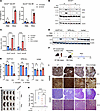 Figure 3
A small-molecule inhibitor of GCN2 sensitizes ALL cells to asparagine depletion. (A) Gcn2+/+ lines 1 and 2, from Figure 2A, were treated with or without PEG (0.01 IU/mL) in the presence or absence of GCN2iB (2.5 μM). Cell viability was recorded on day 3. Statistical significance was determined using 2-way ANOVA. ****P < 0.0001. (B and C) Gcn2+/+ lines 1 and 2 were treated as described in A for 16 hours. ASNS, ATF4, and PARP protein levels were assessed by Western blot (B), and ASNS mRNA was measured by qPCR (C). (D) Human T-ALL cell lines, KOPT-K1, HPB-ALL, and Jurkat were grown with or without exogenous asparagine in the presence or absence of GCN2iB (2.5 μM) for 24 hours. ASNS protein level was detected by Western blot. (E) KOPT-K1, HPB-ALL, and Jurkat cells were treated with GCN2iB (2.5 μM) for 3 days in the presence or absence of exogenous asparagine. Population doublings on day 3 were recorded. Statistical significance was determined using 2-way ANOVA. *P < 0.05; ****P < 0.0001. (F) Schematic illustration of a secondary KrasG12D T-ALL model. KrasG12D;Lck-Cre;Gcn2+/+ primary leukemic spleen cells were transplanted into lethally irradiated mice. GCN2iB was administered daily via oral gavage at 30 mg/kg for 5 days starting at 10 days after transplantation. PEG was administered on day 13 after transplantation and mice were euthanized on day 18. (G) Images of representative spleen from mice in F. (H) Spleen weight of the mice treated in G were recorded, and the Brown-Forsythe ANOVA test was used to determine the P values. *P < 0.05; ****P < 0.0001. (I) IHC staining of ASNS (top) and H&E staining (bottom) of spleen from mice treated in G. Representative microscopy images were obtained at ×5 or ×20 magnification as indicated.
To test efficacy in vivo, primary leukemic cells from Gcn2+/+ mice were transplanted into recipients to generate a more aggressive secondary T-ALL model. The advantage of this model is its short latency of approximately 2 weeks, making it ideal for therapeutic testing of GCN2iB and/or PEG (Figure 3F). While each monotherapy had variable effects on leukemia progression in spleen (no statistical significance of spleen weight between untreated and each monotherapy), combination treatment consistently normalized spleen size (Figure 3, G and H). IHC staining of ASNS showed elevated expression of ASNS in PEG-treated spleen, which was blocked by GCN2iB, correlating with a reduction in leukemia burden (Figure 3I). Together, these results demonstrate that GCN2 inhibition enhances the efficacy of L-asparaginase therapy by suppressing the upregulation of ASNS in T-ALL following treatment.
GCN2-indepenent mechanisms contribute L-asparaginase resistance via ASNS induction. In clinical applications of L-asparaginase, there are reports of ALL resistance that feature enhanced ASNS expression (18), suggesting a selection for variants that boost synthesis of asparagine likely by GCN2 activation and induction of ATF4 that overcome the L-asparaginase–based therapy (19). However, we observed that 2 out of 7 Gcn2–/– T-ALL lines expressed high levels of ASNS (Figure 4A, lines 5 and 7). As a result, lines 5 and 7 were fully resistant to PEG treatment in vitro (Figure 4B). The remaining cell lines are sensitive or partially sensitive to PEG, correlating with ASNS protein expression (Figure 4, A and B). Notably, shRNA-mediated knockdown of ATF4 or ZBTB1 — 2 transcription factors known to regulate ASNS expression (20, 21) — in cell line 7 did not alter ASNS expression and had minimal effects on cell proliferation and survival following PEG treatment (Supplemental Figure 3, A–D), indicating a GCN2/ATF4- and ZBTB1-independent mechanism regulating ASNS expression.
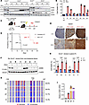 Figure 4
GCN2 inactivation drives GCN2-independent expression of ASNS to confer L-asparaginase resistance. (A) Gcn2–/– T-ALL lines 1, 3, 5, 7, 2, 4, and 10 were treated with PEG for 16 hours. GCN2, ASNS, and ATF4 proteins were assessed by Western blot. (B) T-ALL lines in A were subjected to PEG treatment or untreated (UT) for 3 days, and cell death percentage was recorded by trypan blue staining. (C and D) Schematic illustration of a secondary T-ALL model established by using Gcn2–/– primary KrasG12D;Lck-Cre leukemia cells. PEG was given in 2 doses on days 10 and 34 after transplantation. Kaplan-Meier curve comparing survival between the untreated and PEG-treated mice. Statistical comparison between the 2 groups was done using the log-rank (Mantel-Cox) test. (E) IHC staining of ASNS from the spleen of mice euthanized on day 18 (untreated), day 35 (PEG), and day 42 (PEG). Representative microscopy images were obtained at ×5 or ×20 magnification, as indicated. (F and G) Six single-cell-derived clones from the same untreated spleen tissue in E were subjected to PEG treatment for 16 hours, and protein lysates were subjected to Western blotting analysis for ASNS (F). Cell death percentage following PEG treatment for 3 days were recorded (G). ****P < 0.0001 by 2-way ANOVA. (H) DNA methylation status in the CpG island of the Asns promoter was determined by bisulfite sequencing in Gcn2–/– lines 2, 4, 7, and clone D. Results are shown as heatmaps. The percentages of methylated CpG are indicated on the right of each panel. TSS, transcription start site. (I) Asns mRNA levels from the cells in H were assessed by qPCR.
To determine whether GCN2 inactivation in vivo selects for GCN2-independent mechanisms driving ASNS expression and L-asparaginase resistance, we treated leukemia-bearing mice transplanted with primary Gcn2–/– leukemic cells with PEG (Figure 4C). PEG treatment significantly delayed leukemia-associated lethality (Figure 4D). Analyzing spleen tissues from untreated and PEG-treated mice showed that ASNS expression was markedly increased in PEG-treated animals at the endpoint of morbidity (day 35) (Figure 4E). In contrast, animals euthanized on day 42 remained healthy and exhibited intermediate ASNS induction (Figure 4E). Consistent results were observed in an independent experiment using primary leukemia cells derived from a separate Gcn2–/– donor (Supplemental Figure 3, E and F). In this cohort, all 5 PEG-treated animals reached morbidity (Supplemental Figure 3E), and 2 representative animals (days 25 and 43) displayed elevated expression of ASNS (Supplemental Figure 3F).
To determine whether heterogeneity in ASNS expression contributes to its induction in the in vivo experiment, we established multiple single-cell-derived T-ALL clones from spleen cells harvested from an untreated mouse on day 18 (Figure 4, D and E). Six clones derived from the same spleen exhibited variable ASNS expression (Figure 4F). Furthermore, their sensitivity to PEG treatment was inversely correlated with ASNS protein expression (Figure 4G).
We previously showed that promoter demethylation of human ASNS is required for ATF4-dependent ASNS expression in human T-ALL lines (8). To assess the role of promoter demethylation of the Asns gene in mouse leukemia cells, we performed bisulfite sequencing in 4 Gcn2–/– T-ALL lines expressing variable levels of ASNS. We found that the degree of promoter methylation was inversely correlated with ASNS expression at both protein and mRNA levels (Figure 4, A, F, H, and I), indicating a role for promoter demethylation in driving ASNS expression in Gcn2–/– leukemia cells. These results are consistent with a model where expression of ASNS is a key driver of L-asparaginase resistance. Furthermore, the ASNS induction can occur even when induction of GCN2 and the ISR are disabled.
ASX-173 overcomes resistance caused by GCN2-independent ASNS induction. To further test whether ASNS is the key driver of resistance to L-asparaginase treatment, we generated KrasG12D;Lck-Cre;Asnsfl/fl mice, allowing for concurrent KrasG12D activation and Asns deletion in T cells. Leukemia lines derived from these mice lack ASNS and were highly sensitive to PEG treatment (Figure 5, A and B), confirming ASNS as a key resistance determinant. These results suggest that targeting ASNS will directly overcome resistance to L-asparaginase in ASNShi cancer cells.
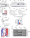 Figure 5
ASX-173 overcomes L-asparaginase resistance in ALL cells due to induced ASNS expression independent of GCN2. (A) KrasG12D;Lck-Cre mice were crossed with AsnsloxP/loxP mice (32). Mouse T-ALL lines from 2 Asns–/– mice (lines 2 and 3) were treated with PEG for 16 hours. A WT mouse T-ALL line was used as a control. ATF4 and ASNS were assessed by Western blot. (B) Mouse T-ALL lines in A were treated with PEG for 3 days and cell death was recorded by trypan blue staining. (C) Basal ASNS expression was measured by Western blot analysis in a panel of human hematological cancer cell lines. ALL (RS4;11, Reh, Nalm-6, HPB-ALL, KOPT-K1, Jurkat), Burkitt lymphoma (BL) (Raji and Ramos), acute myeloid leukemia (AML) (MV4;11, Molm-14, HL-60), and multiple myeloma (MM) (H929, U266). (D) Human T-ALL Jurkat cells and mouse T-ALL Gcn2–/– line 7 from Figure 4A were grown with or without asparagine for 2 days, in the presence of a range of ASX-173 doses (2–1024 nM). IC50 values were measured via MTT assay. (E) IC50 values from D and Supplemental Figure 4A were plotted against basal ASNS expression from C. (F) Jurkat cells were treated with ASX-173 (125 nM) in the presence or absence of exogenous asparagine for 16 hours. Intracellular asparagine levels were measured by liquid chromatograph–mass spectrometry (LC-MS). Statistical significance was determined using 1-way ANOVA. **P < 0.01; ****P < 0.0001. (G) Heatmap showing intracellular levels of amino acids in Jurkat cells treated in F (n = 3). (H) Jurkat, H929, and Ramos cells were cultured with or without asparagine in the presence or absence of ASX-173 (125 nM) for 24 hours. PARP, p-GCN2 (T899), ATF4, and ASNS proteins were assessed via Western blot analysis.
To address the variations in ASNS expression among human hematopoietic cancers, we measured the expression of ASNS protein in lines derived from ALL, Burkitt lymphoma, acute myeloid leukemia, and multiple myeloma. The levels of ASNS as judged by Western blot varied, with ALL lines showing the lowest levels (Figure 5C). These results are consistent with the literature (22, 23), and align with the clinical success of L-asparaginase in the treatment of ALL patients (24, 25).
We next pursued the idea of targeted inhibition of ASNS to expand the utility of L-asparaginase to help alleviate resistance in ALL and potentially expand L-asparaginase among hematopoietic cancers. ASX-173 is a recently reported cell permeable ASNS inhibitor (12, 26). We found that all tested hematopoietic cancer lines responded to ASX-173 when asparagine is depleted from the cell culture media (Figure 5D and Supplemental Figure 4A), with most of these lines showing IC50 values of ASX-173 below 200 nM. None of the tested cell lines responded to ASX-173 even up to 1 μM when asparagine was present in the media (Figure 5D and Supplemental Figure 4A), indicating high specificity of this drug for ASNS. Furthermore, the IC50 values of ASX-173 correlated well with ASNS protein expression in these lines (Figure 5E).
Metabolomic profiling in Jurkat cells confirmed a 70% reduction in intracellular asparagine when exogenous asparagine was removed (Figure 5F); however, this reduction was further exaggerated (>95%) when ASX-173 was given simultaneously (Figure 5F). Notably, there was no reduction in other amino acids, but changes in nucleotides and their precursors when combining ASX-173 with asparagine depletion (Figure 5G and Supplemental Figure 4, B and C). In Jurkat, H929, and Ramos cells, ASX-173 induced PARP cleavage under asparagine-depleted conditions, accompanied with increased GCN2 phosphorylation and ATF4 accumulation (Figure 5H). These results suggest that leukemia cells primarily acquire asparagine through uptake, rendering ASX-173 ineffective when extracellular asparagine is present (Figure 5D); however, upon depletion of exogenous asparagine, cells rely exclusively on ASNS for intracellular asparagine synthesis (Figure 5F). Consequently, ASX-173 triggers ISR and cell death only in the absence of exogenous asparagine (Figure 5H).
ASX-173 suppresses leukemia progression in ASNS-high ALL in vivo. ASX-173 demonstrated promising therapeutic potential in our in vitro experiment when combined with asparagine depletion. To test whether ASX-173 can overcome resistance of L-asparaginase in ASNS-high leukemias in vivo, we established another transplantable secondary T-ALL model. In the experiment in Figure 3, F–I, we retained animals in the GCN2iB plus PEG group for long-term monitoring. Many of the treated animals relapsed (spleen weight > 350 mg) 2 weeks after treatment (Supplemental Figure 5A). Leukemic tissues from relapsed animals expressed high levels of ASNS (Supplemental Figure 5B), which provided a model for therapy-resistant disease. As highlighted in the illustration of our experimental design (Figure 6A), we used CD45.1+ bone marrow cells as the helper cells to differentiate normal hematopoietic cells (CD45.1+) and leukemia cells (CD45.2+) (Figure 6A). ASX-173 was given by oral gavage at 1 dose per day for 5 days, and PEG was administrated after the third dose of ASX-173 (Figure 6A). The combination treatment of ASX-173 with PEG markedly reduced spleen size (Figure 6B) and CD45.2+ leukemia burden in spleen and bone marrow (Figure 6, C and D). Of interest, combining ASX-173 with PEG restored the spleen CD4+/CD8+ cell profile to near normal in recipient mice, which was not seen by monotherapy (Figure 6, E and F). In addition, combining ASX-173 with PEG markedly reduced MYC staining in the spleen tissues (Figure 6G), correlating with a reduction in leukemia burden. Consistently, ASX-173 suppressed MYC protein expression in Jurkat and Gcn2–/– line 7 cells in vitro only when exogenous asparagine was depleted (Figure 6H). PEG treatment resulted in a modest decline in body weight, which was not exaggerated by ASX-173 at the dose used (Supplemental Figure 5C). Histology and IHC studies revealed that combining ASX-173 and PEG markedly reduced leukemia infiltration into the liver and partially restored a normal liver architecture (Supplemental Figure 5D). Together, these findings suggest that ASX-173 enhances the efficacy of PEG-mediated asparagine depletion in suppressing leukemia progression in vivo, with no evidence of prohibitive toxicity.
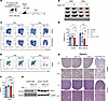 Figure 6
ASX-173 reduces leukemic burden in an ASNS-high ALL model when coupled with L-asparaginase. (A) Schematic illustration of tertiary transplant of leukemia cells from Gcn2+/+ mice relapsed from PEG and GCN2iB combo treatment. Lethally irradiated mice were transplanted with 1.2 × 10⁶ CD45.2+ leukemia cells and 0.5 × 10⁶ CD45.1+ helper bone marrow cells. ASX-173 was administered at 25 mg/kg for 5 days via oral gavage, starting on day 14 after transplantation (blue box). PEG was administered at 2.0 IU/g body weight on day 17 after transplantation. Mice were euthanized on day 23. (B) Images of representative spleen from mice euthanized on day 23 in A. (C and D) CD45.1 and CD45.2 profile of spleen and bone marrow cells measured by flow cytometry and the summary of CD45.2+ cell percentage (n = 3). (E and F) CD4+/CD8+ cell profile of CD45.2+CD3+ T cells from spleens in B and the summary of CD8+ cell percentage (n = 3). Statistical significance was determined using 2-way ANOVA. *P < 0.05; **P < 0.01; ****P < 0.0001. (G) Spleen tissues from B were subjected to IHC staining for c-MYC and H&E staining. Representative microscopy images were obtained at ×5 or ×20 magnification, as indicated. (H) Human Jurkat cells and mouse Gcn2–/– line 7 were subjected to asparagine depletion for 12 hours with or without ASX-173 (125 nM). c-MYC expression was assessed by Western blotting.


