Go to The Journal of Clinical Investigation
  • About
  • Editors
  • Consulting Editors
  • For authors
  • Journal stats
  • Publication ethics
  • Publication alerts by email
  • Transfers
  • Advertising
  • Job board
  • Contact
  • Physician-Scientist Development
  • Current issue
  • Past issues
  • By specialty
    • COVID-19
    • Cardiology
    • Immunology
    • Metabolism
    • Nephrology
    • Oncology
    • Pulmonology
    • All ...
  • Videos
  • Collections
    • In-Press Preview
    • Resource and Technical Advances
    • Clinical Research and Public Health
    • Research Letters
    • Editorials
    • Perspectives
    • Physician-Scientist Development
    • Reviews
    • Top read articles

  • Current issue
  • Past issues
  • Specialties
  • In-Press Preview
  • Resource and Technical Advances
  • Clinical Research and Public Health
  • Research Letters
  • Editorials
  • Perspectives
  • Physician-Scientist Development
  • Reviews
  • Top read articles
  • About
  • Editors
  • Consulting Editors
  • For authors
  • Journal stats
  • Publication ethics
  • Publication alerts by email
  • Transfers
  • Advertising
  • Job board
  • Contact
Top
  • View PDF
  • Download citation information
  • Send a comment
  • Terms of use
  • Standard abbreviations
  • Need help? Email the journal
  • Top
  • Abstract
  • Introduction
  • Results
  • Discussion
  • Methods
  • Manuscript information
  • Supplemental material
  • Acknowledgments
  • Footnotes
  • References
  • Version history
  • Article usage
  • Citations to this article
Advertisement

Research ArticleCell biologyOncology Open Access | 10.1172/jci.insight.205126

RAD52 is a therapeutic vulnerability for acquired PARP inhibitor resistance in ovarian cancer

Yukihide Ota,1,2,3 Maia L. Elizagaray,1,2 Vijayalaxmi Gupta,1,2 Bisiayo E. Fashemi,1,2 Moreniola Akande,1,2 Preedia Babu,1,2 Prasanth Kumar Thuthika,1,2 Elena Lomonosova,1,2 Joshua Brill,1,2 Lulu Sun,4 Brooke Sanders,1 Lindsay M. Kuroki,1 Carolyn K. McCourt,1 Andrea R. Hagemann,1 Ian S. Hagemann,1,5 Premal H. Thaker,1 David G. Mutch,1 Matthew A. Powell,1 Krzysztof Hyrc,6 Priyanka Verma,5 John Krais,5 Benjamin G. Bitler,7 Mary M. Mullen,1,2 and Dineo Khabele1,2

1Division of Gynecologic Oncology, Department of Obstetrics and Gynecology, Alvin J. Siteman Cancer Center, Washington University School of Medicine, St. Louis, Missouri, USA.

2Center for Reproductive Health Sciences, Department of Obstetrics and Gynecology, Washington University, St. Louis, Missouri, USA.

3Department of Obstetrics and Gynecology, Yokohama City University Graduate School of Medicine, Yokohama, Kanagawa, Japan.

4Division of Anatomic and Molecular Pathology, Department of Pathology and Immunology, Washington University School of Medicine, St. Louis, Missouri, USA.

5Division of Oncology, Department of Medicine, Alvin J. Siteman Cancer Center, Washington University School of Medicine, St. Louis, Missouri, USA.

6Department of Neurology, Washington University School of Medicine, St. Louis, Missouri, USA.

7Division of Reproductive Sciences, University of Colorado, Aurora, Colorado, USA.

Address for correspondence. Dineo Khabele, The Mitchell and Elaine Yanow Professor and Chair of the Department of Obstetrics and Gynecology, Department of Obstetrics and Gynecology, Washington University in St. Louis School of Medicine, St. Louis, Missouri 63110, USA. Email: khabeled@wustl.edu.

Find articles by Ota, Y. in: PubMed | Google Scholar

1Division of Gynecologic Oncology, Department of Obstetrics and Gynecology, Alvin J. Siteman Cancer Center, Washington University School of Medicine, St. Louis, Missouri, USA.

2Center for Reproductive Health Sciences, Department of Obstetrics and Gynecology, Washington University, St. Louis, Missouri, USA.

3Department of Obstetrics and Gynecology, Yokohama City University Graduate School of Medicine, Yokohama, Kanagawa, Japan.

4Division of Anatomic and Molecular Pathology, Department of Pathology and Immunology, Washington University School of Medicine, St. Louis, Missouri, USA.

5Division of Oncology, Department of Medicine, Alvin J. Siteman Cancer Center, Washington University School of Medicine, St. Louis, Missouri, USA.

6Department of Neurology, Washington University School of Medicine, St. Louis, Missouri, USA.

7Division of Reproductive Sciences, University of Colorado, Aurora, Colorado, USA.

Address for correspondence. Dineo Khabele, The Mitchell and Elaine Yanow Professor and Chair of the Department of Obstetrics and Gynecology, Department of Obstetrics and Gynecology, Washington University in St. Louis School of Medicine, St. Louis, Missouri 63110, USA. Email: khabeled@wustl.edu.

Find articles by Elizagaray, M. in: PubMed | Google Scholar

1Division of Gynecologic Oncology, Department of Obstetrics and Gynecology, Alvin J. Siteman Cancer Center, Washington University School of Medicine, St. Louis, Missouri, USA.

2Center for Reproductive Health Sciences, Department of Obstetrics and Gynecology, Washington University, St. Louis, Missouri, USA.

3Department of Obstetrics and Gynecology, Yokohama City University Graduate School of Medicine, Yokohama, Kanagawa, Japan.

4Division of Anatomic and Molecular Pathology, Department of Pathology and Immunology, Washington University School of Medicine, St. Louis, Missouri, USA.

5Division of Oncology, Department of Medicine, Alvin J. Siteman Cancer Center, Washington University School of Medicine, St. Louis, Missouri, USA.

6Department of Neurology, Washington University School of Medicine, St. Louis, Missouri, USA.

7Division of Reproductive Sciences, University of Colorado, Aurora, Colorado, USA.

Address for correspondence. Dineo Khabele, The Mitchell and Elaine Yanow Professor and Chair of the Department of Obstetrics and Gynecology, Department of Obstetrics and Gynecology, Washington University in St. Louis School of Medicine, St. Louis, Missouri 63110, USA. Email: khabeled@wustl.edu.

Find articles by Gupta, V. in: PubMed | Google Scholar

1Division of Gynecologic Oncology, Department of Obstetrics and Gynecology, Alvin J. Siteman Cancer Center, Washington University School of Medicine, St. Louis, Missouri, USA.

2Center for Reproductive Health Sciences, Department of Obstetrics and Gynecology, Washington University, St. Louis, Missouri, USA.

3Department of Obstetrics and Gynecology, Yokohama City University Graduate School of Medicine, Yokohama, Kanagawa, Japan.

4Division of Anatomic and Molecular Pathology, Department of Pathology and Immunology, Washington University School of Medicine, St. Louis, Missouri, USA.

5Division of Oncology, Department of Medicine, Alvin J. Siteman Cancer Center, Washington University School of Medicine, St. Louis, Missouri, USA.

6Department of Neurology, Washington University School of Medicine, St. Louis, Missouri, USA.

7Division of Reproductive Sciences, University of Colorado, Aurora, Colorado, USA.

Address for correspondence. Dineo Khabele, The Mitchell and Elaine Yanow Professor and Chair of the Department of Obstetrics and Gynecology, Department of Obstetrics and Gynecology, Washington University in St. Louis School of Medicine, St. Louis, Missouri 63110, USA. Email: khabeled@wustl.edu.

Find articles by Fashemi, B. in: PubMed | Google Scholar

1Division of Gynecologic Oncology, Department of Obstetrics and Gynecology, Alvin J. Siteman Cancer Center, Washington University School of Medicine, St. Louis, Missouri, USA.

2Center for Reproductive Health Sciences, Department of Obstetrics and Gynecology, Washington University, St. Louis, Missouri, USA.

3Department of Obstetrics and Gynecology, Yokohama City University Graduate School of Medicine, Yokohama, Kanagawa, Japan.

4Division of Anatomic and Molecular Pathology, Department of Pathology and Immunology, Washington University School of Medicine, St. Louis, Missouri, USA.

5Division of Oncology, Department of Medicine, Alvin J. Siteman Cancer Center, Washington University School of Medicine, St. Louis, Missouri, USA.

6Department of Neurology, Washington University School of Medicine, St. Louis, Missouri, USA.

7Division of Reproductive Sciences, University of Colorado, Aurora, Colorado, USA.

Address for correspondence. Dineo Khabele, The Mitchell and Elaine Yanow Professor and Chair of the Department of Obstetrics and Gynecology, Department of Obstetrics and Gynecology, Washington University in St. Louis School of Medicine, St. Louis, Missouri 63110, USA. Email: khabeled@wustl.edu.

Find articles by Akande, M. in: PubMed | Google Scholar

1Division of Gynecologic Oncology, Department of Obstetrics and Gynecology, Alvin J. Siteman Cancer Center, Washington University School of Medicine, St. Louis, Missouri, USA.

2Center for Reproductive Health Sciences, Department of Obstetrics and Gynecology, Washington University, St. Louis, Missouri, USA.

3Department of Obstetrics and Gynecology, Yokohama City University Graduate School of Medicine, Yokohama, Kanagawa, Japan.

4Division of Anatomic and Molecular Pathology, Department of Pathology and Immunology, Washington University School of Medicine, St. Louis, Missouri, USA.

5Division of Oncology, Department of Medicine, Alvin J. Siteman Cancer Center, Washington University School of Medicine, St. Louis, Missouri, USA.

6Department of Neurology, Washington University School of Medicine, St. Louis, Missouri, USA.

7Division of Reproductive Sciences, University of Colorado, Aurora, Colorado, USA.

Address for correspondence. Dineo Khabele, The Mitchell and Elaine Yanow Professor and Chair of the Department of Obstetrics and Gynecology, Department of Obstetrics and Gynecology, Washington University in St. Louis School of Medicine, St. Louis, Missouri 63110, USA. Email: khabeled@wustl.edu.

Find articles by Babu, P. in: PubMed | Google Scholar

1Division of Gynecologic Oncology, Department of Obstetrics and Gynecology, Alvin J. Siteman Cancer Center, Washington University School of Medicine, St. Louis, Missouri, USA.

2Center for Reproductive Health Sciences, Department of Obstetrics and Gynecology, Washington University, St. Louis, Missouri, USA.

3Department of Obstetrics and Gynecology, Yokohama City University Graduate School of Medicine, Yokohama, Kanagawa, Japan.

4Division of Anatomic and Molecular Pathology, Department of Pathology and Immunology, Washington University School of Medicine, St. Louis, Missouri, USA.

5Division of Oncology, Department of Medicine, Alvin J. Siteman Cancer Center, Washington University School of Medicine, St. Louis, Missouri, USA.

6Department of Neurology, Washington University School of Medicine, St. Louis, Missouri, USA.

7Division of Reproductive Sciences, University of Colorado, Aurora, Colorado, USA.

Address for correspondence. Dineo Khabele, The Mitchell and Elaine Yanow Professor and Chair of the Department of Obstetrics and Gynecology, Department of Obstetrics and Gynecology, Washington University in St. Louis School of Medicine, St. Louis, Missouri 63110, USA. Email: khabeled@wustl.edu.

Find articles by Thuthika, P. in: PubMed | Google Scholar

1Division of Gynecologic Oncology, Department of Obstetrics and Gynecology, Alvin J. Siteman Cancer Center, Washington University School of Medicine, St. Louis, Missouri, USA.

2Center for Reproductive Health Sciences, Department of Obstetrics and Gynecology, Washington University, St. Louis, Missouri, USA.

3Department of Obstetrics and Gynecology, Yokohama City University Graduate School of Medicine, Yokohama, Kanagawa, Japan.

4Division of Anatomic and Molecular Pathology, Department of Pathology and Immunology, Washington University School of Medicine, St. Louis, Missouri, USA.

5Division of Oncology, Department of Medicine, Alvin J. Siteman Cancer Center, Washington University School of Medicine, St. Louis, Missouri, USA.

6Department of Neurology, Washington University School of Medicine, St. Louis, Missouri, USA.

7Division of Reproductive Sciences, University of Colorado, Aurora, Colorado, USA.

Address for correspondence. Dineo Khabele, The Mitchell and Elaine Yanow Professor and Chair of the Department of Obstetrics and Gynecology, Department of Obstetrics and Gynecology, Washington University in St. Louis School of Medicine, St. Louis, Missouri 63110, USA. Email: khabeled@wustl.edu.

Find articles by Lomonosova, E. in: PubMed | Google Scholar

1Division of Gynecologic Oncology, Department of Obstetrics and Gynecology, Alvin J. Siteman Cancer Center, Washington University School of Medicine, St. Louis, Missouri, USA.

2Center for Reproductive Health Sciences, Department of Obstetrics and Gynecology, Washington University, St. Louis, Missouri, USA.

3Department of Obstetrics and Gynecology, Yokohama City University Graduate School of Medicine, Yokohama, Kanagawa, Japan.

4Division of Anatomic and Molecular Pathology, Department of Pathology and Immunology, Washington University School of Medicine, St. Louis, Missouri, USA.

5Division of Oncology, Department of Medicine, Alvin J. Siteman Cancer Center, Washington University School of Medicine, St. Louis, Missouri, USA.

6Department of Neurology, Washington University School of Medicine, St. Louis, Missouri, USA.

7Division of Reproductive Sciences, University of Colorado, Aurora, Colorado, USA.

Address for correspondence. Dineo Khabele, The Mitchell and Elaine Yanow Professor and Chair of the Department of Obstetrics and Gynecology, Department of Obstetrics and Gynecology, Washington University in St. Louis School of Medicine, St. Louis, Missouri 63110, USA. Email: khabeled@wustl.edu.

Find articles by Brill, J. in: PubMed | Google Scholar

1Division of Gynecologic Oncology, Department of Obstetrics and Gynecology, Alvin J. Siteman Cancer Center, Washington University School of Medicine, St. Louis, Missouri, USA.

2Center for Reproductive Health Sciences, Department of Obstetrics and Gynecology, Washington University, St. Louis, Missouri, USA.

3Department of Obstetrics and Gynecology, Yokohama City University Graduate School of Medicine, Yokohama, Kanagawa, Japan.

4Division of Anatomic and Molecular Pathology, Department of Pathology and Immunology, Washington University School of Medicine, St. Louis, Missouri, USA.

5Division of Oncology, Department of Medicine, Alvin J. Siteman Cancer Center, Washington University School of Medicine, St. Louis, Missouri, USA.

6Department of Neurology, Washington University School of Medicine, St. Louis, Missouri, USA.

7Division of Reproductive Sciences, University of Colorado, Aurora, Colorado, USA.

Address for correspondence. Dineo Khabele, The Mitchell and Elaine Yanow Professor and Chair of the Department of Obstetrics and Gynecology, Department of Obstetrics and Gynecology, Washington University in St. Louis School of Medicine, St. Louis, Missouri 63110, USA. Email: khabeled@wustl.edu.

Find articles by Sun, L. in: PubMed | Google Scholar |

1Division of Gynecologic Oncology, Department of Obstetrics and Gynecology, Alvin J. Siteman Cancer Center, Washington University School of Medicine, St. Louis, Missouri, USA.

2Center for Reproductive Health Sciences, Department of Obstetrics and Gynecology, Washington University, St. Louis, Missouri, USA.

3Department of Obstetrics and Gynecology, Yokohama City University Graduate School of Medicine, Yokohama, Kanagawa, Japan.

4Division of Anatomic and Molecular Pathology, Department of Pathology and Immunology, Washington University School of Medicine, St. Louis, Missouri, USA.

5Division of Oncology, Department of Medicine, Alvin J. Siteman Cancer Center, Washington University School of Medicine, St. Louis, Missouri, USA.

6Department of Neurology, Washington University School of Medicine, St. Louis, Missouri, USA.

7Division of Reproductive Sciences, University of Colorado, Aurora, Colorado, USA.

Address for correspondence. Dineo Khabele, The Mitchell and Elaine Yanow Professor and Chair of the Department of Obstetrics and Gynecology, Department of Obstetrics and Gynecology, Washington University in St. Louis School of Medicine, St. Louis, Missouri 63110, USA. Email: khabeled@wustl.edu.

Find articles by Sanders, B. in: PubMed | Google Scholar

1Division of Gynecologic Oncology, Department of Obstetrics and Gynecology, Alvin J. Siteman Cancer Center, Washington University School of Medicine, St. Louis, Missouri, USA.

2Center for Reproductive Health Sciences, Department of Obstetrics and Gynecology, Washington University, St. Louis, Missouri, USA.

3Department of Obstetrics and Gynecology, Yokohama City University Graduate School of Medicine, Yokohama, Kanagawa, Japan.

4Division of Anatomic and Molecular Pathology, Department of Pathology and Immunology, Washington University School of Medicine, St. Louis, Missouri, USA.

5Division of Oncology, Department of Medicine, Alvin J. Siteman Cancer Center, Washington University School of Medicine, St. Louis, Missouri, USA.

6Department of Neurology, Washington University School of Medicine, St. Louis, Missouri, USA.

7Division of Reproductive Sciences, University of Colorado, Aurora, Colorado, USA.

Address for correspondence. Dineo Khabele, The Mitchell and Elaine Yanow Professor and Chair of the Department of Obstetrics and Gynecology, Department of Obstetrics and Gynecology, Washington University in St. Louis School of Medicine, St. Louis, Missouri 63110, USA. Email: khabeled@wustl.edu.

Find articles by Kuroki, L. in: PubMed | Google Scholar

1Division of Gynecologic Oncology, Department of Obstetrics and Gynecology, Alvin J. Siteman Cancer Center, Washington University School of Medicine, St. Louis, Missouri, USA.

2Center for Reproductive Health Sciences, Department of Obstetrics and Gynecology, Washington University, St. Louis, Missouri, USA.

3Department of Obstetrics and Gynecology, Yokohama City University Graduate School of Medicine, Yokohama, Kanagawa, Japan.

4Division of Anatomic and Molecular Pathology, Department of Pathology and Immunology, Washington University School of Medicine, St. Louis, Missouri, USA.

5Division of Oncology, Department of Medicine, Alvin J. Siteman Cancer Center, Washington University School of Medicine, St. Louis, Missouri, USA.

6Department of Neurology, Washington University School of Medicine, St. Louis, Missouri, USA.

7Division of Reproductive Sciences, University of Colorado, Aurora, Colorado, USA.

Address for correspondence. Dineo Khabele, The Mitchell and Elaine Yanow Professor and Chair of the Department of Obstetrics and Gynecology, Department of Obstetrics and Gynecology, Washington University in St. Louis School of Medicine, St. Louis, Missouri 63110, USA. Email: khabeled@wustl.edu.

Find articles by McCourt, C. in: PubMed | Google Scholar

1Division of Gynecologic Oncology, Department of Obstetrics and Gynecology, Alvin J. Siteman Cancer Center, Washington University School of Medicine, St. Louis, Missouri, USA.

2Center for Reproductive Health Sciences, Department of Obstetrics and Gynecology, Washington University, St. Louis, Missouri, USA.

3Department of Obstetrics and Gynecology, Yokohama City University Graduate School of Medicine, Yokohama, Kanagawa, Japan.

4Division of Anatomic and Molecular Pathology, Department of Pathology and Immunology, Washington University School of Medicine, St. Louis, Missouri, USA.

5Division of Oncology, Department of Medicine, Alvin J. Siteman Cancer Center, Washington University School of Medicine, St. Louis, Missouri, USA.

6Department of Neurology, Washington University School of Medicine, St. Louis, Missouri, USA.

7Division of Reproductive Sciences, University of Colorado, Aurora, Colorado, USA.

Address for correspondence. Dineo Khabele, The Mitchell and Elaine Yanow Professor and Chair of the Department of Obstetrics and Gynecology, Department of Obstetrics and Gynecology, Washington University in St. Louis School of Medicine, St. Louis, Missouri 63110, USA. Email: khabeled@wustl.edu.

Find articles by Hagemann, A. in: PubMed | Google Scholar

1Division of Gynecologic Oncology, Department of Obstetrics and Gynecology, Alvin J. Siteman Cancer Center, Washington University School of Medicine, St. Louis, Missouri, USA.

2Center for Reproductive Health Sciences, Department of Obstetrics and Gynecology, Washington University, St. Louis, Missouri, USA.

3Department of Obstetrics and Gynecology, Yokohama City University Graduate School of Medicine, Yokohama, Kanagawa, Japan.

4Division of Anatomic and Molecular Pathology, Department of Pathology and Immunology, Washington University School of Medicine, St. Louis, Missouri, USA.

5Division of Oncology, Department of Medicine, Alvin J. Siteman Cancer Center, Washington University School of Medicine, St. Louis, Missouri, USA.

6Department of Neurology, Washington University School of Medicine, St. Louis, Missouri, USA.

7Division of Reproductive Sciences, University of Colorado, Aurora, Colorado, USA.

Address for correspondence. Dineo Khabele, The Mitchell and Elaine Yanow Professor and Chair of the Department of Obstetrics and Gynecology, Department of Obstetrics and Gynecology, Washington University in St. Louis School of Medicine, St. Louis, Missouri 63110, USA. Email: khabeled@wustl.edu.

Find articles by Hagemann, I. in: PubMed | Google Scholar |

1Division of Gynecologic Oncology, Department of Obstetrics and Gynecology, Alvin J. Siteman Cancer Center, Washington University School of Medicine, St. Louis, Missouri, USA.

2Center for Reproductive Health Sciences, Department of Obstetrics and Gynecology, Washington University, St. Louis, Missouri, USA.

3Department of Obstetrics and Gynecology, Yokohama City University Graduate School of Medicine, Yokohama, Kanagawa, Japan.

4Division of Anatomic and Molecular Pathology, Department of Pathology and Immunology, Washington University School of Medicine, St. Louis, Missouri, USA.

5Division of Oncology, Department of Medicine, Alvin J. Siteman Cancer Center, Washington University School of Medicine, St. Louis, Missouri, USA.

6Department of Neurology, Washington University School of Medicine, St. Louis, Missouri, USA.

7Division of Reproductive Sciences, University of Colorado, Aurora, Colorado, USA.

Address for correspondence. Dineo Khabele, The Mitchell and Elaine Yanow Professor and Chair of the Department of Obstetrics and Gynecology, Department of Obstetrics and Gynecology, Washington University in St. Louis School of Medicine, St. Louis, Missouri 63110, USA. Email: khabeled@wustl.edu.

Find articles by Thaker, P. in: PubMed | Google Scholar

1Division of Gynecologic Oncology, Department of Obstetrics and Gynecology, Alvin J. Siteman Cancer Center, Washington University School of Medicine, St. Louis, Missouri, USA.

2Center for Reproductive Health Sciences, Department of Obstetrics and Gynecology, Washington University, St. Louis, Missouri, USA.

3Department of Obstetrics and Gynecology, Yokohama City University Graduate School of Medicine, Yokohama, Kanagawa, Japan.

4Division of Anatomic and Molecular Pathology, Department of Pathology and Immunology, Washington University School of Medicine, St. Louis, Missouri, USA.

5Division of Oncology, Department of Medicine, Alvin J. Siteman Cancer Center, Washington University School of Medicine, St. Louis, Missouri, USA.

6Department of Neurology, Washington University School of Medicine, St. Louis, Missouri, USA.

7Division of Reproductive Sciences, University of Colorado, Aurora, Colorado, USA.

Address for correspondence. Dineo Khabele, The Mitchell and Elaine Yanow Professor and Chair of the Department of Obstetrics and Gynecology, Department of Obstetrics and Gynecology, Washington University in St. Louis School of Medicine, St. Louis, Missouri 63110, USA. Email: khabeled@wustl.edu.

Find articles by Mutch, D. in: PubMed | Google Scholar

1Division of Gynecologic Oncology, Department of Obstetrics and Gynecology, Alvin J. Siteman Cancer Center, Washington University School of Medicine, St. Louis, Missouri, USA.

2Center for Reproductive Health Sciences, Department of Obstetrics and Gynecology, Washington University, St. Louis, Missouri, USA.

3Department of Obstetrics and Gynecology, Yokohama City University Graduate School of Medicine, Yokohama, Kanagawa, Japan.

4Division of Anatomic and Molecular Pathology, Department of Pathology and Immunology, Washington University School of Medicine, St. Louis, Missouri, USA.

5Division of Oncology, Department of Medicine, Alvin J. Siteman Cancer Center, Washington University School of Medicine, St. Louis, Missouri, USA.

6Department of Neurology, Washington University School of Medicine, St. Louis, Missouri, USA.

7Division of Reproductive Sciences, University of Colorado, Aurora, Colorado, USA.

Address for correspondence. Dineo Khabele, The Mitchell and Elaine Yanow Professor and Chair of the Department of Obstetrics and Gynecology, Department of Obstetrics and Gynecology, Washington University in St. Louis School of Medicine, St. Louis, Missouri 63110, USA. Email: khabeled@wustl.edu.

Find articles by Powell, M. in: PubMed | Google Scholar

1Division of Gynecologic Oncology, Department of Obstetrics and Gynecology, Alvin J. Siteman Cancer Center, Washington University School of Medicine, St. Louis, Missouri, USA.

2Center for Reproductive Health Sciences, Department of Obstetrics and Gynecology, Washington University, St. Louis, Missouri, USA.

3Department of Obstetrics and Gynecology, Yokohama City University Graduate School of Medicine, Yokohama, Kanagawa, Japan.

4Division of Anatomic and Molecular Pathology, Department of Pathology and Immunology, Washington University School of Medicine, St. Louis, Missouri, USA.

5Division of Oncology, Department of Medicine, Alvin J. Siteman Cancer Center, Washington University School of Medicine, St. Louis, Missouri, USA.

6Department of Neurology, Washington University School of Medicine, St. Louis, Missouri, USA.

7Division of Reproductive Sciences, University of Colorado, Aurora, Colorado, USA.

Address for correspondence. Dineo Khabele, The Mitchell and Elaine Yanow Professor and Chair of the Department of Obstetrics and Gynecology, Department of Obstetrics and Gynecology, Washington University in St. Louis School of Medicine, St. Louis, Missouri 63110, USA. Email: khabeled@wustl.edu.

Find articles by Hyrc, K. in: PubMed | Google Scholar

1Division of Gynecologic Oncology, Department of Obstetrics and Gynecology, Alvin J. Siteman Cancer Center, Washington University School of Medicine, St. Louis, Missouri, USA.

2Center for Reproductive Health Sciences, Department of Obstetrics and Gynecology, Washington University, St. Louis, Missouri, USA.

3Department of Obstetrics and Gynecology, Yokohama City University Graduate School of Medicine, Yokohama, Kanagawa, Japan.

4Division of Anatomic and Molecular Pathology, Department of Pathology and Immunology, Washington University School of Medicine, St. Louis, Missouri, USA.

5Division of Oncology, Department of Medicine, Alvin J. Siteman Cancer Center, Washington University School of Medicine, St. Louis, Missouri, USA.

6Department of Neurology, Washington University School of Medicine, St. Louis, Missouri, USA.

7Division of Reproductive Sciences, University of Colorado, Aurora, Colorado, USA.

Address for correspondence. Dineo Khabele, The Mitchell and Elaine Yanow Professor and Chair of the Department of Obstetrics and Gynecology, Department of Obstetrics and Gynecology, Washington University in St. Louis School of Medicine, St. Louis, Missouri 63110, USA. Email: khabeled@wustl.edu.

Find articles by Verma, P. in: PubMed | Google Scholar

1Division of Gynecologic Oncology, Department of Obstetrics and Gynecology, Alvin J. Siteman Cancer Center, Washington University School of Medicine, St. Louis, Missouri, USA.

2Center for Reproductive Health Sciences, Department of Obstetrics and Gynecology, Washington University, St. Louis, Missouri, USA.

3Department of Obstetrics and Gynecology, Yokohama City University Graduate School of Medicine, Yokohama, Kanagawa, Japan.

4Division of Anatomic and Molecular Pathology, Department of Pathology and Immunology, Washington University School of Medicine, St. Louis, Missouri, USA.

5Division of Oncology, Department of Medicine, Alvin J. Siteman Cancer Center, Washington University School of Medicine, St. Louis, Missouri, USA.

6Department of Neurology, Washington University School of Medicine, St. Louis, Missouri, USA.

7Division of Reproductive Sciences, University of Colorado, Aurora, Colorado, USA.

Address for correspondence. Dineo Khabele, The Mitchell and Elaine Yanow Professor and Chair of the Department of Obstetrics and Gynecology, Department of Obstetrics and Gynecology, Washington University in St. Louis School of Medicine, St. Louis, Missouri 63110, USA. Email: khabeled@wustl.edu.

Find articles by Krais, J. in: PubMed | Google Scholar

1Division of Gynecologic Oncology, Department of Obstetrics and Gynecology, Alvin J. Siteman Cancer Center, Washington University School of Medicine, St. Louis, Missouri, USA.

2Center for Reproductive Health Sciences, Department of Obstetrics and Gynecology, Washington University, St. Louis, Missouri, USA.

3Department of Obstetrics and Gynecology, Yokohama City University Graduate School of Medicine, Yokohama, Kanagawa, Japan.

4Division of Anatomic and Molecular Pathology, Department of Pathology and Immunology, Washington University School of Medicine, St. Louis, Missouri, USA.

5Division of Oncology, Department of Medicine, Alvin J. Siteman Cancer Center, Washington University School of Medicine, St. Louis, Missouri, USA.

6Department of Neurology, Washington University School of Medicine, St. Louis, Missouri, USA.

7Division of Reproductive Sciences, University of Colorado, Aurora, Colorado, USA.

Address for correspondence. Dineo Khabele, The Mitchell and Elaine Yanow Professor and Chair of the Department of Obstetrics and Gynecology, Department of Obstetrics and Gynecology, Washington University in St. Louis School of Medicine, St. Louis, Missouri 63110, USA. Email: khabeled@wustl.edu.

Find articles by Bitler, B. in: PubMed | Google Scholar

1Division of Gynecologic Oncology, Department of Obstetrics and Gynecology, Alvin J. Siteman Cancer Center, Washington University School of Medicine, St. Louis, Missouri, USA.

2Center for Reproductive Health Sciences, Department of Obstetrics and Gynecology, Washington University, St. Louis, Missouri, USA.

3Department of Obstetrics and Gynecology, Yokohama City University Graduate School of Medicine, Yokohama, Kanagawa, Japan.

4Division of Anatomic and Molecular Pathology, Department of Pathology and Immunology, Washington University School of Medicine, St. Louis, Missouri, USA.

5Division of Oncology, Department of Medicine, Alvin J. Siteman Cancer Center, Washington University School of Medicine, St. Louis, Missouri, USA.

6Department of Neurology, Washington University School of Medicine, St. Louis, Missouri, USA.

7Division of Reproductive Sciences, University of Colorado, Aurora, Colorado, USA.

Address for correspondence. Dineo Khabele, The Mitchell and Elaine Yanow Professor and Chair of the Department of Obstetrics and Gynecology, Department of Obstetrics and Gynecology, Washington University in St. Louis School of Medicine, St. Louis, Missouri 63110, USA. Email: khabeled@wustl.edu.

Find articles by Mullen, M. in: PubMed | Google Scholar

1Division of Gynecologic Oncology, Department of Obstetrics and Gynecology, Alvin J. Siteman Cancer Center, Washington University School of Medicine, St. Louis, Missouri, USA.

2Center for Reproductive Health Sciences, Department of Obstetrics and Gynecology, Washington University, St. Louis, Missouri, USA.

3Department of Obstetrics and Gynecology, Yokohama City University Graduate School of Medicine, Yokohama, Kanagawa, Japan.

4Division of Anatomic and Molecular Pathology, Department of Pathology and Immunology, Washington University School of Medicine, St. Louis, Missouri, USA.

5Division of Oncology, Department of Medicine, Alvin J. Siteman Cancer Center, Washington University School of Medicine, St. Louis, Missouri, USA.

6Department of Neurology, Washington University School of Medicine, St. Louis, Missouri, USA.

7Division of Reproductive Sciences, University of Colorado, Aurora, Colorado, USA.

Address for correspondence. Dineo Khabele, The Mitchell and Elaine Yanow Professor and Chair of the Department of Obstetrics and Gynecology, Department of Obstetrics and Gynecology, Washington University in St. Louis School of Medicine, St. Louis, Missouri 63110, USA. Email: khabeled@wustl.edu.

Find articles by Khabele, D. in: PubMed | Google Scholar

Published November 9, 2026 - More info

Published in Volume 11, Issue 21 on November 9, 2026
JCI Insight. 2026;11(21):e205126. https://doi.org/10.1172/jci.insight.205126.
© 2026 Ota et al. This work is licensed under the Creative Commons Attribution 4.0 International License. To view a copy of this license, visit http://creativecommons.org/licenses/by/4.0/.
Published November 9, 2026 - Version history
Received: February 23, 2026; Accepted: September 24, 2026
View PDF
Abstract

Acquired resistance to poly(ADP-ribose) polymerase inhibitors (PARPi) limits their therapeutic benefits in patients with high-grade serous ovarian cancer (HGSC). Identifying therapeutic vulnerabilities that function across distinct forms of acquired PARPi resistance is an urgent need. In BRCA2-deficient PARPi-naive cells, targeting RAD52 can be synthetic lethal with a PARPi, making RAD52 an attractive therapeutic target. However, this approach has not been tested in HGSC models of acquired PARPi resistance. Here, we assessed the effects of RAD52 loss in 2 distinct Brca2-deficient mouse models of HGSC with acquired PARPi resistance. RAD52 loss suppressed homologous recombination and single-strand annealing DNA repair activity in both models. Genetic depletion and pharmacologic inhibition of RAD52 restored PARPi sensitivity both in vitro and in vivo. Furthermore, RAD52 expression was elevated in a newly derived human model of BRCA2-deficient HGSC with acquired PARPi resistance. In this cell line, RAD52 inhibition synergized with a PARPi. Finally, elevated RAD52 expression was associated with poor survival in patients with HGSC. Collectively, these findings establish RAD52 as a clinically relevant therapeutic vulnerability in multiple models of HGSC with acquired PARPi resistance.

Introduction

Ovarian cancer is one of the most lethal gynecologic malignancies. The most prevalent subtype is high-grade serous ovarian carcinoma (HGSC) (1–3), which is often homologous recombination (HR) deficient as a result of deleterious mutations in BRCA1 or BRCA2 (4). In patients with BRCA1/2 mutation, incorporation of poly(ADP-ribose) polymerase inhibitor (PARPi) maintenance therapy after cytoreductive surgery and platinum-based chemotherapy markedly improves outcomes (5–8). However, acquired resistance to PARPi occurs in approximately 50% of patients with BRCA1/2-mutated ovarian cancers (9). Acquired PARPi resistance arises through a variety of mechanisms, including BRCA reversion mutations (10–12), alternative splicing or hypomorphic BRCA1 activity (13, 14), loss of BRCA1 promoter hypermethylation (15), increased drug efflux (16, 17), increased replication fork protection, suppression of replication-coupled gaps (18), and activation of alternative DNA repair pathways (19–22). Identifying therapeutic vulnerabilities that function across these distinct forms of acquired PARPi resistance is an urgent need.

One potential target is RAD52, a multifunctional protein implicated in HR (23–25), single-strand annealing (SSA) (26, 27), and alternative DNA repair mechanisms (24, 28–31). Under normal conditions, RAD52 is dispensable and its loss has minimal effects on HR-mediated DNA repair. In BRCA2-deficient cells, RAD52 can provide compensatory repair capacity. Thus, RAD52 loss can be synthetic lethal with a PARPi in BRCA2-mutant tumors by suppressing RAD52-mediated HR activity and SSA activity (23, 32–35). Increased RAD52 expression has been implicated in resistance to DNA-damaging agents such as platinum compounds and gemcitabine in HR-proficient tumors (36). However, whether RAD52 loss is synergetic lethal with a PARPi in HGSCs with acquired PARPi resistance is unknown.

To address this question, we assessed 2 distinct murine models of Brca2-deficient HGSC with acquired resistance to the PARPi olaparib. Rad52 loss suppressed HR and SSA DNA repair activity in both models. Genetic depletion and pharmacologic inhibition of RAD52 restored PARPi sensitivity in vitro and in vivo. Furthermore, RAD52 expression was elevated in a human model of BRCA2-deficient HGSC with acquired PARPi resistance, and RAD52 inhibition synergized with a PARPi in these cells. Finally, elevated RAD52 expression was associated with poor survival in patients with HGSC. Together, these results establish RAD52 as a therapeutic vulnerability in BRCA2-deficient HGSC with acquired PARPi resistance.

Results

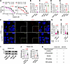
A Brca2-mutated PARPi-resistant mouse ovarian cancer cell line exhibits increased HR and SSA activity and elevated RAD52 expression. We analyzed 2 olaparib-resistant (OR) mouse ovarian cancer cell lines, HGS2-OR and ID8-OR, which were derived from the parental HGS2 (Trp53−/−, Brca2−/−, Pten−/−) and ID8-PB (Trp53−/−, Brca2−/−) cell lines (37, 38). Both OR cell lines showed a significantly higher olaparib IC50 value than their parental counterparts (Figure 1A). Next-generation sequencing confirmed that HGS2-OR retained the >4-kb deletion in Brca2 exon 11, and ID8-OR retained the exon 3 deletion and frameshift (Supplemental Figure 1; supplemental material available online with this article; https://doi.org/10.1172/jci.insight.205126DS1), demonstrating that the acquired PARPi resistance was not due to Brca2 reversion.
PARP inhibitor–resistant HGS2-OR cells exhibit increased HR/SSA repair actiFigure 1

PARP inhibitor–resistant HGS2-OR cells exhibit increased HR/SSA repair activity and RAD52 upregulation. (A) Viability assays of parental (HGS2, ID8-PB) and olaparib-resistant (HGS2-OR, ID8-OR) ovarian cancer cells after 5 days of olaparib treatment. (B and C) HR activity in (B) HGS2-OR and HGS2 cells and (C) ID8-OR, ID8-PB, and ID8-P cells, normalized to the corresponding OR controls. (D and E) SSA activity in (D) HGS2-OR and HGS2 cells and (E) ID8-OR, ID8-PB, and ID8-P cells, normalized to the corresponding OR controls. (F) Representative immunofluorescence images showing RAD51 and γH2AX foci in HGS2 and HGS2-OR cells 4 hours after 10 Gy irradiation. Scale bar size: 10µm. Magnification insets: ×1.66. (G) Quantification of nuclear RAD51 foci. (H) Quantification of nuclear γH2AX foci. (I) Western blot analysis of RAD52 protein. (J) Quantification of RAD52 expression relative to β-actin and normalized to parental cells. (K) Summary of characteristics of cell lines. Data represent mean ± SD from ≥3 independent experiments; individual data points reflect biological replicates. Statistical analyses were performed using 2-sided Student’s t test (A, B, D, and J), 1-way ANOVA followed by Šídák’s correction (C and E), or Mann-Whitney test (G and H). Black and red numbers above graphs are non-significant and significant, respectively, P values. HR, homologous recombination; SSA, single-strand annealing.

To assess the relative contributions of upregulated DNA repair pathways to the development of acquired PARPi resistance in both models, we used reporter assays to measure HR and SSA activity. HGS2-OR cells exhibited significantly higher HR activity than HGS2 cells (Figure 1B and Supplemental Figure 2). ID8-OR cells had similar HR activity as ID8-PB cells and lower HR activity than ID8-P cells, an HR-proficient control (Figure 1C and Supplemental Figure 2). SSA activity was also significantly elevated in HGS2-OR cells but not in ID8-OR cells (Figure 1, D and E). After 10 Gy irradiation, HGS2-OR cells formed more RAD51 foci and fewer γH2AX foci than HGS2 cells, suggesting partial restoration of RAD51-mediated repair activity (Figure 1, F–H, and Supplemental Figure 3). Given that RAD52 can provide compensatory repair capacity through HR and SSA, we investigated RAD52 expression in both models. HGS2-OR cells, but not ID8-OR cells, had elevated RAD52 protein and mRNA (Figure 1, I and J, and Supplemental Figure 3). We assessed other methods of acquired resistance and found that both HGS2-OR and ID8-OR cells displayed elevated expression of P-glycoprotein, a common efflux-based resistance mechanism (Supplemental Figure 3). Conversely, expression of PAF1, which functionally cooperates with RAD52 to promote DNA repair capacity and contributes to therapeutic resistance (36), was non-significantly higher in HGS2-OR cells than in HGS2 cells (Supplemental Figure 3). Thus, although both models of acquired PARPi resistance had upregulation of P-glycoprotein, only HGS2-OR cells exhibited increased HR and SSA activity and elevated RAD52 expression. Characteristics of the cell models are summarized in Figure 1K.

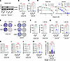
Genetic depletion of Rad52 re-sensitizes Brca2-deficient mouse ovarian cancer cells to olaparib. To further evaluate RAD52 as a therapeutic target, we used short hairpin RNA (shRNA) to generate a stable Rad52 knockdown clone (KD#1) in HGS2-OR cells, and we used CRISPR/Cas9 to generate 2 Rad52-knockout clones (KO#1 and KO#2) in ID8-OR cells (Supplemental Figure 4). Depletion efficacy was confirmed by Western blotting, qRT-PCR, and amplicon sequencing (Figure 2, A–C, and Supplemental Figure 4). Rad52-depleted cells had significantly lower IC50 values and formed fewer colonies during olaparib treatment than scrambled (Scr) controls (Figure 2, D–G). HR and SSA activities were significantly decreased in all Rad52-depleted clones (Figure 2, H–K). After 72 hours of olaparib exposure, Rad52-KO ID8 cells accumulated significantly more γH2AX foci than Scr cells (Figure 2L and Supplemental Figure 5). Western blot analysis further confirmed olaparib-induced γH2AX elevation specifically in Rad52-deficient cells (Supplemental Figure 5). These findings demonstrate that Rad52 loss impairs HR and SSA capacity, leads to accumulation of DNA damage, and restores PARPi sensitivity in both Brca2-deficient, PARPi-resistant mouse HGSC cell lines.
Genetic depletion of Rad52 re-sensitizes BRCA2-deficient ovarian cancer celFigure 2

Genetic depletion of Rad52 re-sensitizes BRCA2-deficient ovarian cancer cells to olaparib. (A) Western blot validation of Rad52 knockdown in HGS2-OR and Rad52 knockout in ID8-OR cells. (B and C) qRT-PCR of Rad52 mRNA in HGS2-OR (B) and ID8-OR (C) cells. (D) Cell viability after 5 days of treatment with 0–20 μM olaparib. (E–G) Clonogenic assay and quantification. (E) Representative images after 8 days of 5 μM olaparib treatment. (J and G) Quantification of clonogenic survival in HGS2-OR (F) and ID8-OR (G) cells. (H and I) HR activity in HGS2-OR (H) and ID8-OR (I) cells. (J and K) SSA activity in HGS2-OR (J) and ID8-OR (K) cells. (L) Quantification of γH2AX foci after 72 hours of 5 μM olaparib. Data represent mean ± SD from ≥3 independent experiments, with individual data points shown. Two-sided Student’s t tests were used for 2-group comparisons (B, D, F, H, and J). One-way ANOVA with Šídák’s multiple-comparison test was used for 3-group comparisons (C, G, I, and K). Kruskal-Wallis followed by Dunn’s test was used for nonparametric data (L). Black and red numbers above graphs are non-significant and significant, respectively, P values. HR, homologous recombination; Ola, olaparib; SSA, single-strand annealing; Veh, vehicle.

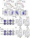
Pharmacological RAD52 inhibition synergizes with olaparib to overcome PARPi resistance in mouse HGSC cells. To test whether pharmacological inhibition of RAD52 can overcome PARPi resistance, we treated cells with the selective small-molecule RAD52 inhibitor D-I03, which blocks RAD52-dependent SSA and D-loop formation (39). D-I03 significantly lowered olaparib IC50 values in both HGS2-OR and ID8-OR cells (Figure 3A and Supplemental Figure 6). Synergy between D-I03 and olaparib was demonstrated in both BLISS and HSA analysis (Figure 3B). Similar results were observed with mitoxantrone, an RPA-RAD52 interaction inhibitor (Supplemental Figure 6) (40). In clonogenic assays, D-I03 potentiated olaparib-induced suppression of colony formation in both resistant cell lines, whereas parental HGS2 and ID8-PB cells were intrinsically sensitive to olaparib alone (Figure 3, C–F). These data indicate that pharmacologic inhibition of RAD52 enhances the cytotoxic effects of olaparib in PARPi-resistant, Brca2-deficient mouse ovarian cancer models in vitro.
Pharmacological RAD52 inhibition synergizes with olaparib to overcome PARPiFigure 3

Pharmacological RAD52 inhibition synergizes with olaparib to overcome PARPi resistance. (A) Viability assay of cells treated with olaparib alone or in combination with D-I03 (2.5 μM for HGS2-OR, 1 μM for ID8-OR) for 5 days. (B) BLISS and HSA model of olaparib and D-I03 in HGS2-OR and ID8-OR cells. (C) Clonogenicity assay of HGS2, and HGS2-OR cells treated with the indicated concentrations of olaparib and D-I03. (D) Quantification of clonogenic assay results. (E) Clonogenicity assay of ID8-PB, and ID8-OR cells treated with the indicated concentrations of olaparib and D-I03. (F) Quantification of clonogenic assay results. Data represent mean ± SD from ≥3 independent experiments, with individual data points shown. One-way ANOVA with Šídák’s multiple-comparison test was used (D and F). Black and red numbers above graphs are non-significant and significant, respectively, P values. Ola, olaparib; Veh, vehicle.

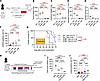
RAD52 inhibition suppresses tumor progression and increases susceptibility to olaparib in mice. To determine whether targeting RAD52 confers benefit in vivo, we intraperitoneally injected ID8-Scr or ID8-KO#1 cells into mice, then treated the mice with daily olaparib or vehicle for 4 weeks (Figure 4A). Mice that received ID8-KO#1 cells developed lower tumor weight, nodule number, and ascites volume in both vehicle and olaparib groups (Figure 4, B–E, and Supplemental Figure 7). ID8-KO#1 tumors also displayed increased γH2AX positivity at baseline and further elevation with olaparib (Figure 4F and Supplemental Figure 7). Survival was significantly longer in mice bearing ID8-KO#1 tumors than in mice with ID8-Scr tumors (Figure 4G).
Genetic and pharmacological RAD52 inhibition suppresses tumor growth in a PFigure 4

Genetic and pharmacological RAD52 inhibition suppresses tumor growth in a PARPi-resistant Brca2-deficient ID8-OR murine model. (A) Experimental scheme for the intraperitoneal xenograft model using ID8-OR–Scr and ID8-OR–Rad52-KO#1 cells. (B–E) Tumor burden metrics: (B) total tumor weight excluding the omentum, (C) weight of the omentum (including the pancreas and surrounding adipose tissue), (D) number of intraperitoneal tumor nodules (≥1 mm), and (E) ascites volume (n = 8 mice/group). (F) Quantification of γH2AX-positive nuclei (intensity > 2+ or 3+) in intraperitoneal tumor sections by immunohistochemistry (IHC). (G) Kaplan-Meier survival analysis comparing Scr-Veh, Scr-Ola, KO#1-Veh, and KO#1-Ola groups. A global log-rank test was followed by pairwise comparisons among 3 groups (Bonferroni-adjusted significance threshold P < 0.0167). Median survival (days): Scr-Veh 49, Scr-Ola 46.5, KO#1-Veh 67, KO#1-Ola not reached. (H) Experimental scheme for pharmacologic RAD52 inhibition. (I) Tumor weights. (J) Quantification of γH2AX-positive nuclei (intensity > 2+ or 3+) in intraperitoneal tumor sections by IHC. Data show mean ± SD of at least 3 independent experiments, with individual dots for each replicate. P values were determined by 1-way ANOVA followed by Šídák’s multiple-comparison test (B–F, and I). Black and red numbers above graphs are non-significant and significant, respectively, P values. i.p, intraperitoneal injection; Ola, olaparib; Veh, vehicle.

To test the effects of pharmacologic RAD52 inhibition in vivo, mice bearing ID8-OR tumors were treated with olaparib, D-I03, the combination, or vehicle for 20 days (Figure 4H). Two early deaths in the D-I03 and combination groups were attributed to treatment-related toxicity (Supplemental Figure 7). At treatment completion, combination therapy resulted in significantly lower tumor burden and greater γH2AX positivity than treatment with vehicle (Figure 4, I and J, and Supplemental Figure 7). Survival was modestly prolonged (Supplemental Figure 7). Together, these findings demonstrate that both genetic and pharmacologic RAD52 inhibition enhance antitumor responses to PARPi treatment in vivo, likely by promoting accumulation of unrepaired DNA damage.

Polq expression and MMEJ activity are upregulated in Rad52-KO mouse HGSC cells treated with olaparib. To examine how Rad52 depletion alters the response to PARPi treatment, we performed bulk RNA-seq of ID8-OR Scr and ID8-KO#1 cells treated with 1 μM olaparib or vehicle for 24 hours (Supplemental Figure 8). ID8-KO#1 cells treated with olaparib exhibited broad transcriptional changes, with 914 upregulated genes and 1,038 downregulated genes relative to Scr controls (Figure 5A). Among DNA-repair-related genes, several transcripts were differentially expressed, including increased Polq, Xrcc3, and Fen1, and decreased Rad51c, Pold4, and Brcc3 (Figure 5A). Z-score analysis across all 4 groups showed that Polq expression was higher in olaparib-treated ID8-KO#1 cells than in olaparib-treated Scr controls (Figure 5B). Consistent with this, qRT-PCR revealed an approximately 1.61-fold elevation in Polq expression in olaparib-treated KO#1 cells (Figure 5C and Supplemental Figure 8). To investigate the functional role of this observation, we performed a microhomology-mediated end-joining/DNA polymerase theta–mediated end joining (MMEJ/TMEJ) and found that ID8-KO#1 cells had higher MMEJ activity than Scr control cells. Using ART558, a POLQ inhibitor, we confirmed that this activity is mediated by this enzyme (Supplemental Figure 9A).

Polq expression is upregulated in Rad52-KO cells treated with olaparib.Figure 5

Polq expression is upregulated in Rad52-KO cells treated with olaparib. (A) Volcano plot showing differentially expressed genes (DEGs) between ID8-OR–Scr and ID8-OR–Rad52-KO#1 cells treated with 5 μM olaparib. Positive log2(fold change) values indicate higher expression in Rad52-KO#1 cells than in Scr cells. DEGs were defined as those with |log2 fold change)| > 0.5 and FDR < 0.05. DNA repair–related DEGs are highlighted. (B) Z-scores of Polq expression in Scr and Rad52-KO#1 cells treated with vehicle or olaparib. (C) qRT-PCR analysis of Polq expression in Scr and Rad52-KO#1 cells following olaparib treatment. Expression was normalized to Actb and presented relative to Scr-olaparib. Red number above the graph in C is a significant P value.

Taken together, these findings indicate that olaparib-treated Rad52-deficient cells upregulate Polq, potentially as a compensatory response.

RAD52 is elevated and targetable in a human BRCA2-mutated PARPi-resistant ovarian cancer model. To test RAD52 inhibition in a human model of HGSC with acquired PARPi resistance, we generated olaparib-resistant PEO1 (PEO1-OR) cells, which exhibited a higher olaparib IC50 than parental PEO1 cells (Figure 6A). Targeted sequencing demonstrated that our parental PEO1 culture contained both the original BRCA2 nonsense mutation (c.4965C>G, p.Y1655*) and a previously described secondary reversion mutation (c.[4964A>T;4965C>G], p.Y1655L), indicating a heterogeneous population. In PEO1-OR cells, both alleles were present, and no additional BRCA2 reversion alleles were detected. PEO1-OR cells also exhibited increased RAD52 expression (Figure 6, B and C). In both BLISS and HSA synergy models, olaparib synergized with D-I03 in PEO1-OR cells (Figure 6D and Supplemental Figure 6).

RAD52 is elevated and targetable in a human BRCA2-mutated PARP inhibitor reFigure 6

RAD52 is elevated and targetable in a human BRCA2-mutated PARP inhibitor resistant ovarian cancer model. (A) Viability assays of parental (PEO1) and olaparib-resistant (PEO1-OR) human ovarian cancer cells after 5 days of olaparib treatment. (B) Western blot analysis of RAD52 protein. (C) Quantification of RAD52 protein relative to β-actin and normalized to parental cells. (D) BLISS and HSA models of olaparib and D-I03 in PEO1-OR. Data represent mean ± SD from ≥3 independent experiments; individual data points reflect biological replicates. Statistical analyses were performed using 2-sided Student’s t test, and the red number above the graph in C is a significant P value.

To test whether our strategy could overcome resistance mediated by restoration of HR, we used the patient-derived PEO4 cell line, which models acquired resistance through a functional BRCA2 reversion mutation (41). Although olaparib and D-I03 IC50 values were higher for PEO4 cells than for PEO1 cells (Supplemental Figure 10), we found minimal synergy with the HSA model and no synergy with the BLISS model (Supplemental Figure 10).

High RAD52 expression is associated with poor overall survival in patients with HGSC. To assess the clinical relevance of RAD52 as a therapeutic vulnerability, we measured RAD52 protein expression by immunohistochemistry in an HGSC tissue microarray (TMA) (n = 72) (Supplemental Figure 11 and Supplemental Table 1). Using the median H-score of 168 as a cutoff, patients with high-RAD52 tumors had significantly worse 4-year overall survival than patients with low-RAD52 tumors (58.3% vs. 80.6%, P = 0.03), although curves converged at later time points (Figure 7, A and B). Analysis of TP53-mutant ovarian serous carcinomas in The Cancer Genome Atlas confirmed that high expression of RAD52 mRNA was associated with shorter overall survival (median overall survival 36.3 vs. 49.5 months, P = 0.02) (Figure 7C). These results demonstrate that elevated RAD52 expression correlates with poor clinical outcomes in patients with HGSC.
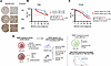
Higher RAD52 expression is associated with poor prognosis in high-grade serFigure 7

Higher RAD52 expression is associated with poor prognosis in high-grade serous ovarian cancer. (A) Representative immunohistochemical images of RAD52 staining in the tissue microarray (TMA). Scale bar: 50 μm. (B) Kaplan-Meier analysis of overall survival (OS) in patients with high-grade serous ovarian cancer represented in the TMA, dichotomized into high (H-score ≥ 168) and low (H-score < 168) RAD52 protein expression groups. The log-rank test was used to analyze survival at 4 years after diagnosis. (C) Kaplan-Meier analysis of OS in patients with serous ovarian cancer with TP53 mutations from The Cancer Genome Atlas (TCGA), stratified by high or low RAD52 mRNA expression. The log-rank test was used to analyze survival at 4 years after diagnosis. (D) Graphical overview summarizing the key findings in this study. Created in BioRender (https://BioRender.com/u905vy9).
Discussion

Here, we demonstrate that RAD52 is a therapeutic vulnerability in BRCA2-deficient ovarian cancers with distinct mechanisms of acquired PARPi resistance (Figure 7D). We examined 2 murine models of Brca2-deficient HGSC with acquired PARPi resistance. Both HGS2-OR and ID8-OR cells had upregulated P-glycoprotein, no Brca2 reversion mutations, and no changes in PAF1 expression. In HGS2-OR cells, but not in ID8-OR cells, PARPi resistance was associated with increased RAD52 expression and elevated HR and SSA activity. Despite these differences, genetic depletion and pharmacologic inhibition of RAD52 restored PARPi sensitivity in both models, indicating that functional dependency on RAD52 may occur even in the absence of overt RAD52 overexpression. Consistent with this dependency, RAD52 loss suppressed both HR and SSA activity. RAD52 expression was also elevated in a newly derived human PARPi-resistant BRCA2-mutant model (PEO1-OR), and RAD52 inhibition synergized with olaparib in these cells. However, RAD52 inhibition did not synergize with olaparib in PEO4 ovarian cancer cells, which harbor a secondary BRCA2 reversion mutation and have functional HR. Finally, we show that high RAD52 expression in human samples correlates with poor survival. Together, our data establishes that RAD52 is a synthetic lethal vulnerability specifically in BRCA2-mutant ovarian cancers that have developed PARPi resistance through mechanisms other than gene reversion.

Our findings show that RAD52 is critical for survival in BRCA2-deficient ovarian cancer cells even after tumors acquire PARPi resistance through divergent mechanisms. Previous studies have demonstrated a synthetic-lethal interaction between BRCA2 loss and RAD52 inhibition primarily in BRCA-deficient, PARPi-naive tumor cell lines. By examining 2 BRCA2-deficient models of acquired PARPi resistance, we found that RAD52 depletion alone was not lethal under basal conditions, yet it re-sensitized resistant cells to PARP inhibition. This dependency on RAD52 persisted despite shared upregulation of P-glycoprotein in both resistant models, indicating that RAD52 is targetable even when drug efflux mechanisms are upregulated.

Mechanistically, we found that Rad52 KO reduced both HR and SSA in our models. The contribution of RAD52 to HR in mammalian cells has long been controversial and has been considered minimal (42). However, our findings suggest that RAD52 can actively promote HR under specific conditions relevant to PARPi resistance. In addition to increased RAD52 expression, other mechanisms may contribute to enhanced HR activity, including RAD52 posttranslational modifications, interactions with phosphorylated RPA, and engagement with cofactors such as DSS1 (33, 43–47). Future studies will be required to determine how these regulatory mechanisms collectively modulate RAD52 function in PARPi-resistant tumors.

In 2 studies, small-molecule RAD52 inhibitors were tested in vivo (35, 48). Here, we used the small-molecule RAD52 inhibitor D-I03 as an initial proof of concept to validate the therapeutic potential of targeting RAD52. However, because RAD52 is not an enzyme, drug design relies on disrupting protein-protein or protein-DNA interactions (49, 50). Such approaches are inherently difficult and likely contribute to the potential off-target effects and notable toxicity observed in our mouse models. Nevertheless, the significant survival benefit we observed (albeit in a single PARPi-resistant Brca2-deficient model) offers genetic evidence that RAD52 represents a bona fide therapeutic vulnerability even in the setting of acquired PARPi resistance. To make targeting RAD52 a clinically feasible approach, drug development efforts should focus on optimizing alternative compounds or targeting upstream and downstream pathway components. A subset of RAD52-deficient clones exhibited increased Polq expression, suggesting that loss of RAD52 promotes a shift toward POLQ-dependent MMEJ/TMEJ. In BRCA2-deficient cells, RAD52 suppresses the premature activation of immature MMEJ/TMEJ and prevents excessive chromosomal fusions (51). Taken together, these findings indicate that RAD52 deficiency both increases cellular dependence on MMEJ through POLQ upregulation and compromises the quality of this repair pathway by allowing MMEJ/TMEJ to operate in inappropriate temporal or structural contexts. In such conditions, BRCA2-deficient tumors may be particularly vulnerable to the combined inhibition of RAD52 and POLQ, which would simultaneously disable RAD52-mediated HR and SSA backup repair mechanisms and POLQ-dependent TMEJ. Further investigation of this potential mechanism of resistance to RAD52 inhibition is warranted.

Although our work primarily focuses on BRCA2-mutant ovarian cancer, targeting RAD52 may have broader clinical relevance. For example, immunohistochemistry analyses of The Cancer Genome Atlas demonstrate that high RAD52 expression correlates with poor prognosis across HGSC, and RAD52 upregulation contributes to cisplatin and gemcitabine resistance even in HR-proficient tumors (36). These observations suggest that RAD52 is a therapeutic vulnerability in a wider spectrum of HGSC and other cancers.

In conclusion, our study provides strong preclinical evidence that RAD52 is a promising therapeutic vulnerability in BRCA2-deficient, PARPi-resistant HGSC. The development of potent, selective, and safe RAD52 inhibitors, as well as agents that target regulatory factors involved in RAD52 function, could advance this approach toward clinical translation.

Methods

Sex as a biological variable. In this study, we exclusively used samples from female mice and patients because ovarian cancer arises exclusively from female reproductive organs.

Cell lines and culture conditions. HGS2-OR and ID8-OR cell lines were previously established from HGS2 (Trp53−/−, Brca2−/−, Pten−/−) and ID8-PB (Trp53−/−, Brca2−/−) cell lines, respectively (37, 38, 52). ID8-P (Trp53−/−, Brca2+/+) and ID8-PB were provided by Ian McNeish (Division of Cancer, Imperial College London, United Kingdom) (53). The HGS2 cell line was thoughtfully provided in-house. The PEO1 and PEO4 cell lines were acquired from Sigma-Aldrich (10032308 lot 12C022 and 10032309 lot 15F025, respectively). The PEO1-OR cell line was generated by continuously exposing PEO1 cells to 2 μM olaparib for 45 days, as previously described (54). HGS2-OR and HGS2 cells were cultured in DMEM/F12 (Gibco, 11320033) plus 10% fetal bovine serum (FBS, Sigma-Aldrich, F0926), penicillin-streptomycin (Gibco, 15140122) (55), and 1× ITS (Gibco, 41400-045) (38). ID8-OR, ID8-PB, and ID8-P cells were cultured in DMEM (Sigma-Aldrich, D6429) supplemented with 4% FBS, penicillin-streptomycin, and 1× ITS) (37). PEO1, PEO1-OR, and PEO4 cells were cultured in RPMI1640 (Gibco, 11875-093) plus 10% FBS and penicillin-streptomycin. All cell lines were incubated at 37°C in 5% CO2. For cell line details, refer to Supplemental Table 2.

Short tandem repeat profiling. PEO1, PEO1-OR, and PEO4 cell pellets (~5 × 10⁶ cells) were submitted to and analyzed by the Genome Engineering and Stem Cell Center (GESC) at the Washington University School of Medicine (56). Percentage match scores were calculated using the Tanabe algorithm by comparing shared alleles across all evaluated loci against the ATCC reference profiles. The PEO1, PEO1-OR, and PEO4 cell lines yielded match scores of 88.46%, 86.79%, and 84.21% respectively, fulfilling the authentication criteria established by ATCC.

RNA interference and lentiviral transduction. Rad52-silenced cell lines were generated with a pool of 3 shRNA lentiviral particles targeting mouse Rad52 (Santa Cruz Biotechnology, sc-37400-V), according to the manufacturer’s instructions. HGS2-OR cells were transfected with lentiviruses and 5 mg/mL polybrene (Santa Cruz Biotechnology, sc-134220) and selected with 8 mg/mL puromycin (Santa Cruz Biotechnology, sc-108071) for 3 days. Cells transfected with scrambled shRNA (Santa Cruz Biotechnology, sc-108080) were used as controls.

CRISPR/CAS9-mediated KO. Predesigned guide RNA for mouse Rad52 and the CRISPR/Cas9 system were used according to the manufacturer’s protocol (Integrated DNA Technologies). The crRNA targeting sequence GTGTTATATTGAAGGTCATC (Mm.Cas9.RAD52.1.AE) and tracrRNA labeled with ATTO 550 dye were annealed to form guide RNA. Then, guide RNA and CAS9 protein were combined to form ribonucleoprotein complexes. Subconfluent ID8-OR cells were transfected with Lipofectamine RNAiMAX (Invitrogen, 13778150). The top 10% of cells with the highest ATTO fluorescence intensity were single-cell sorted by flow cytometry. Single cell clones were isolated, screened by Guide-it Genotype Confirmation kit (Takara Bio, 632611), and validated by Western blotting, qRT-PCR, and next-generation amplicon sequencing (GENEWIZ, Amplicon-EZ) with the following primers: mouse Rad52 forward 5′-GCTCTGTACACCCAGCTACC-3′ and reverse 5′-AGTGTGCCCAGCCATTGTAA-3′. The alleles identified by next-generation sequencing at frequencies greater than 5% are shown (Supplemental Table 7).

Viability assay. Cells were seeded in 96-well plates overnight and then treated with the indicated drugs for 5 days. Medium with drug was replaced on the third day of treatment. Cells were stained and fixed with crystal violet solution consisting of 0.05% crystal violet (Thermo Fisher Scientific, C581-25), 1% formaldehyde (Sigma-Aldrich, 252549), 1× PBS (Corning, 21040-CM), and 1% methanol (Sigma-Aldrich, 1003657780) in distilled H2O, then washed in 70% ethanol (Thermo Fisher Scientific, 04355223). Viability was evaluated by measuring absorbance at 570 nm. Detailed information for each cell line is provided in the Supplemental Table 4.

Clonogenicity assay. Cells were seeded in 12-well plates and incubated overnight. Then, cells were treated with the indicated drugs, and drug-containing medium was refreshed every 3 days for a total of 7–8 days. Colony formation was assessed as described for viability assays. Detailed conditions for each cell line are provided in Supplemental Table 4.

Drug synergy analysis. Cells were seeded in 96-well plates overnight and co-treated for 5 days with olaparib and D-I03. Cell viability was quantified by crystal violet absorbance as described above. Drug interactions were assessed in BLISS independence and HSA models in Combenefit (v2.021) (https://sourceforge.net/projects/combenefit/) and visualized in GraphPad Prism 9. BLISS estimates the expected combined effect under independent drug action, whereas the HSA model compares the observed combination effect with the more effective single agent. In both models, scores above 0 indicate synergy and scores below 0 indicate antagonism.

Western blots. Cells were harvested and lysed in RIPA buffer (Cell Signaling Technology) plus protease and phosphatase inhibitor cocktails (Sigma-Aldrich). Lysates were separated by SDS-PAGE and transferred to PVDF membranes, which were blocked and incubated with primary antibodies followed by horseradish peroxidase–conjugated (HRP-conjugated) secondary antibodies. Chemiluminescent signals were developed using HRP substrate (Millipore) and imaged on a ChemiDoc system (Bio-Rad). Band intensities were quantified with Image Lab software (Bio-Rad). Antibodies against the following proteins were used: RAD52 (Origene, TA890144; 1:2000), γH2AX (Abcam, ab26350; 1:1000), P-glycoprotein (Abcam, ab170904; 1:1000), β-actin (Invitrogen, PA1-183-HRP; 1:2500), anti-mouse IgG-HRP (Cytiva, NA931V; 1:5000), and anti-rabbit IgG-HRP (Cytiva, NA934V; 1:5000).

qRT-PCR. Total RNA was extracted with the RNeasy Protect Mini Kit (Qiagen, 74124) and reverse transcribed into cDNA with the QuantiTect Reverse Transcription Kit (Qiagen, 205313) according to the manufacturer’s instructions. Quantitative PCR was performed with Fast SYBR Green Master Mix (Applied Biosystems, 4385612) on a 7500 Fast Real-Time PCR System (Applied Biosystems). mRNA abundances were normalized to β-actin, and relative expression was calculated by the 2ΔΔCt method. Primer sequences for Rad52, Polq, and Actb are provided in the Supplemental Table 3.

Immunofluorescence. Immunofluorescence was performed as previously described (57). Briefly, cells were seeded on sterilized cover slips in a 12-well plate. For analysis of RAD51 and γH2AX foci in HGS2 and HGS2-OR cells, cells were evaluated either 4 hours after 10 Gy X-ray irradiation or under untreated conditions. For the evaluation of γH2AX foci in ID8-Scr, Rad52-KO#1, and Rad52-KO#2 cells, cells were treated with 5 μM olaparib and analyzed 72 hours after treatment. Cells were washed with cold PBS++ (Leinco Technologies, P364), fixed with 2% paraformaldehyde (Thermo Fisher Scientific, J19943-K2) in PBS++ for 10 minutes, permeabilized with 0.2% Triton X-100 in PBS (Sigma-Aldrich, T8787) for 20 minutes, and then washed and blocked (30 minutes) with staining buffer (PBS, 0.5% BSA, 0.15% glycine, and 0.1% Triton-X-100). Cells were incubated overnight at 4°C with primary antibodies in staining buffer and then stained with secondary antibodies at room temperature for 2 hours. Slides were stained with DAPI (Invitrogen, P37606) and mounted in ProLong Gold (Invitrogen, P36930).

The images were obtained on a confocal microscope (DMI4000B, Leica) equipped with a 63× oil immersion objective lens. Images were acquired in 512 × 512 pixel format in Leica Application Suite X and analyzed with ImageJ software (v1.52a) (58). Color images were converted to 8-bit grayscale, and thresholds were set appropriately and consistently. RAD51 foci and γH2AX foci were counted by using the “analyze particles” function. At least 150 cells were analyzed for each treatment group. The following antibodies and dilutions were used: RAD51 (Abcam, ab133534; 1:1000), γH2AX (MilliporeSigma, 05-636; 1:500), and anti-mouse IgG, Alexa Fluor 555 conjugate (Cell Signaling Technology, 4409; 1:500) (Supplemental Table 5).

Next-generation targeted amplicon sequencing. Targeted amplicon sequencing was performed to evaluate Brca2 reversion mutations in OR cell lines and to characterize CRISPR/Cas9-edited Rad52 alleles. Amplicons spanning Brca2 exon 3, Brca2 exon 11, and the Rad52 CRISPR target region were generated by using gene-specific primers with Illumina overhang adapters. Libraries were prepared in a 2-step PCR protocol and sequenced on an Illumina MiSeq platform (2 × 250 bp) at the Washington University Genome Technology Access Center (GTAC) or via the GENEWIZ Amplicon-EZ service. FASTQ files from the GTAC were analyzed with a custom Python-based workflow to detect insertion/deletion events and in-frame corrections at the Brca2 locus. RAD52-targeted and human BRCA2-targeted amplicons analyzed through the GENEWIZ Amplicon-EZ pipeline provided quantitative variant allele frequencies (VAF), and variants with VAF greater than 5% were included according to our predefined analysis threshold. Detailed PCR conditions, indexing procedures, sequencing specifications, analysis workflows, and complete primer sequences are provided in Supplemental Figure 4 and Supplemental Table 3.

DNA repair assays. HR activity was assessed by using the pDR-GFP reporter system, SSA activity was evaluated by using the hprtSAGFP reporter system, and MMEJ was determined by using the pimEJ2-GFP reporter system, as previously described (59, 60). The reporter constructs used were pDR-GFP (Addgene, 26475), hprtSAGFP (Addgene, 41594), pCBASceI (Addgene, 26477), originally developed by Maria Jasin (61, 62), and EJ2GFP-puro (Addgene, 44025) developed by Jeremy Stark (60). Cells were seeded in 12-well plates and transfected the following day with either pDR-GFP, hprtSAGFP, or pimEJ2GFP together with pCBASceI and Lipofectamine 3000 (Invitrogen, L3000008). For POLQ inhibition, 3 μM ART558 (MedChemExpres, HY-141520) was added right before plasmid transfection. After 24–48 hours, cells were harvested, and GFP-positive cells were quantified by flow cytometry (CYTEK, AURORA, B1 channel). Propidium iodide (Tonbo, SKU 13-6990-T200, YG4 channel) was added immediately before acquisition. Data were analyzed with Spectro Flo software (CYTEK, v3.3.0) and FlowJ v10.10.0 software (Waters Biosciences). Background GFP signals from cells transfected with pDR-GFP, hprtSAGFP, or pimEJ2GFP alone (no pCBASceI) were subtracted from all measurements, as previously described (61).

RNA-seq and differential gene expression analysis. Total RNA was extracted from ID8-OR Scr and ID8-OR Rad52-KO#1 cells treated with 1 μM olaparib or vehicle for 24 hours. RNA integrity was confirmed (RIN > 8). Poly(A)+ RNA–based library preparation and sequencing were performed at the Washington University in GTAC on an Illumina NovaSeq X Plus (2 × 150 bp paired-end). Reads were aligned to the GRCm39 reference genome using STAR software (https://github.com/alexdobin/STAR), and gene-level counts were obtained with featureCounts. Normalization and differential expression analysis were performed with the limma-voom pipeline. Differentially expressed genes were defined as those with |log2FC| of greater than 0.5 and false discovery rate (FDR) of less than 0.05. Gene set enrichment analysis was conducted with Hallmark gene sets.

Mouse experiments. For experiments with ID8-OR Scr or ID8-OR KO#1 cells, 5 × 10⁶ cells were intraperitoneally injected into 6- to 8-week-old female C57BL/6J mice (The Jackson Laboratory). One week after implantation, mice received daily treatment with olaparib (50 mg/kg, oral gavage) or vehicle for 4 weeks (37). Eight mice per group were euthanized the day after the final treatment, and tumor burden was evaluated by measuring total tumor weight (excluding omentum), omental weight, the number of peritoneal tumor nodules larger than 1 mm in diameter, and ascites volume. For survival analysis, 8–12 mice per group were monitored.

For experiments evaluating RAD52 inhibition, 7 × 10⁶ ID8-OR cells were injected intraperitoneally into C57BL/6J mice. Three weeks after implantation, mice were treated daily on weekdays for 20 days with olaparib (75 mg/kg, oral gavage) (63), D-I03 (25 mg/kg, intraperitoneal injection), the combination, or vehicle. Five mice per group were euthanized at the end of treatment, and total tumor weight was measured. For survival analysis, 15–17 mice per group were monitored.

Tumor tissues from both experiments were processed for immunohistochemistry with an anti-γH2AX antibody (Abcam, ab26350; 1:500) followed by VisUCyte HRP Polymer Mouse IgG (R&D Systems, VC001). For each mouse, 3 to 5 fields were imaged at ×20 magnification. Staining intensity was scored as 0, 1, 2, or 3 in QuPath (https://qupath.github.io), and the percentage of cells with scores of 2 or 3 was defined as the positivity rate.

Survival cohorts were monitored after treatment completion in accordance with the NIH Guide for the Care and Use of Laboratory Animals (National Academies Press, 2011). Mice were monitored daily for clinical signs of distress and weight loss. Animals lacking macroscopic tumor engraftment at necropsy were excluded from survival analyses.

TMA. Slides were immunohistochemically stained for RAD52 at the Histology Innovation and Service Center at Washington University in St. Louis. Antigen retrieval was performed by microwave heating in 10 mM citrate buffer for 9 minutes. Sections were incubated with rabbit anti-RAD52 antibody (Abcam, ab117097; 1:400) followed by VisUcyte HRP Polymer Rabbit IgG (R&D Systems, VC003) for signal detection.

TMA cores containing less than 5% tumor cells, as well as samples derived from recurrent tumors or those treated with neoadjuvant chemotherapy, were excluded from the analysis (Supplemental Figure 11 and Supplemental Table 1). Tumor regions were selected under the supervision of a pathologist, and RAD52 expression was quantified via the H-score method (range 0–300) (64) in QuPath (version 0.5.1).

Patients were stratified into “Low” (H-score < 168) and “High” (H-score ≥ 168) expression groups according to the median value. Survival differences were assessed by the log-rank test, focusing on the 4-year time point from diagnosis.

Clinical prognosis analysis from a public database. The Cancer Genome Atlas Ovarian Serous Cystadenocarcinoma dataset (PanCancer Atlas) was accessed through cBioPortal (http://www.cbioportal.org) to obtain mRNA expression data from patients with ovarian cancer. The dataset was last accessed on July 17, 2025. For this analysis, only TP53-mutated samples were included. mRNA abundance was quantified by using RSEM (batch normalized from Illumina HiSeq_RNASeqV2).

Samples were divided into high and low groups according to the median RAD52 mRNA value. Overall survival was assessed by Kaplan-Meier analysis, and differences between groups were evaluated using the log-rank test.

Statistics. Two-sided Student’s t test was used for comparisons between 2 groups. For comparisons among 3 or more groups, 1-way ANOVA followed by Šídák’s multiple-comparison test was performed. For non-parametric comparisons involving 3 or more groups, the Kruskal-Wallis test was used, followed by Dunn’s multiple-comparison test. All data are presented as mean ± standard deviation (SD) unless otherwise stated. A P value of less than 0.05 was considered significant. GraphPad Prism 9 was used for all analyses.

Study approval. All animal procedures were conducted in accordance with the guidelines approved by the Institutional Animal Care and Use Committee (IACUC) at Washington University in St. Louis (protocol 23-0256). Mice were housed under a 12-hour light/12-hour dark cycle at 65°F–75°F with 40%–60% humidity. All efforts were made to minimize discomfort or distress. Euthanasia was performed by cervical dislocation in accordance with American Veterinary Medical Association guidelines. A previously constructed TMA comprising HGSC samples from patients treated at the University of Colorado was provided by the Gynecologic Tissue and Fluid Bank (COMIRB 17-7788) (65, 66).

Data availability. The RNA-seq data generated in this study for ID8-OR-Rad52 and Scr control cells treated with vehicle or olaparib have been deposited in the NCBI Gene Expression Omnibus under accession number GSE302064 (https://github.com/khabele-lab/Rad52). Raw data values associated with the main and supplemental material can be found in Supporting Data Values file.

Manuscript information

Author contributions. YO conceptualized experiments, conducted experiments, acquired and analyzed data, and drafted the manuscript. VG, BEF, MA, PB, and MLE acquired data and contributed to manuscript editing. PKT performed RNA-seq data processing and analysis. EL and JB conducted experiments. LS analyzed and confirmed pathology of samples. BS, LMK, CKM, ARH, ISH, PHT, DGM, and MAP contributed patient samples, provided feedback, and contributed to manuscript editing. KH performed TMA imaging and data acquisition. PV, JK, BGB, and MMM advised on experimental design and contributed to manuscript editing. BGB also provided cell lines (ID8-OR and HGS2-OR) and TMA samples. MMM provided cell lines (PEO1 and PEO1-OR). DK provided funding and reagents, conceptualized experiments, analyzed data, and revised the manuscript.

AI disclosure. No AI was used in the preparation of this article, including for research design, data extraction, analysis and/or synthesis, manuscript writing, or the preparation of figures, graphics, or other content.

Conflict of interest. LS reports consultancy fees from AstraZeneca, and equity from Pairidex, Inc. ISH has received consulting fees from AstraZeneca, GSK, and Daiichi Sankyo outside the scope of the submitted work. PHT reports grants and personal fees from Merck, AstraZeneca, Glaxo Smith Kline, Eisai, AbbVie, Genelux, Corcept, BioNTech, Novocure, Mural Oncology, Pfizer, Immunon, Verastem, and Zentalis. MAP has received consultancy fees from GSK, BioNTech, Immunogen, AstraZenca, Aadi Bioscience, and Merck and Clinical trial support from GSK. BGB serves on OCRA’s SAB. MMM reports personal fees from Valinor Discovery and Bioscent Diagnostics.

Funding. This study was supported by the following:

  • National Cancer Institute grants R01CA243511 and R21CA210210 (to DK).
  • NIH grants R03 TR004017-01 and P50CA244431 (to LMK).
  • American Association of Obstetricians and Gynecologists Foundation Bridge Award (to LMK).
  • Doris Duke Charitable Foundation grant 20152015 (to LMK).
  • NIH grant R35GM160196-01 (to JK).
  • Ovarian Cancer Research Alliance grant ECIG-2025-3-1966 (to JK).Cancer Research Foundation (to JK).
  • Route 66 Endometrial Cancer SPORE CEP (to JK).
  • Alvin J. Siteman Cancer Center through The Foundation for Barnes-Jewish Hospital and the Alvin J. Siteman Cancer Research Fund (to JK).
  • NIH grant R37CA261987 (to BGB).
  • Department of Defense grant (to BGB).
  • Congressionally Directed Medical Research Program (to BGB).
  • Grubstake Foundation (to BGB).
  • Damon Runyon Research Foundation (to MMM).
  • Reproductive Scientist Development Program (RSDP) supported by the Gynecologic Oncology Group Foundation (to MMM).
  • National Cancer Institute Early-Stage Surgeon Scientist Program (to MMM).
  • Victoria Secret Global Fund for Women’s Cancers Career Development Award in partnership with Pelotonia and AACR (to MMM).
  • Pilot Translational and Clinical Studies function of the Washington University Institute of Clinical and Translational Sciences (to MMM).
  • Foundation for Barnes-Jewish Hospital (to MMM).
  • Foundation for Women’s Cancer (to MMM).
  • American Cancer Society (to MMM).

This work is the result of NIH funding, in whole or in part, and is subject to the NIH Public Access Policy. Through acceptance of this federal funding, the NIH has been given a right to make the work publicly available in PubMed Central.

Supplemental material

View Supplemental data

View Unedited blot and gel images

View Supporting data values

Acknowledgments

We especially acknowledge the patients who generously donated their cancer specimens, making this work possible. We thank Deborah Frank for manuscript editing. We acknowledge support from the Gushinkai Fellowship (Support for Young Study Abroad). Imaging of the TMA was performed at the Hope Center Alafi Neuroimaging Lab at Washington University School of Medicine, supported by the NIH Special Instrumentation Grant (S10 OD032121). We also thank the Alvin J. Siteman Cancer Center (supported by National Cancer Institute Cancer Center Support Grant P30 CA091842 and the Institute of Clinical and Translational Sciences grant UL1TR002345 from the National Center for Advancing Translational Sciences of the NIH) at Washington University in St. Louis, for the use of the Flow Cytometry Core (cell sorting), the Histology Innovation and Service Center (immunohistochemistry), and the GTAC (RNA-seq). The authors are solely responsible for the content and interpretation of the work.

Address for correspondence. Dineo Khabele, The Mitchell and Elaine Yanow Professor and Chair of the Department of Obstetrics and Gynecology, Department of Obstetrics and Gynecology, Washington University in St. Louis School of Medicine, St. Louis, Missouri 63110, USA. Email: khabeled@wustl.edu.

Footnotes

Copyright: © 2026, Ota et al. This is an open access article published under the terms of the Creative Commons Attribution 4.0 International License.

Reference information: JCI Insight. 2026;11(21):e205126.https://doi.org/10.1172/jci.insight.205126.

References
  1. Siegel RL, et al. Cancer statistics, 2026. CA Cancer J Clin. 2026;76(1):e70043.
    View this article via: PubMed CrossRef Google Scholar
  2. Gilks CB. Subclassification of ovarian surface epithelial tumors based on correlation of histologic and molecular pathologic data. Int J Gynecol Pathol. 2004;23(3):200–205.
    View this article via: CrossRef PubMed Google Scholar
  3. Seidman JD H. The histologic type and stage distribution of ovarian carcinomas of surface epithelial origin. Int J Gynecol Pathol. 2004;23(1):41–44.
    View this article via: CrossRef PubMed Google Scholar
  4. Konstantinopoulos PA, et al. Homologous recombination deficiency: exploiting the fundamental vulnerability of ovarian cancer. Cancer Discov. 2015;5(11):1137–1154.
    View this article via: CrossRef PubMed Google Scholar
  5. DiSilvestro P, et al. Overall survival with maintenance olaparib at a 7-year follow-up in patients with newly diagnosed advanced ovarian cancer and a BRCA mutation: the SOLO1/GOG 3004 trial. J Clin Oncol. 2023;41(3):609–617.
    View this article via: CrossRef PubMed Google Scholar
  6. Gonzalez-Martin A, et al. Niraparib in patients with newly diagnosed advanced ovarian cancer. N Engl J Med. 2019;381(25):2391–2402.
    View this article via: CrossRef PubMed Google Scholar
  7. Monk BJ, et al. A randomized, phase III trial to evaluate rucaparib monotherapy as maintenance treatment in patients with newly diagnosed ovarian cancer (ATHENA-MONO/GOG-3020/ENGOT-ov45). J Clin Oncol. 2022;40(34):3952–3964.
    View this article via: CrossRef PubMed Google Scholar
  8. Coffman LG, et al. Phase I trial of ribociclib with platinum chemotherapy in ovarian cancer. JCI Insight. 2022;7(18):e160573.
    View this article via: JCI Insight CrossRef PubMed Google Scholar
  9. Banerjee S, et al. Maintenance olaparib for patients with newly diagnosed advanced ovarian cancer and a BRCA mutation (SOLO1/GOG 3004): 5-year follow-up of a randomised, double-blind, placebo-controlled, phase 3 trial. Lancet Oncol. 2021;22(12):1721–1731.
    View this article via: CrossRef PubMed Google Scholar
  10. Edwards SL, et al. Resistance to therapy caused by intragenic deletion in BRCA2. Nature. 2008;451(7182):1111–1115.
    View this article via: CrossRef PubMed Google Scholar
  11. Sakai W, et al. Secondary mutations as a mechanism of cisplatin resistance in BRCA2-mutated cancers. Nature. 2008;451(7182):1116–1120.
    View this article via: CrossRef PubMed Google Scholar
  12. Weigelt B, et al. Diverse BRCA1 and BRCA2 reversion mutations in circulating cell-free DNA of therapy-resistant breast or ovarian cancer. Clin Cancer Res. 2017;23(21):6708–6720.
    View this article via: CrossRef PubMed Google Scholar
  13. Wang Y, et al. The BRCA1-Δ11q alternative splice isoform bypasses germline mutations and promotes therapeutic resistance to PARP inhibition and cisplatin. Cancer Res. 2016;76(9):2778–2790.
    View this article via: CrossRef PubMed Google Scholar
  14. Nesic K, et al. BRCA1 secondary splice-site mutations drive exon-skipping and PARP inhibitor resistance. Mol Cancer. 2024;23(1):158.
    View this article via: CrossRef PubMed Google Scholar
  15. Ter Brugge P, et al. Mechanisms of therapy resistance in patient-derived xenograft models of BRCA1-deficient breast cancer. J Natl Cancer Inst. 2016;108(11)
    View this article via: PubMed CrossRef Google Scholar
  16. Jaspers JE, et al. BRCA2-deficient sarcomatoid mammary tumors exhibit multidrug resistance. Cancer Res. 2015;75(4):732–741.
    View this article via: CrossRef PubMed Google Scholar
  17. Vaidyanathan A, et al. ABCB1 (MDR1) induction defines a common resistance mechanism in paclitaxel- and olaparib-resistant ovarian cancer cells. Br J Cancer. 2016;115(4):431–441.
    View this article via: CrossRef PubMed Google Scholar
  18. Ray Chaudhuri A, et al. Replication fork stability confers chemoresistance in BRCA-deficient cells. Nature. 2016;535(7612):382–387.
    View this article via: CrossRef PubMed Google Scholar
  19. Ceccaldi R, et al. Homologous-recombination-deficient tumours are dependent on Polθ-mediated repair. Nature. 2015;518(7538):258–262.
    View this article via: CrossRef PubMed Google Scholar
  20. Mateos-Gomez PA, et al. Mammalian polymerase θ promotes alternative NHEJ and suppresses recombination. Nature. 2015;518(7538):254–257.
    View this article via: CrossRef PubMed Google Scholar
  21. Zhou J, et al. A first-in-class polymerase theta inhibitor selectively targets homologous-recombination-deficient tumors. Nat Cancer. 2021;2(6):598–610.
    View this article via: CrossRef PubMed Google Scholar
  22. Lavudi K, et al. ALDH1A1 promotes PARP inhibitor resistance by enhancing retinoic acid receptor-mediated DNA polymerase θ expression. NPJ Precis Oncol. 2023;7(1):66.
    View this article via: CrossRef PubMed Google Scholar
  23. Feng Z, et al. Rad52 inactivation is synthetically lethal with BRCA2 deficiency. Proc Natl Acad Sci U S A. 2011;108(2):686–691.
    View this article via: CrossRef PubMed Google Scholar
  24. Bhowmick R, et al. RAD52 facilitates mitotic DNA synthesis following replication stress. Mol Cell. 2016;64(6):1117–1126.
    View this article via: CrossRef PubMed Google Scholar
  25. Yasuhara T, et al. Human Rad52 promotes XPG-mediated R-loop processing to initiate transcription-associated homologous recombination repair. Cell. 2018;175(2):558–570.
    View this article via: CrossRef PubMed Google Scholar
  26. Liang CC, et al. Mechanism of single-stranded DNA annealing by RAD52-RPA complex. Nature. 2024;629(8012):697–703.
    View this article via: CrossRef PubMed Google Scholar
  27. Sugawara N, et al. DNA length dependence of the single-strand annealing pathway and the role of Saccharomyces cerevisiae RAD59 in double-strand break repair. Mol Cell Biol. 2000;20(14):5300–5309.
    View this article via: CrossRef PubMed Google Scholar
  28. Sotiriou SK, et al. Mammalian RAD52 functions in break-induced replication repair of collapsed DNA replication forks. Mol Cell. 2016;64(6):1127–1134.
    View this article via: CrossRef PubMed Google Scholar
  29. Mazina OM, et al. Rad52 inverse strand exchange drives RNA-templated DNA double-strand break repair. Mol Cell. 2017;67(1):19–29.
    View this article via: CrossRef PubMed Google Scholar
  30. Verma P, et al. RAD52 and SLX4 act nonepistatically to ensure telomere stability during alternative telomere lengthening. Genes Dev. 2019;33(3–4):221–235.
    View this article via: CrossRef PubMed Google Scholar
  31. Zhang JM, et al. Alternative lengthening of telomeres through two distinct break-induced replication pathways. Cell Rep. 2019;26(4):955–968.
    View this article via: CrossRef PubMed Google Scholar
  32. Lok BH, et al. RAD52 inactivation is synthetically lethal with deficiencies in BRCA1 and PALB2 in addition to BRCA2 through RAD51-mediated homologous recombination. Oncogene. 2013;32(30):3552–3558.
    View this article via: CrossRef PubMed Google Scholar
  33. Carley AC, et al. Replication protein A phosphorylation facilitates RAD52-dependent homologous recombination in BRCA-deficient cells. Mol Cell Biol. 2022;42(2):e0052421.
    View this article via: CrossRef PubMed Google Scholar
  34. Teng Y, et al. ROS-induced R loops trigger a transcription-coupled but BRCA1/2-independent homologous recombination pathway through CSB. Nat Commun. 2018;9(1):4115.
    View this article via: CrossRef PubMed Google Scholar
  35. Sullivan-Reed K, et al. Simultaneous targeting of PARP1 and RAD52 triggers dual synthetic lethality in BRCA-deficient tumor cells. Cell Rep. 2018;23(11):3127–3136.
    View this article via: CrossRef PubMed Google Scholar
  36. Rauth S, et al. Elevated PAF1-RAD52 axis confers chemoresistance to human cancers. Cell Rep. 2023;42(2):112043.
    View this article via: CrossRef PubMed Google Scholar
  37. Nguyen LL, et al. EHMT1/2 inhibition promotes regression of therapy-resistant ovarian cancer tumors in a CD8 T-cell-dependent manner. Mol Cancer Res. 2024;22(12):1117–1127.
    View this article via: CrossRef PubMed Google Scholar
  38. Nguyen LL, et al. Combining EHMT and PARP inhibition: a strategy to diminish therapy-resistant ovarian cancer tumor growth while stimulating immune activation. Mol Cancer Ther. 2024;23(9):1332–1347.
    View this article via: CrossRef PubMed Google Scholar
  39. Huang F, et al. Targeting BRCA1- and BRCA2-deficient cells with RAD52 small molecule inhibitors. Nucleic Acids Res. 2016;44(9):4189–4199.
    View this article via: CrossRef PubMed Google Scholar
  40. Al-Mugotir M, et al. Selective killing of homologous recombination-deficient cancer cell lines by inhibitors of the RPA:RAD52 protein-protein interaction. PLoS One. 2021;16(3):e0248941.
    View this article via: CrossRef PubMed Google Scholar
  41. Sakai W, et al. Functional restoration of BRCA2 protein by secondary BRCA2 mutations in BRCA2-mutated ovarian carcinoma. Cancer Res. 2009;69(16):6381–6386.
    View this article via: CrossRef PubMed Google Scholar
  42. Rijkers T, et al. Targeted inactivation of mouse RAD52 reduces homologous recombination but not resistance to ionizing radiation. Mol Cell Biol. 1998;18(11):6423–6429.
    View this article via: CrossRef PubMed Google Scholar
  43. Altmannova V, et al. Rad52 SUMOylation affects the efficiency of the DNA repair. Nucleic Acids Res. 2010;38(14):4708–4721.
    View this article via: CrossRef PubMed Google Scholar
  44. Honda M, et al. Tyrosine phosphorylation enhances RAD52-mediated annealing by modulating its DNA binding. EMBO J. 2011;30(16):3368–3382.
    View this article via: CrossRef PubMed Google Scholar
  45. Yasuda T, et al. Novel function of HATs and HDACs in homologous recombination through acetylation of human RAD52 at double-strand break sites. PLoS Genet. 2018;14(3):e1007277.
    View this article via: CrossRef PubMed Google Scholar
  46. Yasuda T, et al. Human SIRT2 and SIRT3 deacetylases function in DNA homologous recombinational repair. Genes Cells. 2021;26(5):328–335.
    View this article via: CrossRef PubMed Google Scholar
  47. Stefanovie B, et al. DSS1 interacts with and stimulates RAD52 to promote the repair of DSBs. Nucleic Acids Res. 2020;48(2):694–708.
    View this article via: CrossRef PubMed Google Scholar
  48. Cramer-Morales K, et al. Personalized synthetic lethality induced by targeting RAD52 in leukemias identified by gene mutation and expression profile. Blood. 2013;122(7):1293–1304.
    View this article via: CrossRef PubMed Google Scholar
  49. Chandramouly G, et al. Small-molecule disruption of RAD52 rings as a mechanism for precision medicine in BRCA-deficient cancers. Chem Biol. 2015;22(11):1491–1504.
    View this article via: CrossRef PubMed Google Scholar
  50. Bhat DS, et al. Therapeutic disruption of RAD52-ssDNA complexation via novel drug-like inhibitors. NAR Cancer. 2023;5(2):zcad018.
    View this article via: CrossRef PubMed Google Scholar
  51. Llorens-Agost M, et al. POLθ-mediated end joining is restricted by RAD52 and BRCA2 until the onset of mitosis. Nat Cell Biol. 2021;23(10):1095–1104.
    View this article via: CrossRef PubMed Google Scholar
  52. Maniati E, et al. Mouse ovarian cancer models recapitulate the human tumor microenvironment and patient response to treatment. Cell Rep. 2020;30(2):525–540.
    View this article via: CrossRef PubMed Google Scholar
  53. Walton J, et al. CRISPR/Cas9-mediated Trp53 and Brca2 knockout to generate improved murine models of ovarian high-grade serous carcinoma. Cancer Res. 2016;76(20):6118–6129.
    View this article via: CrossRef PubMed Google Scholar
  54. Yazinski SA, et al. ATR inhibition disrupts rewired homologous recombination and fork protection pathways in PARP inhibitor-resistant BRCA-deficient cancer cells. Genes Dev. 2017;31(3):318–332.
    View this article via: CrossRef PubMed Google Scholar
  55. Fukumoto T, et al. N(6)-methylation of adenosine of FZD10 mRNA contributes to PARP inhibitor resistance. Cancer Res. 2019;79(11):2812–2820.
    View this article via: CrossRef PubMed Google Scholar
  56. Chen YH, et al. Short tandem repeat profiling via next-generation sequencing for cell line authentication. Dis Model Mech. 2023;16(10):dmm050150.
    View this article via: CrossRef PubMed Google Scholar
  57. Compadre AJ, et al. RAD51 foci as a biomarker predictive of platinum chemotherapy response in ovarian cancer. Clin Cancer Res. 2023;29(13):2466–2479.
    View this article via: CrossRef PubMed Google Scholar
  58. Ota Y, et al. Tissue factor pathway inhibitor-2 inhibits integrin β1 activation and focal adhesion formation and suppresses peritoneal ovarian cancer dissemination in mice. Biochem Biophys Res Commun. 2024;736:150890.
    View this article via: CrossRef PubMed Google Scholar
  59. Jalan M, et al. RAD52 resolves transcription-replication conflicts to mitigate R-loop induced genome instability. Nat Commun. 2024;15(1):7776.
    View this article via: CrossRef PubMed Google Scholar
  60. Bennardo N, et al. Alternative-NHEJ is a mechanistically distinct pathway of mammalian chromosome break repair. PLoS Genet. 2008;4(6):e1000110.
    View this article via: CrossRef PubMed Google Scholar
  61. Stark JM, et al. Genetic steps of mammalian homologous repair with distinct mutagenic consequences. Mol Cell Biol. 2004;24(21):9305–9316.
    View this article via: CrossRef PubMed Google Scholar
  62. Richardson C, et al. Double-strand break repair by interchromosomal recombination: suppression of chromosomal translocations. Genes Dev. 1998;12(24):3831–3842.
    View this article via: CrossRef PubMed Google Scholar
  63. McMahon M, et al. Olaparib, monotherapy or with ionizing radiation, exacerbates DNA damage in normal tissues: insights from a new p21 reporter mouse. Mol Cancer Res. 2016;14(12):1195–1203.
    View this article via: CrossRef PubMed Google Scholar
  64. Ota Y, et al. Tissue factor pathway inhibitor‑2 is specifically expressed in ovarian clear cell carcinoma tissues in the nucleus, cytoplasm and extracellular matrix. Oncol Rep. 2021;45(3):1023–1032.
    View this article via: CrossRef PubMed Google Scholar
  65. Watson ZL, et al. Histone methyltransferases EHMT1 and EHMT2 (GLP/G9A) maintain PARP inhibitor resistance in high-grade serous ovarian carcinoma. Clin Epigenetics. 2019;11(1):165.
    View this article via: CrossRef PubMed Google Scholar
  66. McMellen A, et al. ATF6-mediated signaling contributes to PARP inhibitor resistance in ovarian cancer. Mol Cancer Res. 2023;21(1):3–13.
    View this article via: CrossRef PubMed Google Scholar
Version history
  • Version 1 (November 9, 2026): Electronic publication

Article tools

  • View PDF
  • Download citation information
  • Send a comment
  • Terms of use
  • Standard abbreviations
  • Need help? Email the journal

Metrics

  • Article usage
  • Citations to this article

Go to

  • Top
  • Abstract
  • Introduction
  • Results
  • Discussion
  • Methods
  • Manuscript information
  • Supplemental material
  • Acknowledgments
  • Footnotes
  • References
  • Version history
Advertisement
Advertisement

Copyright © 2026 American Society for Clinical Investigation
ISSN 2379-3708

Sign up for email alerts