
Research ArticleCell biologyVascular biology
Open Access |  10.1172/jci.insight.198202
Steroid hormone antagonism affords vascular protection in a mouse model of vascular Ehlers-Danlos syndrome
Emily E. Juzwiak,1,2 Caitlin J. Bowen,1,2 Rhiannon Edwards,1,3 Leda Restrepo,1 Serena Lee,4 Cassie A. Parks,1,2 Anthony Zeng,1,2 Maya M. Black,1,3 Oscar E. Reyes Gaido,5 Emily E. Bramel,3 Dustin T. Shigaki,6 Michael A. Beer,1,6 Chiara Bellini,4 Harry C. Dietz,1,2 and Elena Gallo MacFarlane1,4,7
1McKusick-Nathans Department of Genetic Medicine, Johns Hopkins University, Baltimore, Maryland USA.
2Howard Hughes Medical Institute, Chevy Chase, Maryland, USA.
3Predoctoral training in Human Genetics and Molecular Biology, Johns Hopkins University, Baltimore, Maryland, USA.
4Department of Bioengineering, Northeastern University, Boston, Massachusetts, USA.
5Department of Dermatology, Brigham and Women’s Hospital, Boston, Massachusetts, USA.
6Department of Biomedical Engineering, Johns Hopkins University, Baltimore, Maryland, USA.
7Department of Surgery, Johns Hopkins University, Baltimore, Maryland, USA.
Address correspondence to: Harry C. Dietz, Miller Research Building Rm #539, 733 N. Broadway Street, Baltimore, Maryland, 21205, USA. Email: hdietz@jhmi.edu. Or to: Elena Gallo MacFarlane, Miller Research Building Rm #529, 733 N. Broadway Street, Baltimore, Maryland, 21205, USA. Email: egallo1@jhmi.edu.
Find articles by Juzwiak, E. in: PubMed | Google Scholar
1McKusick-Nathans Department of Genetic Medicine, Johns Hopkins University, Baltimore, Maryland USA.
2Howard Hughes Medical Institute, Chevy Chase, Maryland, USA.
3Predoctoral training in Human Genetics and Molecular Biology, Johns Hopkins University, Baltimore, Maryland, USA.
4Department of Bioengineering, Northeastern University, Boston, Massachusetts, USA.
5Department of Dermatology, Brigham and Women’s Hospital, Boston, Massachusetts, USA.
6Department of Biomedical Engineering, Johns Hopkins University, Baltimore, Maryland, USA.
7Department of Surgery, Johns Hopkins University, Baltimore, Maryland, USA.
Address correspondence to: Harry C. Dietz, Miller Research Building Rm #539, 733 N. Broadway Street, Baltimore, Maryland, 21205, USA. Email: hdietz@jhmi.edu. Or to: Elena Gallo MacFarlane, Miller Research Building Rm #529, 733 N. Broadway Street, Baltimore, Maryland, 21205, USA. Email: egallo1@jhmi.edu.
Find articles by Bowen, C. in: PubMed | Google Scholar
1McKusick-Nathans Department of Genetic Medicine, Johns Hopkins University, Baltimore, Maryland USA.
2Howard Hughes Medical Institute, Chevy Chase, Maryland, USA.
3Predoctoral training in Human Genetics and Molecular Biology, Johns Hopkins University, Baltimore, Maryland, USA.
4Department of Bioengineering, Northeastern University, Boston, Massachusetts, USA.
5Department of Dermatology, Brigham and Women’s Hospital, Boston, Massachusetts, USA.
6Department of Biomedical Engineering, Johns Hopkins University, Baltimore, Maryland, USA.
7Department of Surgery, Johns Hopkins University, Baltimore, Maryland, USA.
Address correspondence to: Harry C. Dietz, Miller Research Building Rm #539, 733 N. Broadway Street, Baltimore, Maryland, 21205, USA. Email: hdietz@jhmi.edu. Or to: Elena Gallo MacFarlane, Miller Research Building Rm #529, 733 N. Broadway Street, Baltimore, Maryland, 21205, USA. Email: egallo1@jhmi.edu.
Find articles by Edwards, R. in: PubMed | Google Scholar
1McKusick-Nathans Department of Genetic Medicine, Johns Hopkins University, Baltimore, Maryland USA.
2Howard Hughes Medical Institute, Chevy Chase, Maryland, USA.
3Predoctoral training in Human Genetics and Molecular Biology, Johns Hopkins University, Baltimore, Maryland, USA.
4Department of Bioengineering, Northeastern University, Boston, Massachusetts, USA.
5Department of Dermatology, Brigham and Women’s Hospital, Boston, Massachusetts, USA.
6Department of Biomedical Engineering, Johns Hopkins University, Baltimore, Maryland, USA.
7Department of Surgery, Johns Hopkins University, Baltimore, Maryland, USA.
Address correspondence to: Harry C. Dietz, Miller Research Building Rm #539, 733 N. Broadway Street, Baltimore, Maryland, 21205, USA. Email: hdietz@jhmi.edu. Or to: Elena Gallo MacFarlane, Miller Research Building Rm #529, 733 N. Broadway Street, Baltimore, Maryland, 21205, USA. Email: egallo1@jhmi.edu.
Find articles by Restrepo, L. in: PubMed | Google Scholar
1McKusick-Nathans Department of Genetic Medicine, Johns Hopkins University, Baltimore, Maryland USA.
2Howard Hughes Medical Institute, Chevy Chase, Maryland, USA.
3Predoctoral training in Human Genetics and Molecular Biology, Johns Hopkins University, Baltimore, Maryland, USA.
4Department of Bioengineering, Northeastern University, Boston, Massachusetts, USA.
5Department of Dermatology, Brigham and Women’s Hospital, Boston, Massachusetts, USA.
6Department of Biomedical Engineering, Johns Hopkins University, Baltimore, Maryland, USA.
7Department of Surgery, Johns Hopkins University, Baltimore, Maryland, USA.
Address correspondence to: Harry C. Dietz, Miller Research Building Rm #539, 733 N. Broadway Street, Baltimore, Maryland, 21205, USA. Email: hdietz@jhmi.edu. Or to: Elena Gallo MacFarlane, Miller Research Building Rm #529, 733 N. Broadway Street, Baltimore, Maryland, 21205, USA. Email: egallo1@jhmi.edu.
Find articles by Lee, S. in: PubMed | Google Scholar
1McKusick-Nathans Department of Genetic Medicine, Johns Hopkins University, Baltimore, Maryland USA.
2Howard Hughes Medical Institute, Chevy Chase, Maryland, USA.
3Predoctoral training in Human Genetics and Molecular Biology, Johns Hopkins University, Baltimore, Maryland, USA.
4Department of Bioengineering, Northeastern University, Boston, Massachusetts, USA.
5Department of Dermatology, Brigham and Women’s Hospital, Boston, Massachusetts, USA.
6Department of Biomedical Engineering, Johns Hopkins University, Baltimore, Maryland, USA.
7Department of Surgery, Johns Hopkins University, Baltimore, Maryland, USA.
Address correspondence to: Harry C. Dietz, Miller Research Building Rm #539, 733 N. Broadway Street, Baltimore, Maryland, 21205, USA. Email: hdietz@jhmi.edu. Or to: Elena Gallo MacFarlane, Miller Research Building Rm #529, 733 N. Broadway Street, Baltimore, Maryland, 21205, USA. Email: egallo1@jhmi.edu.
Find articles by Parks, C. in: PubMed | Google Scholar
1McKusick-Nathans Department of Genetic Medicine, Johns Hopkins University, Baltimore, Maryland USA.
2Howard Hughes Medical Institute, Chevy Chase, Maryland, USA.
3Predoctoral training in Human Genetics and Molecular Biology, Johns Hopkins University, Baltimore, Maryland, USA.
4Department of Bioengineering, Northeastern University, Boston, Massachusetts, USA.
5Department of Dermatology, Brigham and Women’s Hospital, Boston, Massachusetts, USA.
6Department of Biomedical Engineering, Johns Hopkins University, Baltimore, Maryland, USA.
7Department of Surgery, Johns Hopkins University, Baltimore, Maryland, USA.
Address correspondence to: Harry C. Dietz, Miller Research Building Rm #539, 733 N. Broadway Street, Baltimore, Maryland, 21205, USA. Email: hdietz@jhmi.edu. Or to: Elena Gallo MacFarlane, Miller Research Building Rm #529, 733 N. Broadway Street, Baltimore, Maryland, 21205, USA. Email: egallo1@jhmi.edu.
Find articles by Zeng, A. in: PubMed | Google Scholar
1McKusick-Nathans Department of Genetic Medicine, Johns Hopkins University, Baltimore, Maryland USA.
2Howard Hughes Medical Institute, Chevy Chase, Maryland, USA.
3Predoctoral training in Human Genetics and Molecular Biology, Johns Hopkins University, Baltimore, Maryland, USA.
4Department of Bioengineering, Northeastern University, Boston, Massachusetts, USA.
5Department of Dermatology, Brigham and Women’s Hospital, Boston, Massachusetts, USA.
6Department of Biomedical Engineering, Johns Hopkins University, Baltimore, Maryland, USA.
7Department of Surgery, Johns Hopkins University, Baltimore, Maryland, USA.
Address correspondence to: Harry C. Dietz, Miller Research Building Rm #539, 733 N. Broadway Street, Baltimore, Maryland, 21205, USA. Email: hdietz@jhmi.edu. Or to: Elena Gallo MacFarlane, Miller Research Building Rm #529, 733 N. Broadway Street, Baltimore, Maryland, 21205, USA. Email: egallo1@jhmi.edu.
Find articles by Black, M. in: PubMed | Google Scholar
1McKusick-Nathans Department of Genetic Medicine, Johns Hopkins University, Baltimore, Maryland USA.
2Howard Hughes Medical Institute, Chevy Chase, Maryland, USA.
3Predoctoral training in Human Genetics and Molecular Biology, Johns Hopkins University, Baltimore, Maryland, USA.
4Department of Bioengineering, Northeastern University, Boston, Massachusetts, USA.
5Department of Dermatology, Brigham and Women’s Hospital, Boston, Massachusetts, USA.
6Department of Biomedical Engineering, Johns Hopkins University, Baltimore, Maryland, USA.
7Department of Surgery, Johns Hopkins University, Baltimore, Maryland, USA.
Address correspondence to: Harry C. Dietz, Miller Research Building Rm #539, 733 N. Broadway Street, Baltimore, Maryland, 21205, USA. Email: hdietz@jhmi.edu. Or to: Elena Gallo MacFarlane, Miller Research Building Rm #529, 733 N. Broadway Street, Baltimore, Maryland, 21205, USA. Email: egallo1@jhmi.edu.
Find articles by Reyes Gaido, O. in: PubMed | Google Scholar
1McKusick-Nathans Department of Genetic Medicine, Johns Hopkins University, Baltimore, Maryland USA.
2Howard Hughes Medical Institute, Chevy Chase, Maryland, USA.
3Predoctoral training in Human Genetics and Molecular Biology, Johns Hopkins University, Baltimore, Maryland, USA.
4Department of Bioengineering, Northeastern University, Boston, Massachusetts, USA.
5Department of Dermatology, Brigham and Women’s Hospital, Boston, Massachusetts, USA.
6Department of Biomedical Engineering, Johns Hopkins University, Baltimore, Maryland, USA.
7Department of Surgery, Johns Hopkins University, Baltimore, Maryland, USA.
Address correspondence to: Harry C. Dietz, Miller Research Building Rm #539, 733 N. Broadway Street, Baltimore, Maryland, 21205, USA. Email: hdietz@jhmi.edu. Or to: Elena Gallo MacFarlane, Miller Research Building Rm #529, 733 N. Broadway Street, Baltimore, Maryland, 21205, USA. Email: egallo1@jhmi.edu.
Find articles by Bramel, E. in: PubMed | Google Scholar
1McKusick-Nathans Department of Genetic Medicine, Johns Hopkins University, Baltimore, Maryland USA.
2Howard Hughes Medical Institute, Chevy Chase, Maryland, USA.
3Predoctoral training in Human Genetics and Molecular Biology, Johns Hopkins University, Baltimore, Maryland, USA.
4Department of Bioengineering, Northeastern University, Boston, Massachusetts, USA.
5Department of Dermatology, Brigham and Women’s Hospital, Boston, Massachusetts, USA.
6Department of Biomedical Engineering, Johns Hopkins University, Baltimore, Maryland, USA.
7Department of Surgery, Johns Hopkins University, Baltimore, Maryland, USA.
Address correspondence to: Harry C. Dietz, Miller Research Building Rm #539, 733 N. Broadway Street, Baltimore, Maryland, 21205, USA. Email: hdietz@jhmi.edu. Or to: Elena Gallo MacFarlane, Miller Research Building Rm #529, 733 N. Broadway Street, Baltimore, Maryland, 21205, USA. Email: egallo1@jhmi.edu.
Find articles by Shigaki, D. in: PubMed | Google Scholar
1McKusick-Nathans Department of Genetic Medicine, Johns Hopkins University, Baltimore, Maryland USA.
2Howard Hughes Medical Institute, Chevy Chase, Maryland, USA.
3Predoctoral training in Human Genetics and Molecular Biology, Johns Hopkins University, Baltimore, Maryland, USA.
4Department of Bioengineering, Northeastern University, Boston, Massachusetts, USA.
5Department of Dermatology, Brigham and Women’s Hospital, Boston, Massachusetts, USA.
6Department of Biomedical Engineering, Johns Hopkins University, Baltimore, Maryland, USA.
7Department of Surgery, Johns Hopkins University, Baltimore, Maryland, USA.
Address correspondence to: Harry C. Dietz, Miller Research Building Rm #539, 733 N. Broadway Street, Baltimore, Maryland, 21205, USA. Email: hdietz@jhmi.edu. Or to: Elena Gallo MacFarlane, Miller Research Building Rm #529, 733 N. Broadway Street, Baltimore, Maryland, 21205, USA. Email: egallo1@jhmi.edu.
Find articles by
Beer, M.
in:
PubMed
|
Google Scholar
|

1McKusick-Nathans Department of Genetic Medicine, Johns Hopkins University, Baltimore, Maryland USA.
2Howard Hughes Medical Institute, Chevy Chase, Maryland, USA.
3Predoctoral training in Human Genetics and Molecular Biology, Johns Hopkins University, Baltimore, Maryland, USA.
4Department of Bioengineering, Northeastern University, Boston, Massachusetts, USA.
5Department of Dermatology, Brigham and Women’s Hospital, Boston, Massachusetts, USA.
6Department of Biomedical Engineering, Johns Hopkins University, Baltimore, Maryland, USA.
7Department of Surgery, Johns Hopkins University, Baltimore, Maryland, USA.
Address correspondence to: Harry C. Dietz, Miller Research Building Rm #539, 733 N. Broadway Street, Baltimore, Maryland, 21205, USA. Email: hdietz@jhmi.edu. Or to: Elena Gallo MacFarlane, Miller Research Building Rm #529, 733 N. Broadway Street, Baltimore, Maryland, 21205, USA. Email: egallo1@jhmi.edu.
Find articles by Bellini, C. in: PubMed | Google Scholar
1McKusick-Nathans Department of Genetic Medicine, Johns Hopkins University, Baltimore, Maryland USA.
2Howard Hughes Medical Institute, Chevy Chase, Maryland, USA.
3Predoctoral training in Human Genetics and Molecular Biology, Johns Hopkins University, Baltimore, Maryland, USA.
4Department of Bioengineering, Northeastern University, Boston, Massachusetts, USA.
5Department of Dermatology, Brigham and Women’s Hospital, Boston, Massachusetts, USA.
6Department of Biomedical Engineering, Johns Hopkins University, Baltimore, Maryland, USA.
7Department of Surgery, Johns Hopkins University, Baltimore, Maryland, USA.
Address correspondence to: Harry C. Dietz, Miller Research Building Rm #539, 733 N. Broadway Street, Baltimore, Maryland, 21205, USA. Email: hdietz@jhmi.edu. Or to: Elena Gallo MacFarlane, Miller Research Building Rm #529, 733 N. Broadway Street, Baltimore, Maryland, 21205, USA. Email: egallo1@jhmi.edu.
Find articles by Dietz, H. in: PubMed | Google Scholar
1McKusick-Nathans Department of Genetic Medicine, Johns Hopkins University, Baltimore, Maryland USA.
2Howard Hughes Medical Institute, Chevy Chase, Maryland, USA.
3Predoctoral training in Human Genetics and Molecular Biology, Johns Hopkins University, Baltimore, Maryland, USA.
4Department of Bioengineering, Northeastern University, Boston, Massachusetts, USA.
5Department of Dermatology, Brigham and Women’s Hospital, Boston, Massachusetts, USA.
6Department of Biomedical Engineering, Johns Hopkins University, Baltimore, Maryland, USA.
7Department of Surgery, Johns Hopkins University, Baltimore, Maryland, USA.
Address correspondence to: Harry C. Dietz, Miller Research Building Rm #539, 733 N. Broadway Street, Baltimore, Maryland, 21205, USA. Email: hdietz@jhmi.edu. Or to: Elena Gallo MacFarlane, Miller Research Building Rm #529, 733 N. Broadway Street, Baltimore, Maryland, 21205, USA. Email: egallo1@jhmi.edu.
Find articles by Gallo MacFarlane, E. in: PubMed | Google Scholar
Published April 28, 2026 - More info
JCI Insight. 2026;11(12):e198202. https://doi.org/10.1172/jci.insight.198202.
© 2026 Juzwiak et al. This work is licensed under the Creative Commons Attribution 4.0 International License. To view a copy of this license, visit http://creativecommons.org/licenses/by/4.0/.
Received: July 18, 2025; Accepted: April 20, 2026

-
Results
Increased risk of aortic rupture in VEDS mice correlates with postnatal downregulation of synthetic programs and VSMC differentiation. We previously showed that adolescent male Col3a1G938D/+ mice have increased aortic rupture risk relative to females and that androgen inhibitors are necessary to maintain hydralazine’s protection (initiated at birth) beyond P30–P45 (5, 10). To understand this developmental vulnerability, reproduced in current cohorts (Supplemental Figure 1, A and B; supplemental material available online with this article; https://doi.org/10.1172/jci.insight.198202DS1), we examined transcriptional changes in the descending thoracic aorta of control (Col3a1+/+) and VEDS (Col3a1G938D/+) male mice between P30 (prepuberty) and P60 (postpuberty) using bulk RNA-seq (Figure 1A). Despite robust aortic phenotypes in VEDS mice, most transcriptional changes were age- rather than genotype-driven (|Log2FC| ≥ 0.25, FDR ≤ 0.05) (Figure 1B and Supplemental Table 1), separating into 3 coregulated groups by divisive clustering (Figure 1C).
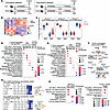 Figure 1
Increased risk of aortic rupture in VEDS mice correlates with postnatal downregulation of synthetic programs and VSMC differentiation. (A) Diagram of experimental conditions for bulk RNA-seq of descending thoracic aorta from untreated control Col3a1+/+ (Ctrl) and Col3a1G938D/+ (VEDS) mice. (B) Heatmap of differentially expressed transcripts between P30 (prepuberty) and P60 (postpuberty) in Ctrl and VEDS samples (FDR ≤ 0.05, abs Log2FC ≥ 0.25). (C) Divisive clustering identified 3 major groups of coregulated genes. (D and E) GO biological process (D) and Reactome (E) enrichment for coregulated transcripts and genotype-dependent genes at P60. (F) STRING networks of enriched terms and transcripts in VEDS versus control aorta at P60. Node colors indicate expression in VEDS: red (upregulated), blue (downregulated). (G) Heatmap and logos of top motifs enriched in accessible chromatin regions (ATAC-seq) in control or VEDS aorta, determined by gkm-PWM algorithm. Heatmap shows Redundancy (R), Weight (W), z score (Z), and Importance (I). (H) GO biological process enrichment for genes near indicated transcription factor binding sites. For D, E, and H: GeneRatio = proportion of genes in pathway/term; q-value = FDR-adjusted P-value (cutoff P ≤ 0.05); top 3 pathways/terms shown.
Age-dependent coregulated transcripts and genotype-specific alterations were examined for GO biological process and Reactome pathway enrichment (Figure 1, D and E). Group 1 transcripts, downregulated from P30 to P60 but less so in VEDS aorta (Figure 1C), were enriched for ECM synthesis/remodeling (Col1a2, Col3a1, Col4a3, Eln, Loxl2, Mmp14, Mmp2, Serpinh1), growth factor receptors promoting VSMC proliferation/migration (Pdgfra), metabolic enzymes for energy production/cellular respiration (Acacb, Acadvl, Aco1, Aco2, Atp5o, Slc25a51, Uqcr10), and metabolic signaling modulators (Adipor2, Ppara) (Figure 1, D and E, and Supplemental Table 1). Group 2 transcripts, related to cell proliferation (histone genes, cell cycle modulators), were also downregulated between P30 and P60 (Figure 1, C–E, and Supplemental Table 1). Group 3 transcripts were developmentally upregulated regardless of genotype, though slightly decreased in VEDS versus controls (Figure 1C), including actomyosin contractile components (Lmod1, Myh11, Myl9, Nebl, Rock1, Rock2, Tpm1), cell-substrate adhesion factors (integrins Itga1, Itga3, Itga5, Itgb1, Itgb5), and VSMC differentiation signals (Tgfb3, Tgfbr1, Rbpms, Rbpms2) (Figure 1, D and E, and Supplemental Table 1). Few transcripts differed between VEDS and control aorta; these included upregulated ECM components/remodeling enzymes (Fbln2, Fbn1, Igfbp6, Lum, Mmp2, Ctsk) and downregulated stress response/tissue repair factors (Jun, Ptgs2, Tlr4, Dnajb1, Atf3) (Figure 1F and Supplemental Table 1).
ATAC-seq chromatin profiling of control and VEDS aortic nuclei at P60 showed concordant developmental transitions and similar transcription factor activity between genotypes (Supplemental Figure 2, A and B, and Supplemental Table 1). Analysis via the gapped k-mer position weight matrixes (gkmPWM) algorithm identified MEF2C and AP-1–related transcription factors (JUN, JUND, FOSL1, FOSL2) motifs as most enriched in accessible chromatin regions (Figure 1G and Supplemental Figure 2B). However, gene loci near predicted MEF2C binding sites were enriched for VSMC differentiation factors in controls (Hes1, Jag1, Rock1, Itga8, Rbpms2) whereas gene loci near MEF2C were enriched for actin filament organization modulators (Arhgap6, Gsn, Svil, Synpo) and stress-activated p38 kinase signaling (Gadd45b, Gadd45g, Mapkapk2, Phlpp1, Zfp36) in VEDS aorta (Figure 1H and Supplemental Table 1). Gene loci with FOSL1:JUND motifs in VEDS aorta (but not FOSL2:JUN in controls) were enriched for stress/starvation response pathways (Foxo1, Foxo3, Prkag2, Prkag3, Cdkn1a) (Figure 1H and Supplemental Table 1). These data suggest increased aortic vulnerability in VEDS associates with physiological downregulation of synthetic/proliferative programs, with VEDS-specific transcriptional alterations at P60 potentially representing compensation or pathogenic deviation.
Coadministration of hydralazine with androgen inhibitors induces upregulation of transcripts promoting ECM deposition. To examine how developmentally regulated transcriptional changes were affected by treatments protecting VEDS mice from aortic rupture, we analyzed aortas collected at P30 and P60 from control and VEDS mice treated with hydralazine since birth, plus VEDS mice receiving hydralazine with either bicalutamide (a potent androgen antagonist) (11–13) or spironolactone (a dual androgen/MR antagonist) (14, 15) starting at P21 (Figure 2A).
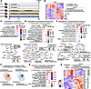 Figure 2
Coadministration of hydralazine with androgen inhibitors induces upregulation of transcripts promoting ECM deposition. (A) Diagram of experimental conditions for bulk RNA-seq of descending thoracic aorta from treated and untreated Col3a1G938D/+ (VEDS) mice and untreated Col3a1+/+ controls (Ctrl). (B) Heatmap of differentially expressed transcripts across all samples (FDR ≤ 0.05). (C–E) Reactome pathway enrichment and STRING networks of transcripts modulated by hydralazine (C), hydralazine/bicalutamide (D), and hydralazine/spironolactone (E). Network nodes colored by expression change in treated versus untreated VEDS: red (upregulated), blue (downregulated). (F) Venn diagram of transcripts modulated by each treatment versus untreated VEDS aorta. (G) Pathway enrichment for transcripts concordantly modulated by both hydralazine/bicalutamide and hydralazine/spironolactone. (H) Heatmap of concordantly modulated transcripts. For C–E, and G: GeneRatio = proportion of genes in pathway/term; q value = FDR-adjusted P value (P ≤ 0.05); top 4 pathways shown.
Pair-wise comparisons between treated and untreated VEDS aortic samples identified transcripts modulated by hydralazine alone and by cotreatment with bicalutamide or spironolactone (Figure 2B and Supplemental Table 2). All treatments upregulated ECM synthesis–related transcripts, including elastogenesis and collagen deposition, though hydralazine alone modulated fewer transcripts (Figure 2, C–E). Hydralazine/bicalutamide and hydralazine/spironolactone cotreatments also downregulated energy metabolism transcripts, including fatty acid metabolism (Figure 2, D and E). A core set of 48 transcripts was modulated in an AR-dependent manner by hydralazine/bicalutamide and hydralazine/spironolactone, but not hydralazine alone (Figure 2F and Supplemental Table 2), largely corresponding to age- rather than genotype-dependent changes (Figure 2, G and H, and Supplemental Table 2). AR inhibition counteracted aging effects by upregulating transcripts for ECM components involved in collagen and elastin deposition, including Col1a2, Eln, Fras1, Itga4, Mfap5, and Fstl1, which encodes a matrix protein recently shown to protect against acute aortic dissection (16) (Figure 2, G and H, and Supplemental Table 2). Other age-dependent processes were either unaffected (downregulation of pro-proliferative pathways) or enhanced (downregulation of fatty acid biosynthesis) by AR antagonism (Figure 2, G and H, and Supplemental Table 2). This transcriptional analysis suggested AR inhibition efficacy associated not with restoring VEDS transcriptional programs to age-matched control levels, but with increasing ECM-related transcript expression beyond healthy control levels.
Isolated genetic and pharmacologic androgen antagonism improves survival in male VEDS mice. Pathway overlap in transcripts modulated by hydralazine/bicalutamide or hydralazine/spironolactone cotreatment, and the modest effect of hydralazine alone, suggested isolated AR antagonism may suffice to improve VEDS mouse survival.
To test this, we examined how bicalutamide treatment or genetic Ar inactivation affected aortic rupture risk in VEDS mice. Mice carrying a conditional Arflox allele (X chromosome) (17) were crossed to VEDS mice expressing β-actin–Cre recombinase, generating globally Ar-deficient VEDS mice. Ar expression, increased in postpubertal aortic walls of both sexes, was effectively abrogated by Cre-mediated deletion in male Arnull/y and female Arnull/null mice (Supplemental Figure 3). In males, global Ar deletion (VEDS Arnull/y) improved survival from 40% to 84%, and pharmacological AR inhibition with bicalutamide improved male VEDS survival from 53% to 74% versus untreated controls (Figure 3, A and B). Unlike hydralazine/bicalutamide cotreatment, which benefits both sexes, neither genetic Ar inactivation nor bicalutamide alone improved female VEDS survival (Figure 3, C and D). No residual sex-dependent survival differences existed between bicalutamide-treated VEDS males and untreated VEDS females (P = 0.53) or between Ar-deficient VEDS males and VEDS females (P = 0.29). Although tissue fragility precludes measurement of blood pressure by tail cuff plethysmography in VEDS mice, assessment in Col3a1+/+ control mice showed that Ar disruption did not affect male blood pressure but slightly increased it in females (Supplemental Figure 4A). Bicalutamide affected neither Ar expression nor blood pressure in either sex (Supplemental Figures 3 and 4). These data indicate that, while AR antagonism protects males, residual risk persists in VEDS mice of both sexes, suggesting the existence of additional AR-independent modulators of aortic rupture risk.
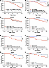 Figure 3
AR antagonism eliminates sexual dimorphism in survival, while MR antagonism protects VEDS mice of either sex from aortic rupture. (A) Kaplan-Meier survival curve comparing control Ar+/y; β-actin–Cre+ (n = 41) and VEDS Arnull/y; β-actin–Cre+ (n = 44) male mice. (B) Kaplan-Meier survival curve comparing untreated male VEDS mice (n = 69) and bicalutamide- (n = 37) or spironolactone- (n = 30) treated (P21-P60) male VEDS mice. (C) Kaplan-Meier survival curve comparing VEDS Ar+/+ (n = 13), VEDS Arnull/+; β-actin–Cre+ (n = 46), and VEDS Arnull/null; β-actin– Cre+ (n = 42) female mice. (D) Kaplan-Meier survival curve comparing untreated female VEDS mice (n = 67) and bicalutamide- (n = 33) or spironolactone-treated (n = 33) (P21–P60) female VEDS mice treated. (E) Kaplan-Meier survival curve comparing untreated male VEDS mice (n = 22) and male VEDS mice treated with spironolactone starting at P60 (n = 24). (F) Kaplan-Meier survival curve comparing untreated female VEDS mice (n = 20) to female VEDS mice treated with spironolactone starting at P60 (n = 23). (G) Kaplan-Meier survival curve comparing untreated male VEDS mice (n = 23) and finerenone-treated (P21–P60) male VEDS mice (n = 36). (H) Kaplan-Meier survival curve comparing untreated female VEDS mice (n = 44), and finerenone-treated (P21–P60) female VEDS mice (n = 30). P values refer to Log-rank Mantel-Cox analysis. For all panels, *P ≤ 0.05, **P ≤ 0.01, ***P ≤ 0.001, and ****P ≤ 0.0001.
Pharmacologic MR antagonism improves survival in both male and female VEDS mice. The elimination of sexual dimorphism by pharmacologic or genetic AR inhibition suggested androgen signaling accounted for most survival differences between male and female VEDS mice. However, bicalutamide has clinical limitations due to sexual side effects in males (11–13, 18). We thus examined the effect of isolated spironolactone treatment, an FDA-approved AR inhibitor with fewer sexual side effects (14, 15). Unlike bicalutamide, spironolactone improved survival in both male and female VEDS mice, resulting in near-complete P60 protection (54% to 93% in males; 69%–97% in females) (Figure 3, B and D). Spironolactone reduced aortic rupture risk even when initiated after puberty (P60), improving P100 survival from 57% to 95% in males and 60% to 95% in females (Figure 3, E and F). Like bicalutamide, spironolactone did not affect Ar expression or blood pressure (Supplemental Figures 3 and 4).
The greater protection by spironolactone versus bicalutamide or genetic Ar inhibition led us to hypothesize that the MR, also inhibited by spironolactone, may be a VEDS therapeutic target (15, 16). To isolate MR inhibition effects, we examined how finerenone, a selective MR antagonist (19) affected VEDS mouse survival. Finerenone improved survival in both males (55% to 82%) and females (65% to 89%) (Figure 3, G and H), without changing blood pressure (Supplemental Figure 4). While finerenone did not affect Ar expression in either sex (Supplemental Figure 3), Nr3c2 (encoding MR) expression increased in finerenone-treated female aortas but remained unchanged in other groups (Supplemental Figure 5). MR inhibition’s beneficial effect was independent of testosterone modulation, as circulating testosterone remained unaffected by AR, AR/MR, or MR inhibition (P = 0.25 for males; P = 0.66 for females) (Supplemental Figure 6). These data suggest that MR antagonism modulates aortic rupture risk in both sexes through an AR-independent mechanism.
AR/MR antagonism increases expression of transcripts promoting ECM deposition and aerobic respiration in multiple cell types. To overcome bulk RNA analysis limitations, potentially confounded by different cell type transcriptional profiles, and examine cell-specific transcriptional changes from isolated AR and MR inhibition (without hydralazine), we performed single-nucleus RNA-seq (snRNA-seq) on descending thoracic aortas of VEDS mice treated with spironolactone, bicalutamide, or finerenone from P21 to P60 (n = 3/group), plus untreated controls and VEDS mice. Clusters corresponding to VSMCs, endothelial cells, fibroblasts, and immune cells were identified using previously described cluster-defining transcripts (Figure 4A) (20), with no genotype- or treatment-specific cell populations or over-/underrepresentation detected between groups (Figure 4, B and C). Immune cells were excluded due to inadequate recovered nuclei depth and lack of inflammatory involvement in VEDS (5).
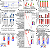 Figure 4
AR/MR inhibition upregulates transcripts promoting ECM deposition and aerobic respiration, and downregulates transcripts related to Rho/Rac signaling and alternative mRNA splicing in multiple cell-types. (A) Dot plot of cluster-defining transcripts for cluster identity. Dot color intensity indicates scaled average expression; dot size shows percentage of nuclei expressing each transcript per cluster. (B) Unsupervised clustering (Louvain resolution 0.1) identifies 4 major aortic cell populations: vascular smooth muscle cells (VSMCs), endothelial cells, fibroblasts, and immune cells, stratified by genotype (control versus VEDS). (C) Stacked bar plot showing nuclei proportion per cluster and no significant differences between treatment groups (2-way ANOVA). (D) Numbers and percentages of transcripts similarly modulated by spironolactone, finerenone, and bicalutamide or preferentially by spironolactone versus one or both other treatments (|Log2FC| ≥ 0.25, Padj ≤ 0.05). (E) Heatmap and hierarchical clustering of transcripts similarly modulated by isolated or dual AR/MR antagonism. Expression in spironolactone (Spiro)-, finerenone- (Fine-), and bicalutamide-treated (Bical-treated) samples shown relative to untreated VEDS aorta. (F) GO biological process (ribosomal transcripts excluded) and Reactome (ribosomal and mitochondrial transcripts excluded) pathway enrichment for transcripts modulated by isolated or dual AR/MR antagonism. GeneRatio = proportion of transcripts in term set; q value = FDR-adjusted P value (P ≤ 0.05). (G) Numbers and percentages of transcripts preferentially modulated by spironolactone versus finerenone, bicalutamide, or both in major aortic cell types. (H) Heatmap of transcripts from G. For E and H: red = upregulated; blue = downregulated versus untreated VEDS.
To identify protection mechanisms common to AR and MR antagonism versus those specific to dual AR/MR inhibition, we focused on transcripts modulated by spironolactone (|Log2 FC| ≥ 0.25 versus untreated VEDS; Padj ≤ 0.05), given its superior aortic rupture prevention efficacy and dual AR/MR inhibitor activity. Spironolactone-modulated transcripts were subdivided into those comparably modulated across all treatments versus those preferentially modulated by spironolactone. We reasoned that dual AR/MR antagonism’s enhanced survival benefit could stem from qualitative (which transcripts are modulated), quantitative (transcriptional change level), and combinatorial effects (concurrent AR/MR target modulation in 1 or more cell types).
Transcripts preferentially modulated by spironolactone were identified as those for which the effect of treatment was, approximately, 20% or more that of bicalutamide, finerenone, or both (corresponding to ~0.25 difference between spironolactone versus untreated VEDS Log2 FC and other treatments versus untreated VEDS). Using this threshold, ~60%–80% of spironolactone-modulated transcripts were comparably modulated by finerenone and bicalutamide across all 3 cell types (Figure 4, D and E, and Supplemental Table 3). Enriched pathways among comparably upregulated transcripts included ECM deposition and aerobic glycolysis (Figure 4F, Supplemental Figure 7, and Supplemental Table 3).
Upregulated transcripts encoded diverse ECM components (Bgn, Col1a1, Sparc, Fn1, Fbln5, Mfap4) and integrin β1 (Itgb1), partially overlapping those age-dependently downregulated by bulk RNA-seq (Supplemental Figure 7 and Supplemental Table 3). Downregulated transcripts across all cell types were related to Rho/Rac1 network modulation, including RhoGEFs and RhoGAPs (Supplemental Figure 7 and Supplemental Table 3).
Although selected contractile factors were upregulated in VSMCs (Acta2, Tpm2, Myl6, Myl9), transcripts associated with differentiated, contractile VSMC phenotype (Mylk, Myocd, Smtn, Pdlim7, Lmod1, Ncoa3, Prdm16) (21–23) were similarly downregulated by all treatments, consistent with a partially “modulated” phenotype in VEDS VSMCs (Supplemental Figure 7 and Supplemental Table 3). Transcripts for factors involved in alternative mRNA processing, an integrin-modulated process associating with VSMC phenotypic transition (24), were also downregulated by treatment in VSMCs (25) (Figure 4F, Supplemental Figure 7, and Supplemental Table 3). In fibroblasts, all treatments promoted contractile protein expression (Acta2, Tpm2, Myl6, Myl9) and downregulated negative TGF-β signaling modulators, including Smurf2 (25), Ldlrad4/C18ORF1 (26), and Mycbp2 (27) (Supplemental Figure 7F). Surprisingly, treatment effects on endothelial cells resembled those in VSMCs, including upregulation of contractile proteins and ECM components, and downregulation of alternative splicing modulators (Figure 4F, Supplemental Figure 7, and Supplemental Table 3).
Overlapping biological processes are modulated by isolated or concomitant AR/MR inhibition with spironolactone. We also examined the 20%–40% of transcripts preferentially modulated by spironolactone versus finerenone, bicalutamide, or both in each cell type (Figure 4D). For most transcripts in this group, spironolactone’s effect mirrored finerenone’s (Figure 4, G and H, and Supplemental Figures 8–10). Fewer transcripts were similarly modulated by bicalutamide and spironolactone, or exclusively by spironolactone (Figure 4, G and H, and Supplemental Figures 8–10). Pathway enrichment for transcripts preferentially up- or downregulated by spironolactone alone overlapped with those modulated by isolated AR or MR inhibition (Figure 4, Supplemental Figures 8–10, and Supplemental Table 3).
Combinatorial effects of dual AR/MR antagonism in endothelial cells included upregulation of both high-affinity adrenomedullin (AM) receptor subunits, encoded by Calcrl (upregulated by spironolactone) and Ramp2 (upregulated by all treatments), which promote endothelial expression of tight junction, adherens junction, and basement membrane molecules (28) (Figure 5A, Supplemental Figures 7 and 8, and Supplemental Table 3). Several transcriptional changes suggested stabilized cell-cell junctions and cell-matrix adhesion, including increased expression of angiopoietin-1 (Angpt1) (29) and vinculin (Vcl) (30) (spironolactone only), collagen V (Col5a2) (31) and fibronectin (Fn1) (spironolactone and finerenone), Nebulette (Nebl) (32) (spironolactone and bicalutamide) (Figure 5A, Supplemental Figure 8, and Supplemental Table 3), integrin β1 (Itgb1), claudin-5 (Cldn5) (33), and basement membrane collagens (Col4a1, Col4a5) (all treatments) (Supplemental Figure 7 and Supplemental Table 3). ECM deposition and barrier integrity stabilization were exemplified by downregulation of calpain-2 (Capn2) (34), phospholipase C β1 and β4 (Plcb1, Plcb4) (35, 36), and protein kinase N3 (Pkn3) (37) (spironolactone and finerenone); VE-statin (Egfl7) (38) (spironolactone only); proline-rich tyrosine kinase 2 (Ptk2) (39); and membrane associated ring-CH-type finger 3 (March3) (40) (all treatments) (Figure 5A, Supplemental Figures 7 and 8, and Supplemental Table 3).
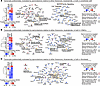 Figure 5
Pathways enriched within transcripts preferentially modulated by spironolactone overlap with those modulated in a comparable manner by isolated AR or MR antagonisms. (A–C) Heatmaps and STRING networks of enriched terms for transcripts preferentially modulated by spironolactone versus finerenone, bicalutamide, or both in endothelial cells (A), VSMCs (B), and fibroblasts (C). Heatmaps show expression in spironolactone- (Spiro-), finerenone- (Fine-,) and bicalutamide-treated (Bical-treated) samples relative to untreated VEDS aorta. Network nodes colored by expression change in spironolactone-treated versus untreated VEDS: red (upregulated), blue (downregulated). Node borders indicate whether spironolactone effect is recapitulated by bicalutamide (dark red), finerenone (blue), or neither (black). Magnified heatmaps are shown in Supplemental Figures 8–10.
In VSMCs, spironolactone and finerenone, but not bicalutamide, upregulated transcripts encoding elastin (Eln), lysyl oxidase (Lox), and microfibril associated protein 5 (Mfap5), whereas fibulin-5 (Fbln5) was also upregulated by bicalutamide, all indicating upregulated elastogenic gene programs (Figure 5B, Supplemental Figures 7 and 9, and Supplemental Table 3) (41). Other matrix components specifically upregulated by spironolactone and finerenone, but not bicalutamide, included insulin-like growth factor binding protein 7 (Igfbp7) (42), cellular communication network factor (Ccn1) (41), LIM and cysteine rich domains 1 (Lmcd1) (43), and basement membrane collagen (Col4a5) (Figure 5B and Supplemental Figure 8). Basement membrane collagens (Col4a1, Col4a5) were also among few transcripts more pronouncedly upregulated by spironolactone versus other treatments in adventitial fibroblasts (Figure 5C and Supplemental Figure 10).
Protection from rupture correlates with induction of a protective transcriptional signature in VSMCs. Transcriptional alterations from AR/MR antagonism in VEDS VSMCs partly resembled the adaptive aortic stress response induced by yes-associated protein (YAP) activation (44). Of 80 “adaptive transcripts” defined by Zhang et al. (44), 64 were detected in our dataset; 8 (Itgb1, Col5a2, Bcl2, Akt3, Tgfb2, Rock1, Rock2, Itga8) were upregulated in VEDS VSMCs versus untreated controls, independently of treatment (Log2 FC ≥ 0.20, Padj ≤ 0.05) (Figure 6A and Supplemental Table 3). AR/MR antagonists further upregulated integrin β1 (Itgb1), collagen (Col1a1, Col3a1, Col1a2, Lox), elastic fiber formation molecules (Mfap4, Eln, Fn1, Emilin1, Fbln5), and oxidative damage-protective enzymes (Gpx1, Sod3) (Figure 6A). However, transcripts encoding integrin α9 (Itga9), α8 (Itga8), and α5 (Itga5) were downregulated by spironolactone, while others (Smad3, Itgb3, Col6a2, Fbn1, Bcl2, Tgfbr2, Tgfbr1, Akt3, Mapk1, Smad2, Tgfb2, Akt2, Smad1, Col15a1, Ern1, Tgfb3, Col18a1) were unaffected (44) (Figure 6A and Supplemental Table 3), suggesting encoded factors may be unnecessary or hindering for protection. Since chronic synthetic pathway activation can provoke VSMC transition toward a deleterious “degradative” phenotype (45), we examined how spironolactone, finerenone, and bicalutamide modulated transcripts previously characterized as defining “contractile,” “synthetic,” “phagocyte-like” (lysosome/protein degradation organelles), “activated,” and “immune-like” VSMC phenotypes (45). Protective treatments upregulated transcripts associated with both contractile and synthetic phenotypes plus those related to proteasome-dependent protein degradation and autophagy; conversely, only B2m (encoding MHC class I–associated beta-2-microglobulin) was upregulated among “activated” VSMC phenotype transcripts (Mmp2, Mmp14, Mmp16, Vcam1, B2m, Spp1, Tnfrs11b, Cx3cl11), and “immune-like” transcripts were undetected in VSMCs (Supplemental Figure 11A). Both bulk-RNA seq and snRNA-seq showed correlation between treatment effectiveness and inhibition of lipogenic enzyme transcripts (Fasn, Scd1, Acly) (Figures 2 and 5). Decreased fatty acid synthase protein levels were confirmed in bulk aortic protein lysates following AR and MR inhibition (Supplemental Figure 11B).
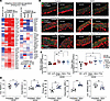 Figure 6
AR/MR antagonism upregulates adaptive transcriptional signature in VSMCs, increases medial collagen and elastin content, and alters the passive mechanical properties of the aorta in VEDS mice. (A) Heatmap of transcripts previously associated with adaptive response to mechanical stress in aneurysmal aorta (44). Expression is shown in VSMCs from VEDS versus control aorta, and spironolactone (Spiro)-, finerenone (Fine)-, and bicalutamide (Bical)-treated versus untreated VEDS mice. Red = upregulated; blue = downregulated. (B) Representative aortic sections from descending aorta of untreated control, untreated VEDS, and VEDS males treated with spironolactone, finerenone, or bicalutamide or with genetic Ar inactivation. Images show elastin (Alexa Fluor 633 hydrazide, green) and collagen (tdTomato-CNA35, red). Scale bar: 20 μm. (C) Quantification of medial collagen and elastic fiber content (normalized to the average value for untreated VEDS) in the descending thoracic aorta of male untreated control (n = 7) and VEDS (n = 7) mice, spironolactone- (Spiro-) (n = 6) or finerenone-treated (Fine-treated) (n = 6) treated VEDS mice, VEDS mice following genetic (Arnull/y, n = 4), or pharmacological (Bical, n = 5) androgen receptor inhibition. Each symbol represents an individual replicate; P value refers to 1-way ANOVA with Šídák post hoc test. (D) Passive biomechanical properties of descending thoracic aortas from untreated (n = 7) and spironolactone-treated (n = 10) VEDS mice under physiological pressure loads. Each symbol represents an individual replicate; light shading represents females. P value refers to Mann-Whitney U test. *P ≤ 0.05, **P ≤ 0.01, ***P ≤ 0.001.
AR/MR inhibition modulates ECM composition and passive biomechanical properties of the aorta. Increased elastogenesis and collagen synthesis gene expression suggested that upregulation of these pathways may compensate for collagen III deficiency and contribute to AR/MR inhibition’s protective effect. We thus examined aortas of Ar-deficient and bicalutamide-, spironolactone-, or finerenone-treated VEDS mice for collagen and elastin content. Histological assessment with picrosirius red staining (PSR) showed expected adventitial collagen content reduction in VEDS aortas, as previously observed (5), but revealed no treatment-dependent changes (Supplemental Figure 12). However, examination of medial collagen and elastic fiber content with pancollagen and elastin-binding probes (46, 47) revealed increases in relative collagen content following AR antagonism and both collagen and elastin following MR inhibition in male samples (Figure 6, B and C). Although similar elastic fiber content increases following spironolactone treatment were detected in female samples, other parameters were unaffected by treatments, possibly due to small sample size (Supplemental Figure 13).
To determine whether observed increases in relative collagen and elastin content associated with changes in aortic biomechanics, we examined passive mechanical responses of aortic tissues from spironolactone-treated and untreated VEDS mice (Figure 6D). Analysis showed spironolactone treatment rendered VEDS aortic tissues more deformable in both circumferential and axial directions across a broad, physiologically relevant stretch range, requiring greater stretch to achieve given stress levels (Supplemental Figure 14A). Accordingly, at representative systolic pressure, spironolactone-treated aortic tissues extended to higher biaxial stretch (Figure 6D) while maintaining stress levels comparable with nontreated mice (Supplemental Figure 14B). Under these loading conditions, spironolactone treatment selectively reduced circumferential tissue stiffness (Figure 6D) while preserving axial stiffness (Supplemental Figure 14B). This behavior conferred greater elastic energy storage during systole, indicating enhanced aortic capacity to function as a pressure reservoir augmenting diastolic blood flow (Figure 6D). Consistent with tissue behavior and preserved wall thickness (Supplemental Figure 14B), spironolactone-treated mice further exhibited increased cyclic distensibility, reflecting reduced structural aortic stiffness as a conduit (Figure 6D).
These data suggest that AR/MR antagonism promotes a partial “synthetic” VSMC phenotype resulting in increased ECM deposition that, together with additional effects on endothelial cell barrier function and adventitial fibroblasts, improves mechanical function and protects VEDS mice from aortic rupture, counteracting physiological downregulation of ECM synthetic pathways occurring at postnatal aortic development completion.


