
Research ArticleEndocrinologyNephrology
Open Access |  10.1172/jci.insight.204385
Not all reference samples are equal in single-cell transcriptomics of human kidney tissue
Rajasree Menon,1 Paul L. Kimmel,2 Edgar A. Otto,3 Lalita Subramanian,3 Christopher L. O’Connor,3 Bradley Godfrey,3 Cathy Smith,3 Fadhl Alakwaa,3 Celine C. Berthier,3 Minnie M. Sarwal,4 E. Steve Woodle,5 Laura Pyle,6 Ye Ji Choi,6 Patricia Ladd,7 John R. Sedor,8 Sylvia E. Rosas,9 Sushrut S. Waikar,10 Abhijit S. Naik,3 Ricardo Melo Ferreira,11 Michael T. Eadon,11 Markus Bitzer,3 Petter Bjornstad,6 Jeffrey B. Hodgin,12 Matthias Kretzler,1,3 and for the Kidney Precision Medicine Project (KPMP)
1Department of Computational Medicine and Bioinformatics, University of Michigan Medical School, Ann Arbor, Michigan, USA.
2Department of Medicine, School of Medicine and Health Sciences, George Washington University, Washington, DC, USA.
3Division of Nephrology, Department of Internal Medicine, University of Michigan Medical School, Ann Arbor, Michigan, USA.
4Division of Transplant Surgery, Department of Surgery, School of Medicine, University of California, San Francisco, California, USA.
5Department of Surgery, University of Cincinnati College of Medicine, Cincinnati, Ohio, USA.
6Departments of Pediatrics and Medicine, Division of Metabolism, Endocrinology and Nutrition, University of Washington, Seattle, Washington, USA.
7Department of Radiology, Division of Renal Diseases and Hypertension, University of Colorado Anschutz School of Medicine, Aurora, Colorado, USA.
8Medical Specialty and Lerner Research Institutes, Cleveland Clinic and Cleveland Clinic Lerner College of Medicine, Case Western Reserve University, Cleveland, Ohio, USA.
9Kidney and Hypertension unit, Joslin Diabetes Center and Harvard Medical School, Boston, Massachusetts, USA.
10Section of Nephrology, Department of Medicine, Boston Medical Center and Boston University Chobanian & Avedisian School of Medicine, Boston, Massachusetts, USA.
11Department of Medicine, Indiana University School of Medicine, Indianapolis, Indiana, USA.
12Department of Pathology, University of Michigan Medical School, Ann Arbor, Michigan, USA.
13KPMP is detailed in Supplemental Acknowledgments.
Address correspondence to: Rajasree Menon, Department of Computational Medicine and Bioinformatics,3B590 MSRB I, 1301 Catherine St., Ann Arbor, Michigan 48109-5624, USA.Email: rajmenon@umich .edu. Or to: Matthias Kretzler, 3B390 MSRB I, 1301 Catherine St., Ann Arbor, Michigan 48109-5624, USA. Phone: 734.615.5757; Email: kretzler@umich.edu.
Find articles by
Menon, R.
in:
PubMed
|
Google Scholar
|

1Department of Computational Medicine and Bioinformatics, University of Michigan Medical School, Ann Arbor, Michigan, USA.
2Department of Medicine, School of Medicine and Health Sciences, George Washington University, Washington, DC, USA.
3Division of Nephrology, Department of Internal Medicine, University of Michigan Medical School, Ann Arbor, Michigan, USA.
4Division of Transplant Surgery, Department of Surgery, School of Medicine, University of California, San Francisco, California, USA.
5Department of Surgery, University of Cincinnati College of Medicine, Cincinnati, Ohio, USA.
6Departments of Pediatrics and Medicine, Division of Metabolism, Endocrinology and Nutrition, University of Washington, Seattle, Washington, USA.
7Department of Radiology, Division of Renal Diseases and Hypertension, University of Colorado Anschutz School of Medicine, Aurora, Colorado, USA.
8Medical Specialty and Lerner Research Institutes, Cleveland Clinic and Cleveland Clinic Lerner College of Medicine, Case Western Reserve University, Cleveland, Ohio, USA.
9Kidney and Hypertension unit, Joslin Diabetes Center and Harvard Medical School, Boston, Massachusetts, USA.
10Section of Nephrology, Department of Medicine, Boston Medical Center and Boston University Chobanian & Avedisian School of Medicine, Boston, Massachusetts, USA.
11Department of Medicine, Indiana University School of Medicine, Indianapolis, Indiana, USA.
12Department of Pathology, University of Michigan Medical School, Ann Arbor, Michigan, USA.
13KPMP is detailed in Supplemental Acknowledgments.
Address correspondence to: Rajasree Menon, Department of Computational Medicine and Bioinformatics,3B590 MSRB I, 1301 Catherine St., Ann Arbor, Michigan 48109-5624, USA.Email: rajmenon@umich .edu. Or to: Matthias Kretzler, 3B390 MSRB I, 1301 Catherine St., Ann Arbor, Michigan 48109-5624, USA. Phone: 734.615.5757; Email: kretzler@umich.edu.
Find articles by Kimmel, P. in: PubMed | Google Scholar
1Department of Computational Medicine and Bioinformatics, University of Michigan Medical School, Ann Arbor, Michigan, USA.
2Department of Medicine, School of Medicine and Health Sciences, George Washington University, Washington, DC, USA.
3Division of Nephrology, Department of Internal Medicine, University of Michigan Medical School, Ann Arbor, Michigan, USA.
4Division of Transplant Surgery, Department of Surgery, School of Medicine, University of California, San Francisco, California, USA.
5Department of Surgery, University of Cincinnati College of Medicine, Cincinnati, Ohio, USA.
6Departments of Pediatrics and Medicine, Division of Metabolism, Endocrinology and Nutrition, University of Washington, Seattle, Washington, USA.
7Department of Radiology, Division of Renal Diseases and Hypertension, University of Colorado Anschutz School of Medicine, Aurora, Colorado, USA.
8Medical Specialty and Lerner Research Institutes, Cleveland Clinic and Cleveland Clinic Lerner College of Medicine, Case Western Reserve University, Cleveland, Ohio, USA.
9Kidney and Hypertension unit, Joslin Diabetes Center and Harvard Medical School, Boston, Massachusetts, USA.
10Section of Nephrology, Department of Medicine, Boston Medical Center and Boston University Chobanian & Avedisian School of Medicine, Boston, Massachusetts, USA.
11Department of Medicine, Indiana University School of Medicine, Indianapolis, Indiana, USA.
12Department of Pathology, University of Michigan Medical School, Ann Arbor, Michigan, USA.
13KPMP is detailed in Supplemental Acknowledgments.
Address correspondence to: Rajasree Menon, Department of Computational Medicine and Bioinformatics,3B590 MSRB I, 1301 Catherine St., Ann Arbor, Michigan 48109-5624, USA.Email: rajmenon@umich .edu. Or to: Matthias Kretzler, 3B390 MSRB I, 1301 Catherine St., Ann Arbor, Michigan 48109-5624, USA. Phone: 734.615.5757; Email: kretzler@umich.edu.
Find articles by
Otto, E.
in:
PubMed
|
Google Scholar
|
1Department of Computational Medicine and Bioinformatics, University of Michigan Medical School, Ann Arbor, Michigan, USA.
2Department of Medicine, School of Medicine and Health Sciences, George Washington University, Washington, DC, USA.
3Division of Nephrology, Department of Internal Medicine, University of Michigan Medical School, Ann Arbor, Michigan, USA.
4Division of Transplant Surgery, Department of Surgery, School of Medicine, University of California, San Francisco, California, USA.
5Department of Surgery, University of Cincinnati College of Medicine, Cincinnati, Ohio, USA.
6Departments of Pediatrics and Medicine, Division of Metabolism, Endocrinology and Nutrition, University of Washington, Seattle, Washington, USA.
7Department of Radiology, Division of Renal Diseases and Hypertension, University of Colorado Anschutz School of Medicine, Aurora, Colorado, USA.
8Medical Specialty and Lerner Research Institutes, Cleveland Clinic and Cleveland Clinic Lerner College of Medicine, Case Western Reserve University, Cleveland, Ohio, USA.
9Kidney and Hypertension unit, Joslin Diabetes Center and Harvard Medical School, Boston, Massachusetts, USA.
10Section of Nephrology, Department of Medicine, Boston Medical Center and Boston University Chobanian & Avedisian School of Medicine, Boston, Massachusetts, USA.
11Department of Medicine, Indiana University School of Medicine, Indianapolis, Indiana, USA.
12Department of Pathology, University of Michigan Medical School, Ann Arbor, Michigan, USA.
13KPMP is detailed in Supplemental Acknowledgments.
Address correspondence to: Rajasree Menon, Department of Computational Medicine and Bioinformatics,3B590 MSRB I, 1301 Catherine St., Ann Arbor, Michigan 48109-5624, USA.Email: rajmenon@umich .edu. Or to: Matthias Kretzler, 3B390 MSRB I, 1301 Catherine St., Ann Arbor, Michigan 48109-5624, USA. Phone: 734.615.5757; Email: kretzler@umich.edu.
Find articles by Subramanian, L. in: PubMed | Google Scholar
1Department of Computational Medicine and Bioinformatics, University of Michigan Medical School, Ann Arbor, Michigan, USA.
2Department of Medicine, School of Medicine and Health Sciences, George Washington University, Washington, DC, USA.
3Division of Nephrology, Department of Internal Medicine, University of Michigan Medical School, Ann Arbor, Michigan, USA.
4Division of Transplant Surgery, Department of Surgery, School of Medicine, University of California, San Francisco, California, USA.
5Department of Surgery, University of Cincinnati College of Medicine, Cincinnati, Ohio, USA.
6Departments of Pediatrics and Medicine, Division of Metabolism, Endocrinology and Nutrition, University of Washington, Seattle, Washington, USA.
7Department of Radiology, Division of Renal Diseases and Hypertension, University of Colorado Anschutz School of Medicine, Aurora, Colorado, USA.
8Medical Specialty and Lerner Research Institutes, Cleveland Clinic and Cleveland Clinic Lerner College of Medicine, Case Western Reserve University, Cleveland, Ohio, USA.
9Kidney and Hypertension unit, Joslin Diabetes Center and Harvard Medical School, Boston, Massachusetts, USA.
10Section of Nephrology, Department of Medicine, Boston Medical Center and Boston University Chobanian & Avedisian School of Medicine, Boston, Massachusetts, USA.
11Department of Medicine, Indiana University School of Medicine, Indianapolis, Indiana, USA.
12Department of Pathology, University of Michigan Medical School, Ann Arbor, Michigan, USA.
13KPMP is detailed in Supplemental Acknowledgments.
Address correspondence to: Rajasree Menon, Department of Computational Medicine and Bioinformatics,3B590 MSRB I, 1301 Catherine St., Ann Arbor, Michigan 48109-5624, USA.Email: rajmenon@umich .edu. Or to: Matthias Kretzler, 3B390 MSRB I, 1301 Catherine St., Ann Arbor, Michigan 48109-5624, USA. Phone: 734.615.5757; Email: kretzler@umich.edu.
Find articles by O’Connor, C. in: PubMed | Google Scholar
1Department of Computational Medicine and Bioinformatics, University of Michigan Medical School, Ann Arbor, Michigan, USA.
2Department of Medicine, School of Medicine and Health Sciences, George Washington University, Washington, DC, USA.
3Division of Nephrology, Department of Internal Medicine, University of Michigan Medical School, Ann Arbor, Michigan, USA.
4Division of Transplant Surgery, Department of Surgery, School of Medicine, University of California, San Francisco, California, USA.
5Department of Surgery, University of Cincinnati College of Medicine, Cincinnati, Ohio, USA.
6Departments of Pediatrics and Medicine, Division of Metabolism, Endocrinology and Nutrition, University of Washington, Seattle, Washington, USA.
7Department of Radiology, Division of Renal Diseases and Hypertension, University of Colorado Anschutz School of Medicine, Aurora, Colorado, USA.
8Medical Specialty and Lerner Research Institutes, Cleveland Clinic and Cleveland Clinic Lerner College of Medicine, Case Western Reserve University, Cleveland, Ohio, USA.
9Kidney and Hypertension unit, Joslin Diabetes Center and Harvard Medical School, Boston, Massachusetts, USA.
10Section of Nephrology, Department of Medicine, Boston Medical Center and Boston University Chobanian & Avedisian School of Medicine, Boston, Massachusetts, USA.
11Department of Medicine, Indiana University School of Medicine, Indianapolis, Indiana, USA.
12Department of Pathology, University of Michigan Medical School, Ann Arbor, Michigan, USA.
13KPMP is detailed in Supplemental Acknowledgments.
Address correspondence to: Rajasree Menon, Department of Computational Medicine and Bioinformatics,3B590 MSRB I, 1301 Catherine St., Ann Arbor, Michigan 48109-5624, USA.Email: rajmenon@umich .edu. Or to: Matthias Kretzler, 3B390 MSRB I, 1301 Catherine St., Ann Arbor, Michigan 48109-5624, USA. Phone: 734.615.5757; Email: kretzler@umich.edu.
Find articles by Godfrey, B. in: PubMed | Google Scholar
1Department of Computational Medicine and Bioinformatics, University of Michigan Medical School, Ann Arbor, Michigan, USA.
2Department of Medicine, School of Medicine and Health Sciences, George Washington University, Washington, DC, USA.
3Division of Nephrology, Department of Internal Medicine, University of Michigan Medical School, Ann Arbor, Michigan, USA.
4Division of Transplant Surgery, Department of Surgery, School of Medicine, University of California, San Francisco, California, USA.
5Department of Surgery, University of Cincinnati College of Medicine, Cincinnati, Ohio, USA.
6Departments of Pediatrics and Medicine, Division of Metabolism, Endocrinology and Nutrition, University of Washington, Seattle, Washington, USA.
7Department of Radiology, Division of Renal Diseases and Hypertension, University of Colorado Anschutz School of Medicine, Aurora, Colorado, USA.
8Medical Specialty and Lerner Research Institutes, Cleveland Clinic and Cleveland Clinic Lerner College of Medicine, Case Western Reserve University, Cleveland, Ohio, USA.
9Kidney and Hypertension unit, Joslin Diabetes Center and Harvard Medical School, Boston, Massachusetts, USA.
10Section of Nephrology, Department of Medicine, Boston Medical Center and Boston University Chobanian & Avedisian School of Medicine, Boston, Massachusetts, USA.
11Department of Medicine, Indiana University School of Medicine, Indianapolis, Indiana, USA.
12Department of Pathology, University of Michigan Medical School, Ann Arbor, Michigan, USA.
13KPMP is detailed in Supplemental Acknowledgments.
Address correspondence to: Rajasree Menon, Department of Computational Medicine and Bioinformatics,3B590 MSRB I, 1301 Catherine St., Ann Arbor, Michigan 48109-5624, USA.Email: rajmenon@umich .edu. Or to: Matthias Kretzler, 3B390 MSRB I, 1301 Catherine St., Ann Arbor, Michigan 48109-5624, USA. Phone: 734.615.5757; Email: kretzler@umich.edu.
Find articles by Smith, C. in: PubMed | Google Scholar
1Department of Computational Medicine and Bioinformatics, University of Michigan Medical School, Ann Arbor, Michigan, USA.
2Department of Medicine, School of Medicine and Health Sciences, George Washington University, Washington, DC, USA.
3Division of Nephrology, Department of Internal Medicine, University of Michigan Medical School, Ann Arbor, Michigan, USA.
4Division of Transplant Surgery, Department of Surgery, School of Medicine, University of California, San Francisco, California, USA.
5Department of Surgery, University of Cincinnati College of Medicine, Cincinnati, Ohio, USA.
6Departments of Pediatrics and Medicine, Division of Metabolism, Endocrinology and Nutrition, University of Washington, Seattle, Washington, USA.
7Department of Radiology, Division of Renal Diseases and Hypertension, University of Colorado Anschutz School of Medicine, Aurora, Colorado, USA.
8Medical Specialty and Lerner Research Institutes, Cleveland Clinic and Cleveland Clinic Lerner College of Medicine, Case Western Reserve University, Cleveland, Ohio, USA.
9Kidney and Hypertension unit, Joslin Diabetes Center and Harvard Medical School, Boston, Massachusetts, USA.
10Section of Nephrology, Department of Medicine, Boston Medical Center and Boston University Chobanian & Avedisian School of Medicine, Boston, Massachusetts, USA.
11Department of Medicine, Indiana University School of Medicine, Indianapolis, Indiana, USA.
12Department of Pathology, University of Michigan Medical School, Ann Arbor, Michigan, USA.
13KPMP is detailed in Supplemental Acknowledgments.
Address correspondence to: Rajasree Menon, Department of Computational Medicine and Bioinformatics,3B590 MSRB I, 1301 Catherine St., Ann Arbor, Michigan 48109-5624, USA.Email: rajmenon@umich .edu. Or to: Matthias Kretzler, 3B390 MSRB I, 1301 Catherine St., Ann Arbor, Michigan 48109-5624, USA. Phone: 734.615.5757; Email: kretzler@umich.edu.
Find articles by Alakwaa, F. in: PubMed | Google Scholar
1Department of Computational Medicine and Bioinformatics, University of Michigan Medical School, Ann Arbor, Michigan, USA.
2Department of Medicine, School of Medicine and Health Sciences, George Washington University, Washington, DC, USA.
3Division of Nephrology, Department of Internal Medicine, University of Michigan Medical School, Ann Arbor, Michigan, USA.
4Division of Transplant Surgery, Department of Surgery, School of Medicine, University of California, San Francisco, California, USA.
5Department of Surgery, University of Cincinnati College of Medicine, Cincinnati, Ohio, USA.
6Departments of Pediatrics and Medicine, Division of Metabolism, Endocrinology and Nutrition, University of Washington, Seattle, Washington, USA.
7Department of Radiology, Division of Renal Diseases and Hypertension, University of Colorado Anschutz School of Medicine, Aurora, Colorado, USA.
8Medical Specialty and Lerner Research Institutes, Cleveland Clinic and Cleveland Clinic Lerner College of Medicine, Case Western Reserve University, Cleveland, Ohio, USA.
9Kidney and Hypertension unit, Joslin Diabetes Center and Harvard Medical School, Boston, Massachusetts, USA.
10Section of Nephrology, Department of Medicine, Boston Medical Center and Boston University Chobanian & Avedisian School of Medicine, Boston, Massachusetts, USA.
11Department of Medicine, Indiana University School of Medicine, Indianapolis, Indiana, USA.
12Department of Pathology, University of Michigan Medical School, Ann Arbor, Michigan, USA.
13KPMP is detailed in Supplemental Acknowledgments.
Address correspondence to: Rajasree Menon, Department of Computational Medicine and Bioinformatics,3B590 MSRB I, 1301 Catherine St., Ann Arbor, Michigan 48109-5624, USA.Email: rajmenon@umich .edu. Or to: Matthias Kretzler, 3B390 MSRB I, 1301 Catherine St., Ann Arbor, Michigan 48109-5624, USA. Phone: 734.615.5757; Email: kretzler@umich.edu.
Find articles by
Berthier, C.
in:
PubMed
|
Google Scholar
|
1Department of Computational Medicine and Bioinformatics, University of Michigan Medical School, Ann Arbor, Michigan, USA.
2Department of Medicine, School of Medicine and Health Sciences, George Washington University, Washington, DC, USA.
3Division of Nephrology, Department of Internal Medicine, University of Michigan Medical School, Ann Arbor, Michigan, USA.
4Division of Transplant Surgery, Department of Surgery, School of Medicine, University of California, San Francisco, California, USA.
5Department of Surgery, University of Cincinnati College of Medicine, Cincinnati, Ohio, USA.
6Departments of Pediatrics and Medicine, Division of Metabolism, Endocrinology and Nutrition, University of Washington, Seattle, Washington, USA.
7Department of Radiology, Division of Renal Diseases and Hypertension, University of Colorado Anschutz School of Medicine, Aurora, Colorado, USA.
8Medical Specialty and Lerner Research Institutes, Cleveland Clinic and Cleveland Clinic Lerner College of Medicine, Case Western Reserve University, Cleveland, Ohio, USA.
9Kidney and Hypertension unit, Joslin Diabetes Center and Harvard Medical School, Boston, Massachusetts, USA.
10Section of Nephrology, Department of Medicine, Boston Medical Center and Boston University Chobanian & Avedisian School of Medicine, Boston, Massachusetts, USA.
11Department of Medicine, Indiana University School of Medicine, Indianapolis, Indiana, USA.
12Department of Pathology, University of Michigan Medical School, Ann Arbor, Michigan, USA.
13KPMP is detailed in Supplemental Acknowledgments.
Address correspondence to: Rajasree Menon, Department of Computational Medicine and Bioinformatics,3B590 MSRB I, 1301 Catherine St., Ann Arbor, Michigan 48109-5624, USA.Email: rajmenon@umich .edu. Or to: Matthias Kretzler, 3B390 MSRB I, 1301 Catherine St., Ann Arbor, Michigan 48109-5624, USA. Phone: 734.615.5757; Email: kretzler@umich.edu.
Find articles by Sarwal, M. in: PubMed | Google Scholar
1Department of Computational Medicine and Bioinformatics, University of Michigan Medical School, Ann Arbor, Michigan, USA.
2Department of Medicine, School of Medicine and Health Sciences, George Washington University, Washington, DC, USA.
3Division of Nephrology, Department of Internal Medicine, University of Michigan Medical School, Ann Arbor, Michigan, USA.
4Division of Transplant Surgery, Department of Surgery, School of Medicine, University of California, San Francisco, California, USA.
5Department of Surgery, University of Cincinnati College of Medicine, Cincinnati, Ohio, USA.
6Departments of Pediatrics and Medicine, Division of Metabolism, Endocrinology and Nutrition, University of Washington, Seattle, Washington, USA.
7Department of Radiology, Division of Renal Diseases and Hypertension, University of Colorado Anschutz School of Medicine, Aurora, Colorado, USA.
8Medical Specialty and Lerner Research Institutes, Cleveland Clinic and Cleveland Clinic Lerner College of Medicine, Case Western Reserve University, Cleveland, Ohio, USA.
9Kidney and Hypertension unit, Joslin Diabetes Center and Harvard Medical School, Boston, Massachusetts, USA.
10Section of Nephrology, Department of Medicine, Boston Medical Center and Boston University Chobanian & Avedisian School of Medicine, Boston, Massachusetts, USA.
11Department of Medicine, Indiana University School of Medicine, Indianapolis, Indiana, USA.
12Department of Pathology, University of Michigan Medical School, Ann Arbor, Michigan, USA.
13KPMP is detailed in Supplemental Acknowledgments.
Address correspondence to: Rajasree Menon, Department of Computational Medicine and Bioinformatics,3B590 MSRB I, 1301 Catherine St., Ann Arbor, Michigan 48109-5624, USA.Email: rajmenon@umich .edu. Or to: Matthias Kretzler, 3B390 MSRB I, 1301 Catherine St., Ann Arbor, Michigan 48109-5624, USA. Phone: 734.615.5757; Email: kretzler@umich.edu.
Find articles by Woodle, E. in: PubMed | Google Scholar
1Department of Computational Medicine and Bioinformatics, University of Michigan Medical School, Ann Arbor, Michigan, USA.
2Department of Medicine, School of Medicine and Health Sciences, George Washington University, Washington, DC, USA.
3Division of Nephrology, Department of Internal Medicine, University of Michigan Medical School, Ann Arbor, Michigan, USA.
4Division of Transplant Surgery, Department of Surgery, School of Medicine, University of California, San Francisco, California, USA.
5Department of Surgery, University of Cincinnati College of Medicine, Cincinnati, Ohio, USA.
6Departments of Pediatrics and Medicine, Division of Metabolism, Endocrinology and Nutrition, University of Washington, Seattle, Washington, USA.
7Department of Radiology, Division of Renal Diseases and Hypertension, University of Colorado Anschutz School of Medicine, Aurora, Colorado, USA.
8Medical Specialty and Lerner Research Institutes, Cleveland Clinic and Cleveland Clinic Lerner College of Medicine, Case Western Reserve University, Cleveland, Ohio, USA.
9Kidney and Hypertension unit, Joslin Diabetes Center and Harvard Medical School, Boston, Massachusetts, USA.
10Section of Nephrology, Department of Medicine, Boston Medical Center and Boston University Chobanian & Avedisian School of Medicine, Boston, Massachusetts, USA.
11Department of Medicine, Indiana University School of Medicine, Indianapolis, Indiana, USA.
12Department of Pathology, University of Michigan Medical School, Ann Arbor, Michigan, USA.
13KPMP is detailed in Supplemental Acknowledgments.
Address correspondence to: Rajasree Menon, Department of Computational Medicine and Bioinformatics,3B590 MSRB I, 1301 Catherine St., Ann Arbor, Michigan 48109-5624, USA.Email: rajmenon@umich .edu. Or to: Matthias Kretzler, 3B390 MSRB I, 1301 Catherine St., Ann Arbor, Michigan 48109-5624, USA. Phone: 734.615.5757; Email: kretzler@umich.edu.
Find articles by Pyle, L. in: PubMed | Google Scholar
1Department of Computational Medicine and Bioinformatics, University of Michigan Medical School, Ann Arbor, Michigan, USA.
2Department of Medicine, School of Medicine and Health Sciences, George Washington University, Washington, DC, USA.
3Division of Nephrology, Department of Internal Medicine, University of Michigan Medical School, Ann Arbor, Michigan, USA.
4Division of Transplant Surgery, Department of Surgery, School of Medicine, University of California, San Francisco, California, USA.
5Department of Surgery, University of Cincinnati College of Medicine, Cincinnati, Ohio, USA.
6Departments of Pediatrics and Medicine, Division of Metabolism, Endocrinology and Nutrition, University of Washington, Seattle, Washington, USA.
7Department of Radiology, Division of Renal Diseases and Hypertension, University of Colorado Anschutz School of Medicine, Aurora, Colorado, USA.
8Medical Specialty and Lerner Research Institutes, Cleveland Clinic and Cleveland Clinic Lerner College of Medicine, Case Western Reserve University, Cleveland, Ohio, USA.
9Kidney and Hypertension unit, Joslin Diabetes Center and Harvard Medical School, Boston, Massachusetts, USA.
10Section of Nephrology, Department of Medicine, Boston Medical Center and Boston University Chobanian & Avedisian School of Medicine, Boston, Massachusetts, USA.
11Department of Medicine, Indiana University School of Medicine, Indianapolis, Indiana, USA.
12Department of Pathology, University of Michigan Medical School, Ann Arbor, Michigan, USA.
13KPMP is detailed in Supplemental Acknowledgments.
Address correspondence to: Rajasree Menon, Department of Computational Medicine and Bioinformatics,3B590 MSRB I, 1301 Catherine St., Ann Arbor, Michigan 48109-5624, USA.Email: rajmenon@umich .edu. Or to: Matthias Kretzler, 3B390 MSRB I, 1301 Catherine St., Ann Arbor, Michigan 48109-5624, USA. Phone: 734.615.5757; Email: kretzler@umich.edu.
Find articles by Choi, Y. in: PubMed | Google Scholar
1Department of Computational Medicine and Bioinformatics, University of Michigan Medical School, Ann Arbor, Michigan, USA.
2Department of Medicine, School of Medicine and Health Sciences, George Washington University, Washington, DC, USA.
3Division of Nephrology, Department of Internal Medicine, University of Michigan Medical School, Ann Arbor, Michigan, USA.
4Division of Transplant Surgery, Department of Surgery, School of Medicine, University of California, San Francisco, California, USA.
5Department of Surgery, University of Cincinnati College of Medicine, Cincinnati, Ohio, USA.
6Departments of Pediatrics and Medicine, Division of Metabolism, Endocrinology and Nutrition, University of Washington, Seattle, Washington, USA.
7Department of Radiology, Division of Renal Diseases and Hypertension, University of Colorado Anschutz School of Medicine, Aurora, Colorado, USA.
8Medical Specialty and Lerner Research Institutes, Cleveland Clinic and Cleveland Clinic Lerner College of Medicine, Case Western Reserve University, Cleveland, Ohio, USA.
9Kidney and Hypertension unit, Joslin Diabetes Center and Harvard Medical School, Boston, Massachusetts, USA.
10Section of Nephrology, Department of Medicine, Boston Medical Center and Boston University Chobanian & Avedisian School of Medicine, Boston, Massachusetts, USA.
11Department of Medicine, Indiana University School of Medicine, Indianapolis, Indiana, USA.
12Department of Pathology, University of Michigan Medical School, Ann Arbor, Michigan, USA.
13KPMP is detailed in Supplemental Acknowledgments.
Address correspondence to: Rajasree Menon, Department of Computational Medicine and Bioinformatics,3B590 MSRB I, 1301 Catherine St., Ann Arbor, Michigan 48109-5624, USA.Email: rajmenon@umich .edu. Or to: Matthias Kretzler, 3B390 MSRB I, 1301 Catherine St., Ann Arbor, Michigan 48109-5624, USA. Phone: 734.615.5757; Email: kretzler@umich.edu.
Find articles by Ladd, P. in: PubMed | Google Scholar
1Department of Computational Medicine and Bioinformatics, University of Michigan Medical School, Ann Arbor, Michigan, USA.
2Department of Medicine, School of Medicine and Health Sciences, George Washington University, Washington, DC, USA.
3Division of Nephrology, Department of Internal Medicine, University of Michigan Medical School, Ann Arbor, Michigan, USA.
4Division of Transplant Surgery, Department of Surgery, School of Medicine, University of California, San Francisco, California, USA.
5Department of Surgery, University of Cincinnati College of Medicine, Cincinnati, Ohio, USA.
6Departments of Pediatrics and Medicine, Division of Metabolism, Endocrinology and Nutrition, University of Washington, Seattle, Washington, USA.
7Department of Radiology, Division of Renal Diseases and Hypertension, University of Colorado Anschutz School of Medicine, Aurora, Colorado, USA.
8Medical Specialty and Lerner Research Institutes, Cleveland Clinic and Cleveland Clinic Lerner College of Medicine, Case Western Reserve University, Cleveland, Ohio, USA.
9Kidney and Hypertension unit, Joslin Diabetes Center and Harvard Medical School, Boston, Massachusetts, USA.
10Section of Nephrology, Department of Medicine, Boston Medical Center and Boston University Chobanian & Avedisian School of Medicine, Boston, Massachusetts, USA.
11Department of Medicine, Indiana University School of Medicine, Indianapolis, Indiana, USA.
12Department of Pathology, University of Michigan Medical School, Ann Arbor, Michigan, USA.
13KPMP is detailed in Supplemental Acknowledgments.
Address correspondence to: Rajasree Menon, Department of Computational Medicine and Bioinformatics,3B590 MSRB I, 1301 Catherine St., Ann Arbor, Michigan 48109-5624, USA.Email: rajmenon@umich .edu. Or to: Matthias Kretzler, 3B390 MSRB I, 1301 Catherine St., Ann Arbor, Michigan 48109-5624, USA. Phone: 734.615.5757; Email: kretzler@umich.edu.
Find articles by Sedor, J. in: PubMed | Google Scholar
1Department of Computational Medicine and Bioinformatics, University of Michigan Medical School, Ann Arbor, Michigan, USA.
2Department of Medicine, School of Medicine and Health Sciences, George Washington University, Washington, DC, USA.
3Division of Nephrology, Department of Internal Medicine, University of Michigan Medical School, Ann Arbor, Michigan, USA.
4Division of Transplant Surgery, Department of Surgery, School of Medicine, University of California, San Francisco, California, USA.
5Department of Surgery, University of Cincinnati College of Medicine, Cincinnati, Ohio, USA.
6Departments of Pediatrics and Medicine, Division of Metabolism, Endocrinology and Nutrition, University of Washington, Seattle, Washington, USA.
7Department of Radiology, Division of Renal Diseases and Hypertension, University of Colorado Anschutz School of Medicine, Aurora, Colorado, USA.
8Medical Specialty and Lerner Research Institutes, Cleveland Clinic and Cleveland Clinic Lerner College of Medicine, Case Western Reserve University, Cleveland, Ohio, USA.
9Kidney and Hypertension unit, Joslin Diabetes Center and Harvard Medical School, Boston, Massachusetts, USA.
10Section of Nephrology, Department of Medicine, Boston Medical Center and Boston University Chobanian & Avedisian School of Medicine, Boston, Massachusetts, USA.
11Department of Medicine, Indiana University School of Medicine, Indianapolis, Indiana, USA.
12Department of Pathology, University of Michigan Medical School, Ann Arbor, Michigan, USA.
13KPMP is detailed in Supplemental Acknowledgments.
Address correspondence to: Rajasree Menon, Department of Computational Medicine and Bioinformatics,3B590 MSRB I, 1301 Catherine St., Ann Arbor, Michigan 48109-5624, USA.Email: rajmenon@umich .edu. Or to: Matthias Kretzler, 3B390 MSRB I, 1301 Catherine St., Ann Arbor, Michigan 48109-5624, USA. Phone: 734.615.5757; Email: kretzler@umich.edu.
Find articles by Rosas, S. in: PubMed | Google Scholar
1Department of Computational Medicine and Bioinformatics, University of Michigan Medical School, Ann Arbor, Michigan, USA.
2Department of Medicine, School of Medicine and Health Sciences, George Washington University, Washington, DC, USA.
3Division of Nephrology, Department of Internal Medicine, University of Michigan Medical School, Ann Arbor, Michigan, USA.
4Division of Transplant Surgery, Department of Surgery, School of Medicine, University of California, San Francisco, California, USA.
5Department of Surgery, University of Cincinnati College of Medicine, Cincinnati, Ohio, USA.
6Departments of Pediatrics and Medicine, Division of Metabolism, Endocrinology and Nutrition, University of Washington, Seattle, Washington, USA.
7Department of Radiology, Division of Renal Diseases and Hypertension, University of Colorado Anschutz School of Medicine, Aurora, Colorado, USA.
8Medical Specialty and Lerner Research Institutes, Cleveland Clinic and Cleveland Clinic Lerner College of Medicine, Case Western Reserve University, Cleveland, Ohio, USA.
9Kidney and Hypertension unit, Joslin Diabetes Center and Harvard Medical School, Boston, Massachusetts, USA.
10Section of Nephrology, Department of Medicine, Boston Medical Center and Boston University Chobanian & Avedisian School of Medicine, Boston, Massachusetts, USA.
11Department of Medicine, Indiana University School of Medicine, Indianapolis, Indiana, USA.
12Department of Pathology, University of Michigan Medical School, Ann Arbor, Michigan, USA.
13KPMP is detailed in Supplemental Acknowledgments.
Address correspondence to: Rajasree Menon, Department of Computational Medicine and Bioinformatics,3B590 MSRB I, 1301 Catherine St., Ann Arbor, Michigan 48109-5624, USA.Email: rajmenon@umich .edu. Or to: Matthias Kretzler, 3B390 MSRB I, 1301 Catherine St., Ann Arbor, Michigan 48109-5624, USA. Phone: 734.615.5757; Email: kretzler@umich.edu.
Find articles by
Waikar, S.
in:
PubMed
|
Google Scholar
|
1Department of Computational Medicine and Bioinformatics, University of Michigan Medical School, Ann Arbor, Michigan, USA.
2Department of Medicine, School of Medicine and Health Sciences, George Washington University, Washington, DC, USA.
3Division of Nephrology, Department of Internal Medicine, University of Michigan Medical School, Ann Arbor, Michigan, USA.
4Division of Transplant Surgery, Department of Surgery, School of Medicine, University of California, San Francisco, California, USA.
5Department of Surgery, University of Cincinnati College of Medicine, Cincinnati, Ohio, USA.
6Departments of Pediatrics and Medicine, Division of Metabolism, Endocrinology and Nutrition, University of Washington, Seattle, Washington, USA.
7Department of Radiology, Division of Renal Diseases and Hypertension, University of Colorado Anschutz School of Medicine, Aurora, Colorado, USA.
8Medical Specialty and Lerner Research Institutes, Cleveland Clinic and Cleveland Clinic Lerner College of Medicine, Case Western Reserve University, Cleveland, Ohio, USA.
9Kidney and Hypertension unit, Joslin Diabetes Center and Harvard Medical School, Boston, Massachusetts, USA.
10Section of Nephrology, Department of Medicine, Boston Medical Center and Boston University Chobanian & Avedisian School of Medicine, Boston, Massachusetts, USA.
11Department of Medicine, Indiana University School of Medicine, Indianapolis, Indiana, USA.
12Department of Pathology, University of Michigan Medical School, Ann Arbor, Michigan, USA.
13KPMP is detailed in Supplemental Acknowledgments.
Address correspondence to: Rajasree Menon, Department of Computational Medicine and Bioinformatics,3B590 MSRB I, 1301 Catherine St., Ann Arbor, Michigan 48109-5624, USA.Email: rajmenon@umich .edu. Or to: Matthias Kretzler, 3B390 MSRB I, 1301 Catherine St., Ann Arbor, Michigan 48109-5624, USA. Phone: 734.615.5757; Email: kretzler@umich.edu.
Find articles by
Naik, A.
in:
PubMed
|
Google Scholar
|
1Department of Computational Medicine and Bioinformatics, University of Michigan Medical School, Ann Arbor, Michigan, USA.
2Department of Medicine, School of Medicine and Health Sciences, George Washington University, Washington, DC, USA.
3Division of Nephrology, Department of Internal Medicine, University of Michigan Medical School, Ann Arbor, Michigan, USA.
4Division of Transplant Surgery, Department of Surgery, School of Medicine, University of California, San Francisco, California, USA.
5Department of Surgery, University of Cincinnati College of Medicine, Cincinnati, Ohio, USA.
6Departments of Pediatrics and Medicine, Division of Metabolism, Endocrinology and Nutrition, University of Washington, Seattle, Washington, USA.
7Department of Radiology, Division of Renal Diseases and Hypertension, University of Colorado Anschutz School of Medicine, Aurora, Colorado, USA.
8Medical Specialty and Lerner Research Institutes, Cleveland Clinic and Cleveland Clinic Lerner College of Medicine, Case Western Reserve University, Cleveland, Ohio, USA.
9Kidney and Hypertension unit, Joslin Diabetes Center and Harvard Medical School, Boston, Massachusetts, USA.
10Section of Nephrology, Department of Medicine, Boston Medical Center and Boston University Chobanian & Avedisian School of Medicine, Boston, Massachusetts, USA.
11Department of Medicine, Indiana University School of Medicine, Indianapolis, Indiana, USA.
12Department of Pathology, University of Michigan Medical School, Ann Arbor, Michigan, USA.
13KPMP is detailed in Supplemental Acknowledgments.
Address correspondence to: Rajasree Menon, Department of Computational Medicine and Bioinformatics,3B590 MSRB I, 1301 Catherine St., Ann Arbor, Michigan 48109-5624, USA.Email: rajmenon@umich .edu. Or to: Matthias Kretzler, 3B390 MSRB I, 1301 Catherine St., Ann Arbor, Michigan 48109-5624, USA. Phone: 734.615.5757; Email: kretzler@umich.edu.
Find articles by Melo Ferreira, R. in: PubMed | Google Scholar
1Department of Computational Medicine and Bioinformatics, University of Michigan Medical School, Ann Arbor, Michigan, USA.
2Department of Medicine, School of Medicine and Health Sciences, George Washington University, Washington, DC, USA.
3Division of Nephrology, Department of Internal Medicine, University of Michigan Medical School, Ann Arbor, Michigan, USA.
4Division of Transplant Surgery, Department of Surgery, School of Medicine, University of California, San Francisco, California, USA.
5Department of Surgery, University of Cincinnati College of Medicine, Cincinnati, Ohio, USA.
6Departments of Pediatrics and Medicine, Division of Metabolism, Endocrinology and Nutrition, University of Washington, Seattle, Washington, USA.
7Department of Radiology, Division of Renal Diseases and Hypertension, University of Colorado Anschutz School of Medicine, Aurora, Colorado, USA.
8Medical Specialty and Lerner Research Institutes, Cleveland Clinic and Cleveland Clinic Lerner College of Medicine, Case Western Reserve University, Cleveland, Ohio, USA.
9Kidney and Hypertension unit, Joslin Diabetes Center and Harvard Medical School, Boston, Massachusetts, USA.
10Section of Nephrology, Department of Medicine, Boston Medical Center and Boston University Chobanian & Avedisian School of Medicine, Boston, Massachusetts, USA.
11Department of Medicine, Indiana University School of Medicine, Indianapolis, Indiana, USA.
12Department of Pathology, University of Michigan Medical School, Ann Arbor, Michigan, USA.
13KPMP is detailed in Supplemental Acknowledgments.
Address correspondence to: Rajasree Menon, Department of Computational Medicine and Bioinformatics,3B590 MSRB I, 1301 Catherine St., Ann Arbor, Michigan 48109-5624, USA.Email: rajmenon@umich .edu. Or to: Matthias Kretzler, 3B390 MSRB I, 1301 Catherine St., Ann Arbor, Michigan 48109-5624, USA. Phone: 734.615.5757; Email: kretzler@umich.edu.
Find articles by
Eadon, M.
in:
PubMed
|
Google Scholar
|
1Department of Computational Medicine and Bioinformatics, University of Michigan Medical School, Ann Arbor, Michigan, USA.
2Department of Medicine, School of Medicine and Health Sciences, George Washington University, Washington, DC, USA.
3Division of Nephrology, Department of Internal Medicine, University of Michigan Medical School, Ann Arbor, Michigan, USA.
4Division of Transplant Surgery, Department of Surgery, School of Medicine, University of California, San Francisco, California, USA.
5Department of Surgery, University of Cincinnati College of Medicine, Cincinnati, Ohio, USA.
6Departments of Pediatrics and Medicine, Division of Metabolism, Endocrinology and Nutrition, University of Washington, Seattle, Washington, USA.
7Department of Radiology, Division of Renal Diseases and Hypertension, University of Colorado Anschutz School of Medicine, Aurora, Colorado, USA.
8Medical Specialty and Lerner Research Institutes, Cleveland Clinic and Cleveland Clinic Lerner College of Medicine, Case Western Reserve University, Cleveland, Ohio, USA.
9Kidney and Hypertension unit, Joslin Diabetes Center and Harvard Medical School, Boston, Massachusetts, USA.
10Section of Nephrology, Department of Medicine, Boston Medical Center and Boston University Chobanian & Avedisian School of Medicine, Boston, Massachusetts, USA.
11Department of Medicine, Indiana University School of Medicine, Indianapolis, Indiana, USA.
12Department of Pathology, University of Michigan Medical School, Ann Arbor, Michigan, USA.
13KPMP is detailed in Supplemental Acknowledgments.
Address correspondence to: Rajasree Menon, Department of Computational Medicine and Bioinformatics,3B590 MSRB I, 1301 Catherine St., Ann Arbor, Michigan 48109-5624, USA.Email: rajmenon@umich .edu. Or to: Matthias Kretzler, 3B390 MSRB I, 1301 Catherine St., Ann Arbor, Michigan 48109-5624, USA. Phone: 734.615.5757; Email: kretzler@umich.edu.
Find articles by
Bitzer, M.
in:
PubMed
|
Google Scholar
|
1Department of Computational Medicine and Bioinformatics, University of Michigan Medical School, Ann Arbor, Michigan, USA.
2Department of Medicine, School of Medicine and Health Sciences, George Washington University, Washington, DC, USA.
3Division of Nephrology, Department of Internal Medicine, University of Michigan Medical School, Ann Arbor, Michigan, USA.
4Division of Transplant Surgery, Department of Surgery, School of Medicine, University of California, San Francisco, California, USA.
5Department of Surgery, University of Cincinnati College of Medicine, Cincinnati, Ohio, USA.
6Departments of Pediatrics and Medicine, Division of Metabolism, Endocrinology and Nutrition, University of Washington, Seattle, Washington, USA.
7Department of Radiology, Division of Renal Diseases and Hypertension, University of Colorado Anschutz School of Medicine, Aurora, Colorado, USA.
8Medical Specialty and Lerner Research Institutes, Cleveland Clinic and Cleveland Clinic Lerner College of Medicine, Case Western Reserve University, Cleveland, Ohio, USA.
9Kidney and Hypertension unit, Joslin Diabetes Center and Harvard Medical School, Boston, Massachusetts, USA.
10Section of Nephrology, Department of Medicine, Boston Medical Center and Boston University Chobanian & Avedisian School of Medicine, Boston, Massachusetts, USA.
11Department of Medicine, Indiana University School of Medicine, Indianapolis, Indiana, USA.
12Department of Pathology, University of Michigan Medical School, Ann Arbor, Michigan, USA.
13KPMP is detailed in Supplemental Acknowledgments.
Address correspondence to: Rajasree Menon, Department of Computational Medicine and Bioinformatics,3B590 MSRB I, 1301 Catherine St., Ann Arbor, Michigan 48109-5624, USA.Email: rajmenon@umich .edu. Or to: Matthias Kretzler, 3B390 MSRB I, 1301 Catherine St., Ann Arbor, Michigan 48109-5624, USA. Phone: 734.615.5757; Email: kretzler@umich.edu.
Find articles by Bjornstad, P. in: PubMed | Google Scholar
1Department of Computational Medicine and Bioinformatics, University of Michigan Medical School, Ann Arbor, Michigan, USA.
2Department of Medicine, School of Medicine and Health Sciences, George Washington University, Washington, DC, USA.
3Division of Nephrology, Department of Internal Medicine, University of Michigan Medical School, Ann Arbor, Michigan, USA.
4Division of Transplant Surgery, Department of Surgery, School of Medicine, University of California, San Francisco, California, USA.
5Department of Surgery, University of Cincinnati College of Medicine, Cincinnati, Ohio, USA.
6Departments of Pediatrics and Medicine, Division of Metabolism, Endocrinology and Nutrition, University of Washington, Seattle, Washington, USA.
7Department of Radiology, Division of Renal Diseases and Hypertension, University of Colorado Anschutz School of Medicine, Aurora, Colorado, USA.
8Medical Specialty and Lerner Research Institutes, Cleveland Clinic and Cleveland Clinic Lerner College of Medicine, Case Western Reserve University, Cleveland, Ohio, USA.
9Kidney and Hypertension unit, Joslin Diabetes Center and Harvard Medical School, Boston, Massachusetts, USA.
10Section of Nephrology, Department of Medicine, Boston Medical Center and Boston University Chobanian & Avedisian School of Medicine, Boston, Massachusetts, USA.
11Department of Medicine, Indiana University School of Medicine, Indianapolis, Indiana, USA.
12Department of Pathology, University of Michigan Medical School, Ann Arbor, Michigan, USA.
13KPMP is detailed in Supplemental Acknowledgments.
Address correspondence to: Rajasree Menon, Department of Computational Medicine and Bioinformatics,3B590 MSRB I, 1301 Catherine St., Ann Arbor, Michigan 48109-5624, USA.Email: rajmenon@umich .edu. Or to: Matthias Kretzler, 3B390 MSRB I, 1301 Catherine St., Ann Arbor, Michigan 48109-5624, USA. Phone: 734.615.5757; Email: kretzler@umich.edu.
Find articles by
Hodgin, J.
in:
PubMed
|
Google Scholar
|
1Department of Computational Medicine and Bioinformatics, University of Michigan Medical School, Ann Arbor, Michigan, USA.
2Department of Medicine, School of Medicine and Health Sciences, George Washington University, Washington, DC, USA.
3Division of Nephrology, Department of Internal Medicine, University of Michigan Medical School, Ann Arbor, Michigan, USA.
4Division of Transplant Surgery, Department of Surgery, School of Medicine, University of California, San Francisco, California, USA.
5Department of Surgery, University of Cincinnati College of Medicine, Cincinnati, Ohio, USA.
6Departments of Pediatrics and Medicine, Division of Metabolism, Endocrinology and Nutrition, University of Washington, Seattle, Washington, USA.
7Department of Radiology, Division of Renal Diseases and Hypertension, University of Colorado Anschutz School of Medicine, Aurora, Colorado, USA.
8Medical Specialty and Lerner Research Institutes, Cleveland Clinic and Cleveland Clinic Lerner College of Medicine, Case Western Reserve University, Cleveland, Ohio, USA.
9Kidney and Hypertension unit, Joslin Diabetes Center and Harvard Medical School, Boston, Massachusetts, USA.
10Section of Nephrology, Department of Medicine, Boston Medical Center and Boston University Chobanian & Avedisian School of Medicine, Boston, Massachusetts, USA.
11Department of Medicine, Indiana University School of Medicine, Indianapolis, Indiana, USA.
12Department of Pathology, University of Michigan Medical School, Ann Arbor, Michigan, USA.
13KPMP is detailed in Supplemental Acknowledgments.
Address correspondence to: Rajasree Menon, Department of Computational Medicine and Bioinformatics,3B590 MSRB I, 1301 Catherine St., Ann Arbor, Michigan 48109-5624, USA.Email: rajmenon@umich .edu. Or to: Matthias Kretzler, 3B390 MSRB I, 1301 Catherine St., Ann Arbor, Michigan 48109-5624, USA. Phone: 734.615.5757; Email: kretzler@umich.edu.
Find articles by
Kretzler, M.
in:
PubMed
|
Google Scholar
|
1Department of Computational Medicine and Bioinformatics, University of Michigan Medical School, Ann Arbor, Michigan, USA.
2Department of Medicine, School of Medicine and Health Sciences, George Washington University, Washington, DC, USA.
3Division of Nephrology, Department of Internal Medicine, University of Michigan Medical School, Ann Arbor, Michigan, USA.
4Division of Transplant Surgery, Department of Surgery, School of Medicine, University of California, San Francisco, California, USA.
5Department of Surgery, University of Cincinnati College of Medicine, Cincinnati, Ohio, USA.
6Departments of Pediatrics and Medicine, Division of Metabolism, Endocrinology and Nutrition, University of Washington, Seattle, Washington, USA.
7Department of Radiology, Division of Renal Diseases and Hypertension, University of Colorado Anschutz School of Medicine, Aurora, Colorado, USA.
8Medical Specialty and Lerner Research Institutes, Cleveland Clinic and Cleveland Clinic Lerner College of Medicine, Case Western Reserve University, Cleveland, Ohio, USA.
9Kidney and Hypertension unit, Joslin Diabetes Center and Harvard Medical School, Boston, Massachusetts, USA.
10Section of Nephrology, Department of Medicine, Boston Medical Center and Boston University Chobanian & Avedisian School of Medicine, Boston, Massachusetts, USA.
11Department of Medicine, Indiana University School of Medicine, Indianapolis, Indiana, USA.
12Department of Pathology, University of Michigan Medical School, Ann Arbor, Michigan, USA.
13KPMP is detailed in Supplemental Acknowledgments.
Address correspondence to: Rajasree Menon, Department of Computational Medicine and Bioinformatics,3B590 MSRB I, 1301 Catherine St., Ann Arbor, Michigan 48109-5624, USA.Email: rajmenon@umich .edu. Or to: Matthias Kretzler, 3B390 MSRB I, 1301 Catherine St., Ann Arbor, Michigan 48109-5624, USA. Phone: 734.615.5757; Email: kretzler@umich.edu.
Find articles by (KPMP), f. in: PubMed | Google Scholar
Published May 20, 2026 - More info
JCI Insight. 2026;11(14):e204385. https://doi.org/10.1172/jci.insight.204385.
© 2026 Menon et al. This work is licensed under the Creative Commons Attribution 4.0 International License. To view a copy of this license, visit http://creativecommons.org/licenses/by/4.0/.
Received: January 8, 2026; Accepted: May 8, 2026

-
Introduction
The heterogeneous nature of kidney diseases has hindered our understanding of disease pathogenesis. Advances in genomics technologies have begun to unveil the molecular mechanisms underlying kidney diseases (1–4), yet these discoveries have relied heavily on comparisons between diseased and reference tissue. Given the challenges of obtaining disease-free kidney tissue from healthy humans, a wide variety of sources for reference tissue have been used. Moreover, factors such as sample procurement, preservation method, tissue processing technology, sex, age, and underlying physiological condition of the donor may affect reference tissues and corresponding gene expression profiles (5).
As molecular analysis tools continue to advance, an understanding of the advantages and limitations of different reference tissue sources has become critical in the context of comparative analyses. In this study, we compared single-cell gene expression data from 3 reference tissue sources: percutaneous kidney research biopsies from healthy controls (HC group), unaffected parts of tumor-nephrectomies (TN group), and perioperative living kidney donor biopsies (LD group).
We analyzed single-cell gene expression data from percutaneous kidney research biopsies of patients with type 2 diabetes with diabetic kidney disease (DKD), patients with type 1 diabetes for over 25 years without clinical evidence of kidney disease (hereafter, diabetes mellitus resilient [DM-R]), and youth with early onset type 2 diabetes and kidney disease (early DKD) as disease comparators to determine how reference tissue selection affects identification of molecular mechanisms driving DKD progression (Figure 1).

-
Results
Single-cell and Visium data analysis. In our down-sampled integrated single-cell dataset, comprising 119,193 cells from 74 samples across 6 sample groups, we annotated 23 distinct cell types (Figure 2A). Within these types, we identified 2 cell states for proximal tubule cells, healthy and adaptive/maladaptive (PT and aPT). The aPT state was specifically marked by overexpression of VCAM1, a known indicator of adaptive/maladaptive proximal cells (1). For the thick ascending loop of Henle cells (TAL), we identified 3 states: TAL1, TAL2, and aTAL, where the aTAL state was characterized by high expression of prominin 1 (PROM1) (1). Importantly, no batch effects were detected in the integrated dataset (Figure 2B, Supplemental Figure 1; supplemental material available online with this article; https://doi.org/10.1172/jci.insight.204385DS1). Supplemental Table 1 provides the number of cells per cell type identified from all the samples used in this study. Figure 2C shows the specific expression of markers used to annotate the cell types.
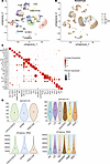 Figure 2
Single-cell data. (A) UMAP showing the annotated cell types from the integrated analysis of a total of 119,193 cells from 6 sample types: 20,000 cells each from LD (n = 9), TN (n = 9), DM-R (n = 18), early DKD (n = 9), and DKD (n = 17) and 19,193 cells from HC (n = 12). (B) UMAP of the integrated dataset showing successful integration of HC, LD, TN, DM-R, early DKD, and DKD. (C) Dot plot showing the specific markers for the annotated cell types. (D) Violin plots showing the quality control features: percentage of mitochondrial reads per cell (percent.mt) and number of features per cell (nFeature_RNA) in the 3 cohorts: CROCODILE/IMPROV-T2D, PRECISE, and KPMP and the 6 sample groups studied. The background color in the violin density plot of the sample groups indicates the cohort from which the samples were procured. POD, podocyte; PEC, parietal epithelial cell; PT, proximal tubule; aPT, adaptive/maladaptive; DTL, descending thin loop of Henle; ATL, ascending thin loop of Henle; TAL, thick ascending loop of Henle; aTAL, adaptive/maladaptive thick ascending loop of Henle; DCT, distal convoluted tubule; CNT, connecting tubule; PC, principal cell; IC-A, intercalated type A; IC-B, intercalated type B; tPC-IC, transient between PC and IC; EC, endothelial cell; EC-AEA, efferent and afferent arteriolar endothelial cells; EC-GC, glomerular endothelial cell; FIB, fibroblast; VSMC, vascular smooth muscle cell; P, pericyte; MC, mesangial cell; B, B cell; T, T cell; NK T/C, natural killer T cell.
Single-cell analysis of kidney tissue, compared with other tissues, is known to have a high mitochondrial read content (6). In this study, we included only cells with 50% or fewer reads of the 13 protein-coding mitochondrial genes. The proportion of reads mapping to mitochondrial genes within a cell serves as an essential quality control metric and identifies stressed and damaged cells. Figure 2D presents violin plots of commonly used single-cell quality control features: mitochondrial read percentage per cell and number of genes per cell. Quality control metrics were largely comparable across the 6 sample groups representing the 3 cohorts (CROCODILE, KPMP, and PRECISE).
Uniform manifold approximation and projection (UMAP) visualization from the integrated analysis of 54 10X Genomics Visium datasets — including 13 DKD, 15 DM-R, 4 early DKD, 5 HC, 15 LD, and 2 TN samples — is presented in Supplemental Figure 2A. The merged dataset comprised 45,739 spots. Given that only DM-R, DKD, and LD groups had sufficient sample numbers, differential expression analyses with and without adjustment for confounders were conducted exclusively on these cohorts. Supplemental Figure 2B displays UMAPs for these 3 sample groups.
Early stress and injury/disease markers
Early stress response gene expression can be activated within minutes after stimulation (7, 8) and can serve as a sensitive biomarker of cellular stress. Activating transcription factor 3 (ATF3), dual specificity phosphatase 1 (DUSP1), FK506-binding protein 5 (FKBP5), fos proto-oncogene (FOS), JUNB, and JUN were lowest in HC and highest in TN among the reference tissues (Figure 3A). Both the LD and TN groups had higher expressions of these genes than the DM-R and early DKD groups.
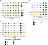 Figure 3
Early stress response and injury/disease markers. (A) Dot plot illustrating scaled expression of early stressor genes ATF3, DUSP1, FKBP5, FOS, JUNB, and JUN across 6 sample types. Expression was highest in TN and LD samples. (B) Dot plot showing expression of early CKD/injury markers in 6 sample types. Among reference groups, HC exhibited the lowest levels of HAVCR1, LCN2, and IL18. (C) Dot plot displaying expression of progressive/late disease markers across 6 sample types. Reference groups had relatively low expression compared with DKD, with little difference observed among reference groups.
In addition to the nonspecific stress markers studied above, kidney tissue–specific markers of cellular stress and damage have been developed and were assessed next. The expression levels of the following early kidney injury/disease markers are shown in Figure 3B: α-2 macroglobulin (A2M), hepatitis A virus cellular receptor 1 (HAVCR1, encoding kidney injury marker 1, KIM-1), IL-18 (IL18), lipocalin-2 (LCN2, encoding NGAL) and N-acetylglutamate synthase (NAGS). Among the reference groups, HC had the lowest expression for most of these genes. Although expressed at low levels, HAVCR1 and NAGS markers of kidney damage were detected in greater percentage of cells with higher expression in LD compared with the other 2 reference groups (Figure 3B). IL18, an early indicator of acute kidney injury, showed relatively high expression levels in the TN group. No expression of LCN2, another early AKI marker, was observed in any of the reference groups.
Finally, chronic kidney disease (CKD) markers of chronic tissue stress and damage were evaluated. No differences in the late CKD markers, including CXCL2 (CCL2), collagen A1 (COL1A1), cystatin C (CST3), MMP7 (MMP7), TGF-β1 (TGFB1), TNF (TNF), and VCAM1, were observed among the reference tissue groups (Figure 3C).
Cell type proportion. Figure 4A and Supplemental Figure 3 illustrate the proportions of cell types across sample groups. Relative to DKD, the reference groups exhibited lower proportions (adjusted P < 0.05) of aPT, aTAL, lymphoid, and myeloid cells, as shown in the heatmap (Figure 4B) generated from mean cell type proportions. The observed heterogeneity among aPT, aTAL, and immune cells is consistent with findings from recent single-cell studies (1, 9, 10); for example, only a very small B cell population was found in HC compared with LD and TN (Figure 4C).
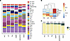 Figure 4
Immune cells and tubular epithelial cell states. (A) Proportion of cell types found in each of the 6 sample types: HC (n = 12), LD (n = 9), TN (n = 9), DM-R (n = 18), early DKD (n = 9), DKD (n = 17). (B) The mean proportion of PT, aPT, TAL1, TAL2, aTAL, lymphoid, and myeloid cells per sample groups studied is shown in the heatmap. The scaled expression levels show the most expression in DKD compared with reference groups. (C) The proportions of T and B cells within the lymphoid cell population were assessed across 6 sample groups. B cell proportion was lowest in the HC group and highest in the DKD group.
Effect of postoperative tissue procurement (preprocessing effect). Differential expression analysis between the postoperative procured sample types (LD and TN) to the percutaneous biopsy procurement (HC, all DKD samples), along with manual curation, identified a set of 25 genes (Supplemental Table 2) that exhibited significantly elevated expression in the postoperative reference samples (Figure 5, A and B). These genes were expressed in all cell types (Figure 5C). Network analysis using STRING (11) revealed direct interactions among 19 of these 25 genes (Figure 5D), with enrichment in the JNK and p38 MAPK pathways (Figure 5E). Notably, all LD and TN samples showed higher expression (adjusted P < 0.05) of the gene set score (computed from the 25 genes), with little intersample variability, compared with percutaneous biopsy tissues from HC and diabetic samples (Figure 5B). In the Visium integrated data, we observed high levels of preprocessing effects in the proximal cells of LD samples compared with that of DM-R and DKD samples (Figure 5F).
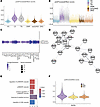 Figure 5
Preprocess effect. Using pseudo-bulk mRNA analysis, a gene set that was upregulated in postoperative tissue procurement method compared with percutaneous needle biopsies was identified. LD (n = 9) and TN (n = 9) samples were acquired by postoperative biopsy procedure and HC (n = 12), DM-R (n = 18), early DKD (n = 9), and DKD (n = 17) samples were by percutaneous needle biopsies. (A) Violin density plot showing the score calculated at cell level for the 25 genes that were upregulated in postoperative tissue biopsies. (B) Violin plot showing the elevated expression of the score in LD and TN samples compared with needle biopsy samples. (C) Dot plot showing the expression of preprocess effect score calculated based on the expression of the 25 genes in the cell types identified. (D) String interaction network showing direct interaction of 19/25 genes. (E) Top 5 enriched Gene Ontology biological processes for the gene set. (F) Violin plot showing preprocess effect score in Visium data from LD (n = 15), DM-R (n = 15), and DKD (n = 13).
Age effect. Figure 6A shows the age distribution per sample group. The HC and early DKD groups were considerably younger than all other sample groups. Using Pearson’s correlation analysis of pseudo-bulk read counts from both PT and aPT cells across 27 LD samples (in addition to 9 LD samples used in the integrated dataset, we used 18 LD samples from the Transplant Transcriptomic Atlas transplant study cohort, resulting in 27 samples in this analysis) (Supplemental Table 3), we identified 23 genes positively associated and 30 genes negatively associated with participant age (P value < 0.05) (Supplemental Table 4). Figure 6, B and C, show positive and negative correlations between age and mean sample-level Age_Gene_Scores (positive: P < 0.00001; negative: P < 0.0002). Cell-level Age_Gene_Scores were calculated from single-cell mRNA expression data for these age-correlated genes (Figure 6D). Genes positively correlated with age were enriched in healthy PT cells, and genes inversely correlated with age were more highly enriched in aPT cells. Significant (adjusted P < 0.05) positively and negatively enriched Hallmark pathways for the age-correlated genes are shown in Figure 6E. MTORC1 signaling, TNFA signaling via NF-κB, the p53 pathway, apoptosis, and peroxisome were significantly enriched for positive age-correlated genes. IFN-γ and allograft rejection were enriched pathways for the negative age-correlated genes.
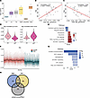 Figure 6
Effect of age and sex. (A) Bar plot showing the age distribution in each of the 6 sample groups: HC (n = 12), LD (n = 9), TN (n = 9), DM-R (n = 18), early DKD (n = 9), and DKD (n = 17). Early DKD and HC groups were much younger compared with other groups. (B) Plot showing the significant correlation between the scores from the positively age-associated genes and age of the living donors. (C) Plot showing the correlation between the scores from the negatively age-associated genes and age of the living donors. (D) Violin density plot showing the module scores calculated for the genes expressed in the proximal cells that are positively and negatively associated with age. The gene scores from the negatively associated genes were enriched in aPT cells, and the gene scores from the positively associated genes were higher in the PT cell state. (E) Top significantly enriched Hallmark MsigDB pathways for the age-associated gene set. (F) Violin plot illustrating higher expression scores, derived from significantly upregulated genes, in females compared with males across all 74 samples. (G) Top significantly enriched Hallmark MsigDB pathways for the sex-associated gene set. (H) The Venn diagram shows no overlap between gene sets associated with age, preprocess effect, and sex.
Sex effect. Differential expression analysis of samples from the 43 female and 31 male participants in the single-cell cohort, adjusted for age and preprocessing effects, identified 29 genes (adjusted P < 0.05) significantly enriched in females (Figure 6F and Supplemental Table 5). Gene scores calculated from the mRNA expression of these genes were consistently higher in samples from females versus males. Hallmark pathway enrichment analysis indicated that oxidative phosphorylation and MYC target pathways were significantly negatively enriched (adjusted P < 0.05) (Figure 6G). Figure 6H presents a Venn diagram that highlights the lack of overlap among genes correlated with preprocessing effects, age, and sex.
Pathway analysis. Hierarchical clustering of aggregated pseudo read counts at the sample group level without adjusting for confounding factors is shown in Figure 7A. After correcting for the confounding age, sex, and preprocess effect factors, the 2 kidney disease groups, DKD and early DKD, were grouped together, and DM-R clustered with the reference groups.
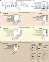 Figure 7
Analysis of the top pathways. (A) Hierarchical clustering on the right generated using aggregate read counts at the sample type level. Hierarchical clustering on the left utilized aggregate read counts with batch effect correction applied via the edgeR package. For HC, early DKD, DM-R, and DKD, age and sex were used as the covariate in batch effect correction; for TN and LD, age, process effect score, and sex served as covariates. (B) Plot displaying the top significant MsigDB Hallmark pathways (adjusted P < 0.05) associated with differentially expressed genes among the 3 reference groups. (C) DKD versus LD and DKD versus TN. DKD versus HC comparison was excluded due to nonoverlapping age distributions between these groups. Differential expression analysis was performed using DESeq2, both with and without adjustment for confounding variables: age, preprocess effect, and sex were included for DKD versus LD and DKD versus TN analyses. (D) DKD (n = 13) and LD (n = 15) in Visium data. The observed trends were consistent with those from single-cell analyses. (E) DM-R versus each of the 3 reference groups. Age and sex were used as the confounders for the DM-R versus HC comparison; age, sex, and preprocess effect were included for DM-R versus LD and DM-R versus TN. (F) DM-R (n = 15) versus LD (n = 15) in Visium data. (G) Early DKD versus HC. Comparisons of early DKD with other reference groups were excluded due to the absence of overlapping age distribution in these groups. Age and sex were used as the confounders for the early DKD versus HC comparison. (H) Violin plots depict pathway scores for selected MsigDB pathways, calculated from genes differentially expressed (P < 0.05) between early DKD (n = 4) and HC (n = 5) samples in Visium transcriptomic data.
Pathway enrichment based on differential gene expression of proximal cells among reference samples. To characterize the molecular programs specific to different reference tissues, we performed pathway enrichment analysis on genes differentially expressed among the 3 reference groups. Differential expression analyses were conducted both with and without adjustments for age, sex, and preprocess effect. Figure 7B presents a heatmap of pathway scores for the top Hallmark pathways (adjusted P < 0.05) from the Molecular Signatures Database (MsigDB) across binary group comparisons.
After adjusting for confounding factors, oxidative phosphorylation and fatty acid metabolism were enriched in the HC group compared with the LD and TN groups (Figure 7B). There was no significant difference in fatty acid metabolism between the LD and TN groups, whereas there was significant reduction (adjusted P < 0.05) of oxidative phosphorylation in the LD group compared with the TN group (Figure 7B). Both LD and TN samples, when compared with HC, showed enrichment in apoptosis, epithelial-mesenchymal transition (EMT), TNFA signaling via NF-κB, and IL-2/STAT5 signaling pathways — even after adjustment. In the TN group compared with both LD and HC groups, pathways such as EMT, TNFA signaling via NF-κB, and IFN-γ response were enriched.
Overall, these pathway findings closely align with individual stress marker profiles, demonstrating the highest tissue differentiation and lowest stress/inflammation in HC samples, with stress and injury pathways observed in LD biopsies and, more prominently, in TN samples.
Pathway enrichment based on differential gene expression analysis of proximal cells between reference groups and DKD samples. We next assessed how the choice of reference tissue influenced the enrichment of pathways shown in Figure 7B in disease versus reference comparisons.
Figure 7C illustrates significant (adjusted P < 0.05) Hallmark pathway enrichment for genes differentially expressed in DKD compared with reference groups (LD and TN). We excluded the DKD versus HC comparison due to the lack of age overlap between these groups. Oxidative phosphorylation and fatty acid metabolism were consistently lower in the DKD group versus the TN group, regardless of adjustment for confounding factors; however, this difference was not observed for oxidative phosphorylation between the DKD and LD groups after confounder adjustment. EMT and IFN-γ response were enriched in the DKD group relative to both reference groups, and TNFA signaling via NF-κB was reduced in the DKD group compared with the LD and TN groups, highlighting the influence of reference tissue selection on interpreting key inflammatory pathways in DKD (Figure 7C).
Figure 7D displays pathway enrichment analysis using Visium data highlighting significant (adjusted P < 0.05) differences between the DKD and LD groups, both before and after adjustment for confounding variables. Pathway enrichment patterns were consistent across both adjusted and unadjusted analyses in single-cell and Visium datasets. IFN-γ response remained elevated in the DKD group, corroborating findings from the single-cell data. TNFA signaling via NF-κB showed significant reduction in the DKD compared with LD group in both adjusted and unadjusted analyses (Figure 7D and Supplemental Figure 4A). Notably, differences in oxidative phosphorylation and fatty acid metabolism between the DKD and LD groups were diminished when confounding variables were considered.
Figure 7E illustrates significant (adjusted P < 0.05) Hallmark pathway enrichment for differentially expressed genes between the DM-R and reference groups (HC, LD, and TN). After adjusting for confounders, oxidative phosphorylation was significantly higher in DM-R than in LD. IFN-γ response enrichment in DM-R versus HC was no longer evident after adjusting for confounding factors, age, and sex. IL-2/STAT5 and TNFA signaling via NF-κB remained consistently lower in the DM-R group compared with LD and TN groups, regardless of adjustments. Results from the analysis of DM-R versus LD using Visium data further support the observations from single-cell data (Figure 7F, Supplemental Figure 3C, and Supplemental Figure 4B). Interestingly, the aPT cell state appeared to exhibit higher oxidative phosphorylation than healthy PT cells (Supplemental Figure 3C). Apoptosis, EMT, and IFN-γ response pathways were enriched in the TN group compared with other reference groups (Supplemental Figure 5) using Visium data.
Figure 7G displays the hallmark pathway enrichment for genes differentially expressed between the early DKD and HC groups. We excluded comparisons with other reference groups since there was no age overlap between early DKD and these groups. All selected pathways showed significantly higher levels (adjusted P < 0.05) in early DKD compared with HC. Violin density plots of the Visium data also indicated higher activity of these pathways in early DKD than in HC, though this finding should be interpreted cautiously given the small sample sizes in both groups. No-overlap between age, gene and preprocess effect were observed (Figure 7H).
-
Methods
Sex as a biological variable. Both male and female participants who were enrolled in all the cohorts were included in the analyses.
Human data. Key clinical and demographic characteristics of participants who contributed kidney biopsy samples in the 3 reference and 3 diabetes-related groups are summarized in Table 1. For this study, we have used data from kidney tissue collected by the Kidney Precision Medicine Project (KPMP, see https://www.kpmp.org for details), which focuses on molecular mechanisms in acute kidney injury and CKD. From the KPMP cohort, 9 LD, 17 DKD, and 18 DM-R biopsy samples were used in this study. All KPMP biopsy samples were procured at multiple repositories using a standardized protocol (30).
The TN samples comprised tumor-free kidney cortical tissue from nephrectomy specimens rapidly sourced in the operating room for molecular analyses in the PRECISE cohort, University of Michigan (31). We selected 9 TN samples from the PRECISE cohort with minimal glomerular pathology, characterized by low segmental glomerular sclerosis, global glomerular sclerosis, and imploding glomeruli. All TN samples were from participants without diabetes, with eGFR values greater than 80, and serum creatinine levels of 0.85 ± 0.15.
The 9 early DKD samples were from the Impact of Metabolic Surgery on Pancreatic, Renal and Cardiovascular Health in Youth with Type 2 Diabetes (IMPROVE-T2D) study and the Renal Hemodynamics, Energetics and Insulin Resistance in Youth Onset Type 2 Diabetes Study (Renal HEIR) and 12 control samples from research kidney biopsies performed on healthy volunteers in the Control of Renal Oxygen Consumption, Mitochondrial Dysfunction, and Insulin Resistance study (CROCODILE), as previously described (24).
HC samples and all disease biopsy samples used in the study were collected via percutaneous research needle biopsies. LD and TN samples were obtained through postoperative surgical biopsies, with LD biopsies performed on the explanted kidney before implantation into the transplant recipient.
For identifying genes associated with age using correlation analysis, we used single-cell data from tissue obtained from living donors. In addition to the 9 KPMP LD samples, we included 18 samples from the University of Michigan living donor cohort generated for the Human Kidney Transplant Transcriptomic Atlas. Immediately after procurement, tissue was transferred to a CryoStor and stored in liquid nitrogen.
Single-cell data generation. As a KPMP tissue interrogation site, the University of Michigan generates single-cell data from KPMP biopsy samples, which are procured at multiple repositories using a standardized protocol before being sent to Michigan. Non-KPMP datasets used in this study — including PRECISE, University of Michigan Transplant LD, and CROCODILE/ IMPROVE-T2D cohort samples — were also processed at the University of Michigan using the same protocol (https://doi.org/10.17504/protocols.io.7dthi6n) as KPMP samples. Briefly, single-cell dissociation was carried out using Liberase TL at 37°C, as described in the detailed protocol (dx.doi.org/10.17504/protocols.io.7dthi6n). The resulting single-cell suspension was immediately transferred to the University of Michigan Advanced Genomics Core for further processing. Single-cell RNA-seq was performed using 10X Genomics technology. Sequence files were aligned to the reference genome (GRCh38/hg38), and feature-barcode matrices were generated using the Cell Ranger software included in the 10X Genomics analysis suite.
Single-cell data analysis. The read count matrices were initially processed using SoupX (v1.5.0) to correct for ambient mRNA contamination. Data processing was performed using the Seurat R package (version 5.0). Quality control was enforced with cutoffs of greater than 500 and less than 5,000 genes per cell, and less than 50% mitochondrial reads per cell. Each sample group underwent normalization, variable gene identification, and principal component analysis (PCA) independently. Subsequently, the integration of all 6 sample types was achieved via reciprocal PCA (RPCA). Dimensionality reduction using UMAP and unsupervised clustering (resolution 0.4) were conducted on the integrated dataset. Cell clusters were annotated based on markers of major nephron segments, as well as endothelial, interstitial, and immune cell types. Further analyses were performed on a down-sampled integrated object containing 20,000 randomly selected cells from each sample type and the integrated dataset containing a total of 119,193 cells from all 6 sample types together.
Preprocess effect analysis. The gene set upregulated in postoperative sample types (LD and TN) compared with other groups (HC, DM-R, early DKD, and DKD) was identified via differential expression analysis using edgeR (32) on pseudo-bulk RNA read counts. The final gene set was selected after manually confirming overexpression across all samples in LD and TN groups to avoid sample-to-sample variation. The interaction network of this gene set was constructed using STRING (11) and visualized as a network in Cytoscape (33). Gene Ontology biological process enrichment (34) was performed on the preprocess effect gene set.
Age effect. To evaluate the impact of age within a reference group while minimizing confounding factors, we performed a correlation analysis between gene expression and donor age using 27 LD samples, as the HC and TN groups were underpowered. In addition to the 9 LD samples used in the integrated dataset, we used additional 18 LD samples from Transplant Transcriptomic Atlas transplant study cohort. Supplemental Table 3 provides age information for all samples used in the correlation analysis. We identified the top 2,500 highly variable genes in proximal cells from normalized pseudo read counts (aggregated by sample and processed with edgeR (32) and assessed their correlation with age using Pearson’s correlation test. For the positively and negatively age-correlated genes, we calculated an “Age_Gene_Score” in the integrated Seurat object using the AddModuleScore function.
Sex effect. To identify a set of genes that are specific to a sex, we performed a differential expression analysis on the pseudo read counts from the proximal cells from 43 female versus 31 male participants. The DESeq2 R package was used for analysis. Age and preprocess scores were used as confounding variables.
Adjusting for age, sex, and preprocess effect in differential expression analyses. Differential expression analyses were performed between the sample groups using the DESeq2 R package (35) on pseudo-bulk mRNA counts, generated by aggregating raw read counts from proximal cells for each sample. The analyses we conducted were both unadjusted and adjusted for age, sex, and preprocess effect as confounding factors. To account for a preprocess effect, we calculated a composite score by averaging z scores of 25 genes positively associated with postoperative tissue procurement. Participant age and sex at biopsy were included as covariates in the adjusted models.
Pathway enrichment analysis. Pathway activity enrichment was assessed using decoupleR (36) on the 2-tailed Wald-statistic values from the above-mentioned differential expression analysis of aggregated pseudo read counts from proximal cells of sample groups. Hallmark pathways from MsigDB were used as the resource for enrichment analysis. Pathways with enrichment adjusted P values below 0.05 were deemed significant.
Pathway enrichment analysis was initially conducted on differential expression results from the reference group comparisons. The most significantly enriched pathways (adjusted P < 0.05) were identified and subsequently assessed for enrichment in disease versus reference group comparisons.
Validation of pathway activity using Visium transcriptomic data. To assess pathway enrichment of differentially expressed genes between disease and reference groups, we analyzed transcriptomic datasets generated using 10X Genomics Visium technology. Data for LD, TN, DM-R, and DKD samples were obtained from the KPMP repository (https://atlas.kpmp.org/repository/), and HC and early DKD datasets were generated locally for an unrelated project. Human kidney tissue was prepared and imaged according to the Visium Spatial Gene Expression (10X Genomics) manufacturer’s protocol (CG000240, Visium Tissue Preparation Guide) and as previously described (1, 37).
There were 15 samples each for LD and DM-R, 13 for DKD, 5 for HC, 4 for early DKD, and 2 for TN. Because of the limited sample sizes in the HC, early DKD, and TN groups, we excluded these groups from pathway enrichment validation analyses. Instead, these groups were utilized solely for generating density plots for selected pathway gene sets.
Visium data integration was performed using Seurat. Briefly, each dataset underwent Single-Cell Transform (SCT) normalization, followed by dimensionality reduction with PCA. Integration anchors were identified using RPCA, and the datasets were merged into a single integrated object. Additional dimensionality reduction was then performed on the integrated object using both PCA and UMAP. Spot annotation was carried out with Seurat’s label transfer method, using the integrated and down-sampled single-cell dataset as the reference. For the validation of the observations from the proximal single-cell data, we focused on the Visium spots annotated as proximal (PT and aPT).
Statistics. To identify preprocess effect genes, we first used Seurat’s FindMarkers function to detect genes that were differentially expressed in LD and TN compared with other groups, applying an adjusted P value threshold of less than 0.05. Next, edgeR was performed on the pseudo-bulk read counts of these significant genes. Genes with a P value less than 0.05 and a log fold-change greater than 0 were then manually assessed and classified as preprocess effect genes.
Pathway activity enrichment was assessed using decoupleR on genes with significant differential expression (adjusted P < 0.05, using the Benjamini-Hochberg method), as identified by DESeq2 from aggregated pseudo read counts comparing proximal cells in the reference and disease groups. Pathways with enrichment adjusted P value less than 0.05 were considered significant.
Study approval. All samples used in this study were obtained with patient consent and with the approval of IRBs of participating institutions, described below. The KPMP study is reviewed and approved under a protocol by the KPMP single IRB of the University of Washington (IRB 20190213). The PRECISE cohort and the Human Kidney Transplant Transcriptomic Atlas studies were approved by the IRB of the University of Michigan. CROCODILE, Renal HEIR, and IMPROVE-T2D were approved by the Colorado Multiple IRB. Written informed consent was received from each participant and/or their parents, as appropriate for age, prior to enrollment.
Data availability. The count matrices of all KPMP samples used in this study are available from https://atlas.kpmp.org/repository/ The single-cell count matrices for the Human Kidney Transplant Transcriptomic Atlas are available from NCBI’s Gene Expression Omnibus (GEO GSE169285). Healthy control and early chronic disease sample read count matrices are available at GEO GSE220939 and GSE279086. The tumor nephrectomy data are at https://cellxgene.cziscience.com/collections/a98b828a-622a-483a-80e0-15703678befd Please see the Supporting Data Values file for values used to generate the figures in the main text and the supplemental figures.


